Acute respiratory distress syndrome (ARDS)
Noncardiogenic acute hypoxaemic respiratory failure: definitions, Berlin and 2023 global criteria, imaging, ventilation and outcomes.
- Acute respiratory distress syndrome (ARDS) is a clinical syndrome of acute, diffuse, inflammatory lung injury that produces noncardiogenic pulmonary oedema, severe hypoxaemia and stiff lungs, usually within a week of an insult such as pneumonia, sepsis or trauma.
- The current bedside definition is the Berlin definition: acute onset within one week, bilateral opacities not explained by cardiac failure, and a PaO2/FiO2 of 300 or less measured with at least 5 cm H2O of positive end-expiratory pressure (PEEP), with severity mild, moderate or severe at thresholds of 300, 200 and 100.
- A newer 2023 global consensus extends this to patients on high-flow nasal oxygen (at least 30 litres per minute) or noninvasive ventilation with 5 cm H2O, accepts pulse oximetry when saturation is 97% or less, adds ultrasound, and provides modified criteria that do not require PEEP or blood gases for resource-limited settings.
- The histological prototype is diffuse alveolar damage, which moves through overlapping exudative, proliferative and fibrotic phases; radiology follows the same time course from diffuse ground-glass to organising change.
- Care is supportive and lung-protective: low tidal volume (about 6 mL/kg of predicted body weight), plateau pressure under 30 cm H2O, judicious PEEP, a conservative fluid strategy, prone positioning for at least 12 to 16 hours in moderate to severe disease, early neuromuscular blockade in severe disease, systemic corticosteroids and, in selected patients, venovenous extracorporeal membrane oxygenation (VV-ECMO).
By the end of this page you should be able to:
- State the conceptual model of ARDS and explain why it is a syndrome rather than a single disease.
- Apply the Berlin definition and the 2023 global definition, including the oxygenation cut-offs, timing and imaging criteria, and the resource-limited modification.
- Link common risk factors to the mechanisms of diffuse alveolar damage and its exudative, proliferative and fibrotic phases.
- Interpret chest radiograph and computed tomography (CT) findings at different stages of ARDS and distinguish ARDS from cardiogenic oedema and diffuse alveolar haemorrhage.
- Plan a ventilatory and supportive strategy for a patient with ARDS, including tidal volume, plateau pressure, PEEP, fluids, prone positioning, neuromuscular blockade, corticosteroids and ECMO selection.
- State the complications and prognosis of ARDS and explain how care differs in low-resource settings.
DefinitionAcute respiratory distress syndrome (ARDS)
ARDS is a clinical syndrome of acute, diffuse, inflammatory lung injury that is precipitated by a predisposing risk factor such as pneumonia, nonpulmonary infection, trauma, transfusion, burn, aspiration or shock. The injury increases pulmonary vascular and alveolar epithelial permeability, producing noncardiogenic pulmonary oedema and gravity-dependent atelectasis, and presents with arterial hypoxaemia and bilateral radiographic opacities, increased shunting, increased alveolar dead space and decreased lung compliance.
ARDS is deliberately defined as a syndrome. Many different insults converge on the same bedside picture, so the diagnosis describes a final common pathway, not a single disease.[1][5] Histological findings are variable: intra-alveolar oedema, inflammation, hyaline membrane formation and alveolar haemorrhage, often together called diffuse alveolar damage, but these features are not always present and are not required to make the clinical diagnosis.[1]
The Berlin definition is the current standard used in most critical care research and in high-resource practice.[4][3] It requires:
- Timing. Acute onset or worsening of hypoxaemic respiratory failure within one week of the predisposing insult or of new or worsening respiratory symptoms.
- Imaging. Bilateral opacities on chest radiograph or CT that are not fully explained by pleural effusion, lobar or lung collapse, or nodules or masses. Either modality may be used.
- Origin of the oedema. Respiratory failure not fully explained by cardiac failure or fluid overload. A formal wedge pressure is no longer required; clinical assessment, and echocardiography if no risk factor is apparent, is accepted.
- Oxygenation. A PaO2/FiO2 of 300 or less measured with at least 5 cm H2O of PEEP (or continuous positive airway pressure, CPAP). If no ARDS risk factor is present, cardiogenic oedema must be excluded objectively, for example with echocardiography.
Severity is then classified by the PaO2/FiO2 measured with PEEP:
| Severity | PaO2/FiO2 |
|---|---|
| Mild | Greater than 200 and up to 300 |
| Moderate | Greater than 100 and up to 200 |
| Severe | 100 or less |
The Berlin definition replaced the earlier American-European Consensus Conference (AECC) definition and removed the old umbrella term "acute lung injury." It made "acute" mean seven days or less, made bilateral opacities necessary and explicit, and dropped the pulmonary capillary wedge pressure cut-off in favour of a clinical assessment.[4] Its predictive value for mortality is modest (area under the curve 0.577), but it is feasible and reliable at the bedside.[4]
A global consensus conference in 2023 built on the Berlin definition to incorporate changes in practice and to make the diagnosis usable in resource-limited settings.[1] The same core elements were retained: an acute onset within one week, bilateral opacities not fully explained by effusion, atelectasis or nodules or masses, and noncardiogenic pulmonary oedema. Three categories were then created:
- Intubated ARDS retains the Berlin severity categories (mild 200 to 300, moderate 100 to 200, severe 100 or less) and still requires at least 5 cm H2O PEEP for all categories.
- Non-intubated ARDS includes patients on high-flow nasal oxygen (HFNO) at flow rates of at least 30 litres per minute, or on noninvasive ventilation or CPAP with at least 5 cm H2O end-expiratory pressure, when the PaO2/FiO2 is 300 or less.
- ARDS in resource-limited settings uses the Kigali modification (see Low-resource settings), with neither PEEP nor a minimum oxygen flow rate required.
Pulse oximetry (SpO2/FiO2 315 or less when saturation is 97% or less) may stand in for an arterial blood gas in all three categories, and lung ultrasound may replace chest radiography or CT when the operator is trained to recognise the loss of lung aeration.[1]
Clinical pearlWhy the 2023 definition matters
Up to half of patients with mild ARDS and a fifth of those with severe ARDS were not recognised under the Berlin definition in the LUNG SAFE study. Extending the definition to high-flow nasal oxygen and to resource-limited settings captures patients earlier and lets trials of early treatment enrol them.[1][2]
ARDS is common in intensive care but often under-recognised. LUNG SAFE found that patients with ARDS account for about 10% of intensive care admissions, and about 3 million cases occur worldwide each year.[3] Reported incidence varies widely with region and healthcare system, from about 7 cases per 100,000 person-years in Spain to about 79 in United States cohorts.[4] In a 50-country observational study the distribution of severity was roughly one quarter mild, half moderate and one quarter severe, with a stepwise rise in mortality from 27% in mild to 45% in severe disease.[7] In the United States the annual burden is roughly 200,000 cases and 74,500 deaths.[3] Risk is not uniform. Nonmodifiable risk factors include alcohol abuse, obesity and admission severity of illness; potentially modifiable ones include the volume of red blood cell transfusion, hypoproteinaemia, delayed resuscitation in shock and inadequate antibiotics in septic shock.[4] Diabetic patients appear to have about half the risk of at-risk non-diabetic patients.[4] About 10% of patients admitted with a predisposing condition develop ARDS, from 2.7% of those with pancreatitis to 27% of those with smoke inhalation.[4]
ARDS follows direct (pulmonary) injury or indirect (systemic) injury. Direct insults attack the alveolar epithelium first; indirect insults usually injure the pulmonary endothelium first, so the radiographic and pathophysiological patterns differ.
| Direct (pulmonary) | Indirect (extrapulmonary) |
|---|---|
| Pneumonia - bacterial, viral (including SARS-CoV-2) and fungal | Sepsis |
| Aspiration of gastric contents or drowning | Non-thoracic major trauma |
| Lung contusion | Pancreatitis |
| Toxic inhalation | Cardiopulmonary bypass |
| Vasculitis | Transfusion of blood products |
| Pulmonary burns | Major burns injury |
The leading specific causes are therefore sepsis, pneumonia, aspiration, trauma, pancreatitis, transfusion, drowning and, since 2020, COVID-19. These map one-to-one onto the predisposing risk factors listed in both the Berlin and the global definitions.[1][5][3]
Red flagDo not stop at the first insult
A predisposing risk factor must be present and new, but ARDS is often multifactorial. Obesity, shock, hypoproteinaemia and large transfusions all add to risk, and delayed antibiotics or inadequate resuscitation convert a vulnerable patient into one with ARDS.
The central process is an acute inflammatory injury that damages the alveolar-capillary barrier. Severe inflammation in the lung, systemically, or both, releases mediators that injure the epithelium, the endothelium and the extracellular matrix. The results are alveolar flooding, interstitial oedema with increased extravascular lung water, and thromboembolic obstruction of pulmonary microvessels. At the bedside these become hypoxaemia, loss of compliance, increased dead space and the radiological opacities of ARDS.[5]
More specifically, the insult activates alveolar macrophages, which release pro-inflammatory cytokines such as TNF, IL-1, IL-6 and IL-8. These attract neutrophils, which damage the alveolar and capillary epithelium by releasing toxic mediators. The alveoli then fill with bloody, protein-rich fluid and surfactant can no longer keep them open.[3]
Four "prototype" injuries are useful for reasoning about individual patients. Epithelial-dominant injury, typical of pneumonia or aspiration, floods the alveoli directly. Endothelial-dominant injury, typical of sepsis, produces interstitial oedema and increased extravascular lung water. Extracellular-matrix-dominant injury, typical of ventilator-induced lung injury, perpetuates inflammatory signalling from stressed matrix. Coagulopathy-dominant injury, typical of COVID-19, is driven by microthrombi in pulmonary microvessels.[5]
The histological prototype of ARDS is diffuse alveolar damage (DAD). About half of patients who meet the clinical definition of ARDS have DAD on biopsy or autopsy, and DAD is more common in severe disease, so the clinical diagnosis is a proxy for the pathology rather than the pathology itself.[5][6] Where DAD is present, its changes run through three overlapping phases.
Exudative (early) phase. This is the acute inflammatory stage, most prominent in the first week. Interstitial and intra-alveolar oedema, haemorrhage and fibrin deposition appear first. Hyaline membranes - the hallmark of this phase - peak at three to seven days. Neutrophilic infiltrates, fibrin thrombi in small pulmonary arteries and minimal interstitial inflammation are present. Type II pneumocyte hyperplasia begins late in this phase. Respiratory failure here is driven by protein-rich fluid in distal airspaces and by loss of surfactant production from type II cells.[4][6]
Proliferative (subacute) phase. This begins about one week after injury and is dominated by repair: proliferation of type II pneumocytes, fibroblast infiltration into the interstitium and alveolar spaces, early fibrotic change and myointimal thickening of alveolar capillaries.[4][6] Interstitial mononuclear infiltrates are prominent. Hyaline membranes are resorbed. Squamous metaplasia of terminal airways may occur and can look alarming, even mimicking malignancy.
Fibrotic (late) phase. In a subset of patients repair fails and fibroproliferation dominates, with collagen deposition, macrophage infiltration and resolution of the exudative phase. This produces reduced compliance, worsening ventilation-perfusion mismatch and, in some cases, honeycomb remodelling.[5][6] Some lungs resolve almost completely, which is why lung function usually recovers toward baseline within a year in survivors.[4]
Overlap between phases is considerable: a biopsy may show exudation in one area and organisation in another, because different lung regions are injured at different times.[6]
The chest radiograph and CT reflect the same timeline. Plain film shows bilateral, often symmetric, diffuse opacities that are not explained by effusions or collapse. CT separates three morphological patterns: a focal basal-dorsal pattern of consolidation, a patchy pattern of consolidation and ground-glass separated by spared areas, and a diffuse pattern involving all lobes without a gradient.[5] Patchy and diffuse patterns are usually grouped as a nonfocal phenotype, which is easier to recruit and less prone to overdistension than the focal phenotype. CT also measures gas volumes and condensed tissue weight accurately, and it is the gold standard for recruiting assessment, although it is not required to make the diagnosis.[5]
Lung ultrasound shows multiple bilateral B lines and consolidations and correlates with extravascular lung water; it is especially valuable when CT or radiography is unavailable, provided the operator is trained.[1][5]
Quantitative scoring is sometimes used. The Radiographic Assessment of Lung Edema (RALE) score sums consolidation and density for each quadrant to a maximum of 48 and has prognostic value, while the global lung ultrasound score tracks re-aeration after recruitment.[5]
The Berlin concept of onset reflects this heterogeneity. Some patients progress from mild to severe disease within 24 hours, while others follow a protracted course, as in COVID-19 ARDS. Most patients who start with mild ARDS later progress, and roughly one third of initially mild cases become moderate or severe.[4] Yet once the process is established, the underlying illness, not only the lung, determines outcome.
ARDS is usually recognised after an interval rather than at first contact. A predisposing condition is usually present, but the formal diagnostic threshold may be crossed well after admission.
The patient's illness progresses over hours. Symptoms are those of hypoxaemia and reduced compliance:
- Increasing breathlessness, initially on exertion and then at rest, often with tachypnoea and accessory muscle use.
- Reduced oxygen saturation that rises little or not at all with a higher concentration of supplemental oxygen - the hallmark of shunt.
- Cough, sometimes with frothy sputum.
- Chest tightness or discomfort, without the pleuritic pain of a focal pneumonia.
Where the insult is systemic, the history is dominated by the sepsis, trauma or pancreatitis, and the lung failure is one component of a multisystem process.
- Tachypnoea, tachycardia and hypotension if shock is present.
- Cyanosis in severe cases.
- Diffuse, fine end-inspiratory crackles on auscultation, often most marked at the bases and lung periphery, without the focal bronchial breathing of lobar consolidation.
- Decreased chest wall excursion and increased work of breathing.
- Signs of the underlying trigger, such as an abdominal source of sepsis or multiple injuries.
Formal auscultation and percussion add little beyond confirming a diffuse process; the severity is better gauged by the ventilator settings needed to support oxygenation.
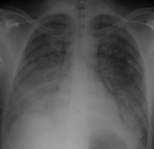
Figure 1Chest Xray 40 yr old male acute respiratory distress syndrome as a complication of murine typhus. 13-1421-F1
Long description
Chest X-ray of a 40-year-old man with ARDS (murine typhus): diffuse bilateral airspace opacities

Early films may look almost normal or show only subtle ground-glass change. Over 24 to 72 hours bilateral diffuse opacities predominate. In the minority who survive prolonged disease, late films show coarse reticular change and cystic remodelling. CT confirms the spared ventral lung, the dependent consolidation and, in chronic cases, fibrosis with traction bronchiectasis.
ARDS remains a clinical diagnosis; no investigation on its own confirms it.
- Arterial blood gas. Defines hypoxaemia precisely and provides the PaO2/FiO2. Measures of gas exchange, including alveolar dead space and the degree of shunt, belong here. Blood gas and oximetric measurements should be made at rest, at least 30 minutes after any change in position, FiO2 or flow rate.[1]
- Pulse oximetry with a reliable waveform can substitute for an arterial gas when saturation is 97% or less, using SpO2/FiO2 of 315 or less as the diagnostic threshold. It is not reliable in suspected methaemoglobinaemia or carboxyhaemoglobinaemia.[1]
- Chest radiograph (frontal) is the standard imaging modality.
- CT of the chest is reserved for resolving diagnostic doubt, characterising morphology or guiding an uncertain management decision. It is the gold standard for quantifying aerated and condensed lung tissue.
- Lung ultrasound identifies bilateral loss of aeration and suits bedside and resource-limited use, with the proviso of training.
- Echocardiography distinguishes cardiogenic oedema by showing left ventricular dysfunction, elevated filling pressures or valve disease, and it is used whenever no risk factor for ARDS is evident.
- Laboratory tests support the search for the trigger: blood cultures, sputum or lower respiratory sampling, screening tests for atypical infection, inflammatory markers and tests for the complications of shock.
- Bronchoscopy is reserved for detecting haemorrhage, infection or an obstructed airway when the diagnosis is uncertain. It changes management in 60% to 80% of selected cases but carries a complication rate of about 10% in critically ill patients.[4]
- Right heart catheterisation for pulmonary capillary wedge pressure is rarely needed now that echocardiography is widely available.
The commonest confusers are listed below. Each is separated by its own features, and several can coexist with genuine ARDS.
| Condition | Features that separate it from ARDS |
|---|---|
| Cardiogenic pulmonary oedema | Raised left atrial pressure: elevated B-type natriuretic peptide, systolic or diastolic dysfunction or valve disease on echocardiography, response to diuretics and nitrates, often a pre-existing cardiac history. ARDS is not excluded by a higher wedge pressure if a risk factor is present, but cardiogenic oedema as the sole cause keeps the patient out of the syndrome.[1][4] |
| Diffuse alveolar haemorrhage | Haemoptysis, a falling haemoglobin, diffuse bilateral infiltrates, progressively bloodier returns at bronchoscopy and haemosiderin-laden macrophages in bronchoalveolar lavage. It is a disease to treat, not a syndrome, and the distinction matters for immunosuppression. |
| Acute interstitial pneumonitis (Hamman-Rich) | A fulminant diffuse alveolar damage picture of unknown cause in a previously healthy person, with both acute and organising change on biopsy. |
| Acute eosinophilic pneumonia | Rapid respiratory failure with fever, diffuse infiltrates and eosinophilia in lavage or biopsy, and a prompt response to corticosteroids. |
| Cryptogenic organising pneumonia | Subacute course over weeks, patchy peripheral consolidations, and a contrasting response to steroids. |
| Metastatic or infective mimics | An obstructing tumour or airway disease, or unresolved infection, that explains the whole picture. |
ARDS mimics account for a substantial share of cases in clinical practice, and only about half of patients who meet the clinical criteria turn out to have diffuse alveolar damage at autopsy.[5][6]
The diagnostic criteria are those of the Berlin and global definitions above. Staging follows the PaO2/FiO2, with or without noninvasive support in the global definition. Scores commonly used alongside the diagnosis include:
- Lung Injury Prediction Score (LIPS). Predicts the development of ARDS in at-risk patients (area under the curve 0.80) from the insult and risk modifiers such as obesity, hypoproteinaemia and tachypnoea.[4]
- Sequential Organ Failure Assessment (SOFA). Quantifies the multisystem failure that usually drives death.
- Murray Lung Injury Score. Combines radiographic and physiological variables; it is sometimes used to select patients for ECMO.
- Driving pressure (plateau pressure minus PEEP) is not a diagnostic score, but values above 14 cm H2O are associated with increased mortality and can be used to guide settings.[7] Severity categories track prognosis: hospital mortality is about 27% in mild, 32% in moderate and 45% in severe ARDS, with an overall figure of about 40%.[4] Only 13% to 19% of patients die directly from refractory respiratory failure, usually from multisystem organ failure.[4]
The strategy has three parts: treat the cause; support oxygenation while doing as little further lung damage as possible; and, for the most hypoxaemic patients, escalate to prone positioning, pharmacological adjuncts and, in selected cases, ECMO.
Approach to a patient with confirmed ARDS
- Treat the causeAntibiotics for sepsis or pneumonia, drainage for an abdominal source, transfusion protocol, remove the precipitant
- Lung-protective ventilationTidal volume about 6 mL/kg predicted body weight (range 4 to 8), plateau pressure under 30 cm H2O, permissive hypercapnia as needed
- Set PEEPHigher rather than lower in moderate to severe disease; oxygenation-based or mechanics-based titration; no prolonged recruitment manoeuvres
- Question: Severity by PaO2/FiO2 and clinical course?
Mild: greater than 200 to 300
- Continue lung-protective ventilationJudicious lower PEEP; higher PEEP may trend to harm in mild disease
- Consider high-flow nasal oxygen or noninvasive supportAvoid intubation if safe and the patient is not failing
Moderate: greater than 100 to 200
- Higher PEEP without recruitment manoeuvresTitrate to oxygenation or compliance; monitor haemodynamics
- Systemic corticosteroidsEspecially within the first 14 days of moderate to severe disease
- Conservative fluid managementIf not in shock; target euvolaemia with a diuretic and reduced crystalloid
Severe: 100 or less
- Prone positioning at least 12 to 16 hours per dayStart within 36 hours of onset whenever possible
- Neuromuscular blockade for up to 48 hoursEarly severe disease, within 48 hours of onset, if dyssynchronous or difficult to ventilate
- Dexamethasone or methylprednisoloneEarly treatment shows reduced ventilation time and mortality in selected trials
- Consider VV-ECMORefractory hypoxaemia (PaO2/FiO2 under 80) or acidosis (pH under 7.25 with PaCO2 at least 60) despite optimal conventional care
- Reassess dailyAdjust settings to the least invasive effective level; screen for complications and non-pulmonary organ failure
- Airway and sedation. Intubate when hypoxaemia or ventilatory failure exceeds what noninvasive support can safely manage. Provide analgesia and sedation, aiming for the lightest effective level; deep sedation should not be routine.
- Oxygenation. Use the lowest FiO2 that keeps saturation at the target; avoid prolonged hyperoxia.
- Physiological targets. Keep oxygen saturation and carbon dioxide at the targets set in the local intensive care protocol, and tolerate permissive hypercapnia rather than abandon lung-protective settings.- Haemodynamics. Treat shock with the usual fluids and vasopressors first; only move to a conservative fluid strategy (below) once the patient is euvolaemic and off vasopressors.
| Goal | Setting to change | How, and the limit that applies |
|---|---|---|
| Raise oxygenation | Inspired oxygen (FiO2) | Use the lowest FiO2 that keeps saturation at target; avoid prolonged hyperoxia. |
| Raise oxygenation | PEEP | Higher PEEP in moderate to severe disease, titrated to oxygenation or compliance while plateau pressure stays under 30 cm H2O; it may trend to harm in mild disease.[2][7] |
| Lower PaCO2 | Respiratory rate | Raise it as far as is safe, for example when plateau pressure forces the tidal volume down, before accepting hypercapnia.[2][7] |
| Lower PaCO2 | Tidal volume | Not raised freely: about 6 mL/kg predicted body weight (range 4 to 8) with plateau pressure under 30 cm H2O; permissive hypercapnia is accepted instead.[2][7] |
Lung-protective ventilation. Mechanical ventilation is the backbone of care, but it is also the main iatrogenic threat. ARDS produces a "baby lung": only part of the lung is aerated and compliant, so a normal tidal volume overstretching that small lung causes ventilator-induced lung injury, while cyclic collapse and reopening tears injured units. Hence the strong recommendation to limit tidal volume (4 to 8 mL/kg of predicted body weight, targeting about 6) and plateau pressure (under 30 cm H2O).[2][4][7] This strategy reduced 28-day mortality from 38.8% to 31.0% in the ARDS Network trial, and the benefit is attributed to reduced systemic inflammation and fewer organ failures.[4]
The ARDS Network tidal volume regimen targets 6 mL/kg predicted body weight; plateaus are checked during an inspiratory hold. If plateau pressure exceeds 30, tidal volume is reduced toward 4 mL/kg and respiratory rate is raised as far as safe before accepting hypercapnia. Plateau pressure can be estimated only in volume-controlled modes, so when pressure-controlled modes are used the peak and mean pressures must be read with this in mind.
PEEP. PEEP keeps injured alveoli open, improves oxygenation and can recruit collapsed units. It is also a double-edged sword: excess PEEP overdistends healthy lung and raises pulmonary vascular resistance. Higher PEEP improves outcomes in moderate to severe disease (meta-analysis relative mortality reduction of about 10%, with higher PEEP in severe ARDS cutting mortality from 39% to 34%) but may trend to harm in mild disease, where high PEEP may overdistend healthy lung.[4][7]
Practical strategies include the ARDS Network PEEP/FiO2 tables (lower PEEP and higher PEEP versions), titrating PEEP to the highest level that holds plateau pressure below 30, and titration to best compliance or to the lowest achievable driving pressure. [2][7] A reasonable target is a moderate level of around 8 to 10 cm H2O in clinical practice, moving higher only in severe hypoxaemia. [7]
Recruitment manoeuvres. Sustained inflation alveolar recruitment can improve oxygenation, but aggressive staircase recruitment combined with decremental PEEP titration increased 28-day mortality in a large trial and is not routinely recommended. Shorter recruitment manoeuvres or sighs may be considered as rescue therapy for refractory hypoxaemia under cover of careful haemodynamic monitoring.[7][2]
Prone positioning. Prone positioning is one of the most effective interventions in moderate to severe ARDS. It unloads the ventral lung and mediastinal compression, makes ventilation more uniform, recruits dorsal alveoli and matches perfusion better. Trials have shown reduced 28-day (16.0% versus 32.8%) and 90-day (23.6% versus 41.0%) mortality when prone positioning is applied within 36 hours and for at least 16 hours a day in patients with a PaO2/FiO2 below 150 with PEEP above 5.[3][7] Current guidelines strongly recommend prone positioning for at least 12 hours a day in severe ARDS and long daily sessions in moderate to severe disease.[2][7] An appropriate practical target is at least 12 to 16 hours a day. Contraindications include facial or neck trauma, spinal instability, recent sternotomy, large ventral burns, raised intracranial pressure, massive haemoptysis and a high risk of needing resuscitation. Complications are tube and line displacement, pressure ulcers, raised intra-abdominal pressure and brachial plexus injury. [3][7]
Conservative fluid management. Because hydrostatic forces worsen oedema even in noncardiogenic lung injury, ARDS patients who are not in shock should be managed with a conservative fluid strategy, using central venous pressure, urine output and, sometimes, pulmonary artery occlusion pressure to guide diuresis. FACTT showed no mortality difference (25.5% versus 28.4% at 60 days) but more ventilator-free days and better lung function without extra adverse events.[3][4][7] A practical approach reduces the overall fluid balance by 500 to 1000 mL per day with diuretics and reduced crystalloid, aiming for central venous pressure around 4 mm Hg in a patient with adequate urine output of at least 0.5 mL/kg/hour.[4][7]
Systemic corticosteroids. Corticosteroids reduce inflammation and have become standard adjuncts. An updated guideline suggests systemic corticosteroids for all patients with ARDS (conditional recommendation, moderate certainty), and pooled data show they probably reduce mortality (relative risk 0.84) and may shorten mechanical ventilation by about four days.[2] Corticosteroid regimens vary across trials. In a randomised trial in moderate to severe ARDS, dexamethasone given within 30 hours of onset improved 60-day mortality (21% versus 36%) and ventilator-free days, using 20 mg daily for days one to five and 10 mg daily for days six to ten.[7] A systematic review of glucocorticoids also shows lower mortality in trials of COVID-19-related ARDS, where dexamethasone for 10 days has established benefit; whether this translates to non-COVID ARDS remains uncertain and should be judged by the likely cause.[7]
Early administration within 14 days is advised; starting after two weeks may be harmful. [3][7] Hydrocortisone is an alternative in septic shock. Methylprednisolone 1 to 2 mg/kg per day is suggested for moderate to severe disease, with a slow six to 14 day taper. Monitoring must include surveillance for hyperglycaemia, gastrointestinal bleeding and superinfection, particularly in endemic regions.[3][2]
Neuromuscular blockade. Deep sedation is not routine, but a 48-hour infusion of cisatracurium in early severe ARDS (PaO2/FiO2 150 or less within the previous 48 hours) reduced 90-day mortality (31.6% versus 40.7%) in one trial and improved oxygenation, without increased weakness in that study.[3] A later trial of cisatracurium versus lighter sedation in 1006 patients showed no mortality difference, so current practice reserves neuromuscular blockade for early severe disease (PaO2/FiO2 100 or less within 48 hours of onset) with refractory dyssynchrony, very high driving pressure or inability to protect the lung. Limit its use to about 48 hours.[2][3]
VV-ECMO. VV-ECMO supports oxygenation and carbon dioxide removal through an extracorporeal membrane and allows ultra-protective ventilation. It is suggested (conditional recommendation, low certainty) for selected patients with severe ARDS who have reversible etiologies and very severe hypoxaemia (PaO2/FiO2 under 80 mmHg) or hypercapnia (pH under 7.25 with PaCO2 at least 60 mmHg) despite optimal conventional care, within the first seven days. It increases ventilator-free and organ support-free days but raises the risk of haemorrhage, so less invasive therapies must be tried first and it should be provided in high-volume centres. Absolute contraindications include uncontrolled bleeding, intracranial haemorrhage or life expectancy under six months. [2][3][7]
Inhaled vasodilators improve short-term oxygenation but have not improved survival and are not recommended routinely, though they may bridge refractory hypoxaemia to ECMO.[3]
Other drugs. Pulmonary surfactant, beta-agonists, statins, ketoconazole, activated protein C and many other agents have not shown benefit and are not recommended. [3]
Survivors usually recover lung function to near baseline within a year, but many have long-term neuromuscular, cognitive and psychological sequelae, and they use more healthcare after discharge. Follow-up should detect deconditioning, post-intensive care syndrome and the complications of prolonged mechanical ventilation.
- Barotrauma and volutrauma. Pneumothorax, pneumomediastinum and, later, cystic change from high pressures or tidal volumes.
- Ventilator-associated pneumonia and other hospital-acquired infection, especially when paralysis and sedation are used.
- Critical illness neuromyopathy from prolonged ventilation, sepsis and corticosteroids or neuromuscular blockade.
- Renal failure, hepatic dysfunction and shock as part of the multisystem failure that usually causes death.
- Pulmonary fibrosis in a minority of patients who progress to the fibrotic phase.
- Long-term physical and cognitive impairment after discharge.
Mortality remains high: about 35% to 46% in US cohorts and about 40% overall, varying by severity and cause. Trauma-related ARDS carries a better prognosis (mortality about 10%) than medical causes. Survivors often reach near-baseline lung function within a year, but with significant neuromuscular and cognitive burden.[3][4]
There is no screening test for ARDS; the emphasis is prevention:
- Treat sepsis early and give adequate antibiotics and resuscitation within the first hours.
- Use low tidal volumes (4 to 6 mL/kg predicted body weight) in any mechanically ventilated patient at risk, which reduced the incidence of ARDS from 13.5% to 2.6% in one study.[4]
- Restrict blood product transfusion to evidence-based triggers, because each unit adds risk.[4]
- Elevate the head of the bed and use aspiration precautions during enteral feeding.
- Apply lung-protective settings in high-risk operations and minimise fluid overload.
Pregnancy complicated by ARDS is uncommon and usually reflects sepsis, aspiration or COVID-19. Lung-protective ventilation and proning remain applicable. Fetal monitoring, plans for delivery and team management (obstetrics, neonatology, intensive care) are dictated by gestation and maternal status rather than by a specific ARDS protocol. Outside this setting no dedicated trials alter standard care.
The Kigali modification, now adopted in the global definition, exists precisely for settings where chest radiography, arterial blood gas analysis and mechanical ventilation are not routinely available. Its working features are: [1]
- No need for PEEP or a minimum oxygen flow rate to count as ARDS - although if advanced support is available the standard criteria apply.
- Pulse oximetry alone may establish the diagnosis and severity when saturation is 97% or less (SpO2/FiO2 315 or less).
- Lung ultrasound with a trained operator may replace chest radiography or CT for the bilateral opacities criterion.
- The same one-week onset and noncardiogenic origin criteria, with clinical judgement substituting for objective testing when the latter is unavailable.
Practical points for these settings:
- Recognise the syndrome clinically: acute hypoxaemia with bilateral crackles in a patient with pneumonia, sepsis, trauma, aspiration or pancreatitis, in whom cardiac failure does not explain the whole picture.
- Use high-flow nasal oxygen or CPAP with at least 5 cm H2O where available, and escalate by clinical severity rather than by blood gas numbers.
- If mechanical ventilation is not available, manage with high-flow or CPAP, careful fluid restriction, proning if staffing permits, and early transfer to a centre with intensive care and dialysis.
- Ultrasound and portable oximeters are the highest-yield tools to introduce.
- ARDS is acute, diffuse, inflammatory, noncardiogenic pulmonary oedema with hypoxaemia and bilateral opacities, usually within a week of a predisposing insult.
- Common causes are sepsis, pneumonia, aspiration, trauma, pancreatitis, transfusion, drowning and COVID-19.
- The Berlin definition gives the current standard, with severity cut-offs at PaO2/FiO2 300, 200 and 100 measured on at least 5 cm H2O PEEP.
- The 2023 global definition adds non-intubated ARDS, resource-limited ARDS, pulse oximetry (SpO2/FiO2 315 or less), high-flow nasal oxygen (flow at least 30 L/min) and lung ultrasound.
- Diffuse alveolar damage is the histological prototype, progressing through exudative (first week, hyaline membranes at three to seven days), proliferative (from about a week) and fibrotic phases.
- Imaging moves from bilateral ground-glass and consolidation to reticular and cystic change in the few who develop late fibrosis.
- ARDS mimics are cardiogenic oedema and diffuse alveolar haemorrhage; clinicians separate them by raised left-sided pressures, bronchoscopic blood and falling haemoglobin.
- Management rests on treating the cause, lung-protective ventilation (4 to 8 mL/kg predicted body weight, plateau under 30 cm H2O) and judicious PEEP.
- Prone positioning for at least 12 to 16 hours a day reduces mortality in moderate to severe disease.
- Systemic corticosteroids probably reduce mortality; dexamethasone regimens in early moderate to severe disease look effective.
- Neuromuscular blockade is reserved for early severe, dyssynchronous patients; VV-ECMO is for selected patients with PaO2/FiO2 under 80 or severe acidosis despite optimal care.
- Conservative fluids, early sepsis treatment and low tidal volumes in at-risk patients are the main preventive measures.
- Mortality is 35% to 46% overall but about 10% when ARDS follows trauma; lung function usually returns to near baseline within a year in survivors.
Answer each one in your head first, then open the answer.
A patient with severe trauma develops worsening hypoxaemia on day 3. The PaO2/FiO2 is 120 on PEEP 10. The chest film shows bilateral diffuse opacities. BNP is normal and echocardiography shows normal left ventricular function. Is this ARDS by the Berlin definition, and how severe?
Show answerHide answer
Yes - onset within one week, bilateral opacities, no cardiac explanation, and PaO2/FiO2 120 on adequate PEEP, which is moderate. Trauma is a recognised indirect risk factor for ARDS.
A patient arrives in a district hospital with no gas analyser. Saturation is 90% on high-flow nasal oxygen at 40 L/min, and ultrasound shows bilateral B lines. How can the global definition help, and is a diagnosis possible?
Show answerHide answer
Yes. This fits non-intubated or resource-limited ARDS: HFNO at least 30 L/min, SpO2/FiO2 225 (90/0.40), below 315, with ultrasound evidence of bilateral noncardiogenic pulmonary oedema.
It is day 5 of ARDS. A biopsy shows organising change with fibroblast proliferation and type II cell hyperplasia, but hyaline membranes are being resorbed. Which phase is this, and what does it predict for the patient?
Show answerHide answer
The proliferative phase, beginning around one week. It carries risk of progression to fibrosis with a longer duration of ventilation, but also of recovery; management is unchanged and remains lung-protective.
The plateau pressure is measured at 34 cm H2O with a tidal volume of 420 mL, PEEP 10 cm H2O, in a 70 kg adult. What should be done first?
Show answerHide answer
Reduce the tidal volume toward 4 mL/kg of predicted body weight, increase the respiratory rate, and tolerate hypercapnia while keeping plateau pressure under 30; check the calculation to predicted rather than actual body weight.
A patient is two days into severe ARDS (PaO2/FiO2 85) and remains dyssynchronous with the ventilator despite deep sedation. What two adjuncts are most strongly supported?
Show answerHide answer
Prone positioning for at least 12 to 16 hours, and neuromuscular blockade for up to 48 hours in early severe dyssynchronous disease. Corticosteroids should also be considered.
Which patient with ARDS is the better candidate for VV-ECMO: one with PaO2/FiO2 70 on optimised ventilation but a pH of 7.35, or one with a pH of 7.20 and PaCO2 65?
Show answerHide answer
Both qualify consideration - the first by very severe hypoxaemia (under 80) and the second by severe acidosis (pH under 7.25 with PaCO2 at least 60). Intubation duration, reversibility and comorbidity decide transfer. Conventional therapy including proning must be tried first.
Pneumonia, sepsis, acute kidney injury, corticosteroids, mechanical ventilation and critical illness rehabilitation are the natural follow-on topics for a reader of this page.
Hypoxaemia and respiratory failure
How hypoxaemia develops, how to read the blood gas and A-a gradient, and how to give oxygen and ventilatory support safely.Pulmonary embolism
Acute pulmonary embolism: risk factors, pathophysiology, diagnosis, risk stratification, treatment with doses, and complications.
Another topic links here.
- Matthay MA, Arabi Y, Arroliga AC, et al. A new global definition of acute respiratory distress syndrome. Am J Respir Crit Care Med 2024;209(1):37-47. doi:10.1164/rccm.202303-0558WS. Accessed .
- Qadir N, Sahetya S, Munshi L, et al. An update on management of adult patients with acute respiratory distress syndrome: an official American Thoracic Society clinical practice guideline. Am J Respir Crit Care Med 2024;209(1):24-36. doi:10.1164/rccm.202311-2011ST. Accessed .
- Banavasi H, Nguyen P, Osman H, Soubani AO. Management of ARDS - what works and what does not. Am J Med Sci 2021;362(1):13-23. doi:10.1016/j.amjms.2020.12.019. Accessed .
- Walkey AJ, Summer R, Ho V, Alkana P. Acute respiratory distress syndrome: epidemiology and management approaches. Clin Epidemiol 2012;4:159-169. doi:10.2147/CLEP.S28800. Accessed .
- Sinha P, Bos LD. Pathophysiology of the acute respiratory distress syndrome: insights from clinical studies. Crit Care Clin 2021;37(4):795-815. doi:10.1016/j.ccc.2021.05.005. Accessed .
- Cheung OY, Graziano P, Leslie KO. Acute lung injury (chapter 5). Practical Pulmonary Pathology; 2011. Accessed .
- Menk M, Estenssoro E, Sahetya SK, et al. Current and evolving standards of care for patients with ARDS. Intensive Care Med 2020;46(12):2157-2167. doi:10.1007/s00134-020-06299-6. Accessed .
Figure credits
Educational content for medical students and doctors. It is not a substitute for clinical judgement or local guidelines.