Pulmonary embolism
Acute pulmonary embolism: risk factors, pathophysiology, diagnosis, risk stratification, treatment with doses, and complications.
- Pulmonary embolism (PE) is a blood clot lodged in a pulmonary artery, usually a clot that formed in a leg vein and broke loose. It is a complication of venous thromboembolism (VTE), not a separate disease.
- It is the third commonest cause of cardiovascular death after coronary artery disease and stroke, and the leading cause of death in patients with cancer and in pregnant or postpartum women. [3]
- Without treatment, about a third of patients who survive an initial episode die of a later embolus. Anticoagulation cuts the risk of death to less than 5 percent. [3]
- Diagnosis rests on pre-test probability (Wells or revised Geneva score): likely probability leads to CT pulmonary angiography, unlikely probability leads to D-dimer, and a normal high-sensitivity D-dimer in a low-risk patient excludes PE.
- Risk stratification separates high-risk (hypotensive), intermediate (normotensive with right-ventricular strain) and low-risk PE, and this decides whether thrombolysis, anticoagulation alone, or home treatment is used.
- First-line anticoagulation in most patients is a direct oral anticoagulant (DOAC) or low-molecular-weight heparin (LMWH), given for at least 3 months (at least 6 months in active cancer). Systemic thrombolysis with alteplase (10 mg over 1-2 minutes, then 90 mg over 2 hours, total about 100 mg) is reserved for high-risk PE without a high bleeding risk. [1]
By the end of this page you should be able to:
- Explain the Virchow triad and recognise provoked, unprovoked, cancer-associated and thrombophilic risks for PE.
- Trace the pathophysiology from clot in the leg to right-ventricular overload and respiratory failure.
- Recognise the common presentations and the limits of the "classical" picture.
- Apply pre-test probability scores, the PERC rule, D-dimer (including age-adjusted testing) and imaging to diagnose PE.
- Stratify PE as high-, intermediate-low, intermediate-high or low risk and choose the appropriate first treatment.
- Prescribe anticoagulation with route, drug, dose and duration, and know when to use thrombolysis, catheter therapy, surgery or an inferior vena cava (IVC) filter.
- Manage PE in pregnancy, cancer, renal impairment and antiphospholipid syndrome, and in low-resource settings.
- Recognise chronic thromboembolic pulmonary hypertension (CTEPH) and post-PE syndrome.
DefinitionPulmonary embolism
Pulmonary embolism is a blockage of a pulmonary artery, most often by a thrombus that has travelled from the deep veins of the legs. It is not a disease in its own right but the most serious expression of venous thromboembolism (VTE), the continuum from deep vein thrombosis (DVT) to PE. A "central" or "high-risk" PE blocks a main pulmonary artery or causes haemodynamic compromise; "peripheral" emboli are smaller and more distal.
After the clot lodges, the sudden rise in pulmonary vascular resistance strains the right ventricle, while the affected lung tissue shows ventilation-perfusion mismatch. That strain, not the clot itself, is what makes PE dangerous. [9]
A review of current data estimates an annual incidence of 39 to 120 cases of PE per 100,000 people, and describes PE as the third commonest cause of cardiovascular death after coronary artery disease and stroke. [1] In the United States the American Heart Association estimated 1,182,065 hospital discharges for VTE in 2022, including 469,115 cases of PE, and PE as the primary cause of nearly 8,800 deaths in 2023 with a contributing role in more than 46,000. [3]
Important caveats: PE is present in 60 to 80 percent of patients with DVT, yet more than half of those patients have no symptoms, and at post-mortem about 60 percent of hospital deaths have PE that was missed in as many as 70 percent of cases. [3] Overall 30-day mortality has stayed around 16 percent (range 9 to 44 percent) despite rising incidence, because diagnosis and treatment have improved. [1]
The incidence rises sharply with age: after the fourth decade it reaches roughly 1 in 100 older people. [1] Death rates from PE are 20 to 30 percent higher in men, but under the age of 55 years PE is more common in women, reflecting pregnancy and oral contraceptive use. [3] Incidence is higher in Black people than in White people, and lower again in people of Asian, Native American and Pacific Islander ancestry. [3]
PE in children is genuinely rare (estimated post-mortem incidence 0.05 to 3.7 percent). When it occurs it usually has a clear cause, such as a central venous catheter, serious medical illness or inherited thrombophilia. [3]
Clot formation in the veins is explained by three interacting influences, the Virchow triad: [3]
- Endothelial injury - damage to the vein lining, for example by trauma, surgery or a central line.
- Stasis or turbulence of blood flow - slow or static blood, as in prolonged bed rest, long-haul travel or heart failure, lets platelets and fibrin accumulate instead of being washed away.
- Blood hypercoagulability - an excess of pro-coagulant forces or a lack of natural anticoagulants.
Thrombi usually start as a platelet nidus in the venous valve pockets of the calves and grow by accretion of platelets and fibrin; a piece can then break off and travel to the lungs. [3]
Risk factors are best grouped as transient (reversible), persistent and unprovoked. [6][1] The distinction matters more than almost anything else, because it sets the duration of treatment.
- Transient, major risk factors - surgery within the previous 12 weeks, immobilisation for 3 days or more, lower-limb fracture, major trauma, hospitalisation for heart failure or myocardial infarction, the puerperium. PE risk stays elevated for at least 12 weeks after surgery. [1][7]
- Transient, minor risk factors - travel of 4 hours or more, minor leg injury, short hospitalisation, general anaesthesia without surgery.
- Persistent risk factors - active cancer, chronic heart or lung disease, inflammatory bowel disease, obesity, smoking, increasing age.
- Unprovoked PE - no identifiable risk factor. About 40 percent of patients with PE have no predisposing factor found. [1]
The PIOPED II study found that 94 percent of patients with PE had one or more risk factors, the commonest being immobilisation. [3] A similarly large 2025 European review estimated that 96 percent of VTE patients have at least one recognised predisposing factor and 76 percent have two or more. [1]
Malignancy is found in 17 percent of patients with VTE, and the neoplasms most often associated are pancreatic, bronchogenic, and genitourinary, colonic, gastric and breast carcinomas. [3] Cancer is both a strong provoking factor and a reason to treat longer: PE is the leading cause of death in cancer patients after the cancer itself. [3]
The risk of VTE is increased throughout pregnancy and rises five-fold in the postpartum period compared with pregnancy itself. Reported incidence ranges from one case in 200 deliveries to one case in 1,400 deliveries, with fatal events in one to two per 100,000 pregnancies. [3] PE is a leading cause of maternal death, including in the first two trimesters, though risk is highest in the third trimester and the first 6 weeks after delivery. [2]
Oestrogen-containing contraceptives increase VTE in healthy women, with a relative risk around 3 and an absolute risk of 20 to 30 cases per 100,000 women per year. The effect rises with oestrogen dose and with age, and with postmenopausal hormone replacement. [3]
Hereditary thrombophilia should be suspected in young patients, those with recurrent VTE, or those with a family history. The main inherited states are: [3]
- Factor V Leiden, causing resistance to activated protein C (the commonest genetic risk factor, present in up to 5 percent of the general population and the commonest cause of familial thromboembolism). [3]
- Deficiencies of protein C, protein S or antithrombin III (these three account for about 10 percent of venous thrombosis in younger people). [3]
- Prothrombin gene mutation, raised factor VIII, dysfibrinogenaemia and disorders of plasminogen/plasminogen activator.
Acquired thrombophilias include antiphospholipid syndrome, myeloproliferative disease, paroxysmal nocturnal haemoglobinuria and heparin-induced thrombocytopenia. Antiphospholipid syndrome deserves special attention: triple-positive disease (lupus anticoagulant plus anticardiolipin plus anti-beta-2-glycoprotein I) is treated with warfarin rather than a DOAC. [7]
| Risk factor | Notes |
|---|---|
| Recent major surgery or trauma | Highest after hip, pelvic, spinal surgery and leg fractures; risk persists for 12 weeks. [3] |
| Immobilisation or hospitalisation | Bed rest, casts, critical care; the commonest contextual factor in PE. [3] |
| Active cancer | 17 percent of VTE; pancreatic, bronchogenic and genitourinary tumours predominate. [3] |
| Pregnancy and the puerperium | Risk peaks postpartum; a leading cause of maternal death. [3] |
| Oral contraceptives and HRT | Relative risk about 3; dose and age related. [3] |
| Hereditary thrombophilia | Factor V Leiden, protein C/S or antithrombin III deficiency, prothrombin mutation. [3] |
| Long-haul travel, obesity, smoking, age | Modifiable or persistent contributors. [1] |
Most PE start in the calf veins, propagate to the proximal (femoro-ilio-popliteal) veins, and embolise in up to 15 to 32 percent of cases; upper-extremity thrombi (usually catheter associated) embolise far less often, in about 6 percent. [9] The embolus travels through the right atrium and ventricle and lodges in the pulmonary arterial tree, most often producing multiple, bilateral defects with a lower-lobe predominance.
The normal right ventricle is built for a low-pressure circuit. When a clot blocks pulmonary arteries, pulmonary vascular resistance rises abruptly and produces pressure overload. [3][9]
- The right ventricle dilates and its systolic function falls; as it distends it pushes the interventricular septum into the left ventricle, reducing left-ventricular filling and cardiac output. [9]
- Hypoxaemia comes from ventilation-perfusion (V/Q) mismatch, intrapulmonary shunting and, in large PE, increased alveolar dead space. Tachypnoea and tachycardia are compensatory. [3]
- Humoral mediators (including serotonin and thromboxane) add vasoconstriction, so even a relatively small clot burden can acutely overload the right ventricle. [9]
- Stress (troponin release) and stretch (brain natriuretic peptide, BNP) reflect the severity of right-ventricular insult and carry prognostic weight. [5]
If the initial clot does not lyse, or if embolisation recurs, chronic thromboembolic pulmonary hypertension (CTEPH) and chronic right heart failure can follow. [3][9]
Small peripheral emboli without infarction cause pleuritic pain, local inflammation and sometimes a small effusion; large central emboli cause syncope, shock and sudden death. True pulmonary infarction with a wedge-shaped, pleural-based opacity (Hampton's hump) is uncommon and is only one of the classic, rarely seen radiographic signs. [5]
The single most useful teaching point is that the "classical" presentation (sudden pleuritic chest pain, breathlessness and hypoxia all at once) is uncommon: studies of unexpected deaths found nagging symptoms for weeks beforehand, and 40 percent had seen a doctor in the preceding weeks. [3][4] PE is the great masquerader: it can present as fever, syncope, abdominal or flank pain, wheeze, or new atrial fibrillation, especially in older people and in those with dementia. [4]
Common symptoms, in roughly descending order: [4]
- Dyspnoea (73 percent of confirmed PE) and tachypnoea.
- Pleuritic chest pain (66 percent; occurs in as many as 84 percent in some series). It suggests a peripheral embolus.
- Cough (37 percent), sometimes with haemoptysis (13 percent).
- Syncope, presyncope or palpitations (may be the only symptom, particularly in massive PE).
- Subjective fever, anxiety, or a "feeling of impending doom".
Dyspnoea in PE is out of keeping with the chest examination; a normal chest examination with marked dyspnoea and hypoxia should make you think of PE before almost anything else.
Clinical signs in recognised PE (PIOPED data): [4]
- Tachypnoea (respiratory rate above 20): 96 percent.
- Rales: 58 percent.
- Accentuated second heart sound: 53 percent.
- Tachycardia (heart rate above 100): 44 percent.
- Fever (temperature above 37.8 C): 43 percent (fever above 39.5 C is not from PE).
- Diaphoresis: 36 percent; S3 or S4 gallop: 34 percent.
- Signs suggesting thrombophlebitis: 32 percent; lower-limb oedema: 24 percent.
- Cyanosis: 19 percent.
Signs of right heart strain include a right-ventricular heave, loud pulmonary component of the second sound, elevated jugular venous pressure, a right-sided gallop and hypotension; in massive PE the picture is shock with cool, sweaty, oliguric extremities. [4]
Clinical examination is neither sensitive nor specific. Pulse oximetry is especially unreliable: it is normal in most patients with PE and should not direct the work-up. [5]
The two common questions are: what else causes breathlessness, chest pain or haemoptysis; and, once a mass or clot is seen, what else could it be. Emergency mimics include acute coronary syndrome, pneumonia, pneumothorax, heart failure, pericarditis and musculoskeletal chest pain.
| Condition | Clue that argues against PE |
|---|---|
| Acute coronary syndrome | Ischaemic ECG changes, troponin rise without right-ventricular loading, response to nitrates. |
| Pneumonia | Fever above 39.5 C, productive cough, focal consolidation, leucocytosis, response to antibiotics. [4] |
| Pneumothorax | Sudden pleuritic pain, unilateral hyper-resonance, absent breath sounds, chest X-ray. |
| Heart failure | Chronic orthopnoea, elevated jugular pressure with left-sided signs, diffuse pulmonary oedema. |
| Pericarditis | Positional pain, diffuse ST elevation, friction rub. |
| Musculoskeletal pain | Reproducible chest-wall tenderness, no risk factors, normal imaging. |
Investigate with intent: most patients suspected of PE do not have it, and the job of the work-up is to exclude PE safely while not irradiating the rest. The core tools are D-dimer, probability scoring, duplex ultrasound of the legs and imaging of the pulmonary arteries, supported by troponin, BNP, chest radiograph and ECG.
Evidence supports deciding the clinical probability of PE before any test. [4] Two validated two-level scores are in common use.
The modified Wells score (endorsed by the 2019 ERS/ESC guideline): [4]
- Previous PE or DVT (+1.5); heart rate over 100 (+1.5); surgery or immobilisation within 30 days (+1.5); clinical signs of DVT (+3); alternative diagnosis less likely than PE (+3); haemoptysis (+1); cancer treated within 6 months (+1). [4]
- Low 0-1, intermediate 2-6, high 6 or more. [4]
The revised Geneva score: [4]
- Age over 65 (+1); previous DVT or PE (+3); surgery under general anaesthesia or lower-limb fracture within 1 month (+2); active malignancy (+2); unilateral lower-limb pain (+3); haemoptysis (+2). [4]
- Clinical signs: heart rate 75-94 (+3), 95 or more (+5); pain on lower-limb deep venous palpation with unilateral oedema (+4). [4]
- Low 0-3, intermediate 4-10, high 11 or more. [4]
The PERC rule is for emergency department patients with a very low clinical probability (below 15 percent) for whom testing should not be started at all. All eight criteria must be met to avoid testing: age under 50, heart rate under 100, oxygen saturation at least 95 percent on room air, no unilateral leg swelling, no haemoptysis, no recent surgery or trauma within 4 weeks, no history of VTE, no exogenous oestrogen. If any one is present, test. [4]
The YEARS algorithm is a three-item clinical rule designed to reduce CT use: clinical signs of DVT, haemoptysis, and "PE most likely diagnosis". If none are present, a D-dimer below 1,000 ng/mL excludes PE; if one or more are present, the cutoff is 500 ng/mL. [2][4]
D-dimer is highly sensitive but poorly specific, because it rises with age, pregnancy, inflammation, cancer and infection. A normal high-sensitivity D-dimer in a patient with low or unlikely probability safely excludes PE; it should not be used when probability is high. [5][7] A review of the evidence puts the 30-day exclusion safety of a negative D-dimer in low-probability patients at the accepted standard, and NICE recommends considering an age-adjusted threshold in people older than 50. [7] One review states the practical formula directly: age multiplied by 10 μg/L for patients aged over 50, against the usual 500 micrograms per litre cutoff. [1]
Figure 1Pulmonary embolism
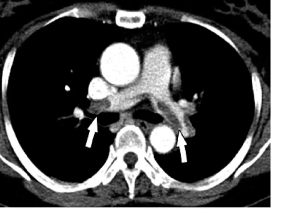
Long description
Chest spiral CT (with and without contrast agent) showing multiple filling defects of principal branches due to acute and chronic pulmonary embolism.

- CT pulmonary angiography (CTPA) is the first-line imaging test for a stable patient with suspected PE, with excellent specificity and the bonus of finding alternative diagnoses (pneumonia, aortic dissection, malignancy). [5][9] It can assess right-ventricular dysfunction (right-to-left ventricular diameter ratio 0.9 or more on multidetector CTPA has about 92 percent sensitivity for RV dysfunction). [5]
- V/Q scanning (preferably V/Q SPECT) is the alternative when CTPA is unavailable or contraindicated (allergy, severe renal impairment with creatinine clearance under 30 mL/min, pregnancy). Planar V/Q scans are more often indeterminate (about 42 percent versus 7 percent for CTPA). [2][5]
- Duplex ultrasound of the legs finding a proximal DVT is sufficient to diagnose and treat VTE in a patient with suspected PE. A negative scan does not exclude PE: in about two thirds of PE patients the clot is not visible on ultrasound. [5]
- Echocardiography is neither sensitive nor specific for PE (transoesophageal echo 82 percent sensitivity for central PE, overall 59 percent), but right-ventricular dysfunction on bedside echo supports the diagnosis and carries prognostic weight (mortality roughly 10 percent with RV dysfunction versus about 0 percent without). [5][9]
- Pulmonary angiography remains the invasive reference standard, with less than 0.5 percent mortality and less than 5 percent morbidity. [5]
The chest X-ray is usually normal in PE; when abnormal the findings are nonspecific (atelectasis, small effusion, raised hemidiaphragm). The two classic but rarely seen signs are Hampton's hump (a wedge-shaped, pleural-based triangular opacity pointing at the hilum, due to pulmonary infarction) and Westermark's sign (focal oligoemia, a truncated hilar artery with a pale lung field beyond). [5] A normal chest film in a hypoxic, dyspnoeic patient without bronchospasm is itself a pointer to PE. [5]
The most common ECG findings in PE are sinus tachycardia and nonspecific ST-T changes; the ECG can also suggest an alternative such as acute coronary syndrome. Classic right-heart strain is present in only about 20 percent of proven PE and includes: [5]
- S1Q3T3 pattern (deep S in lead I, Q wave and inverted T in lead III).
- Right bundle branch block.
- Right axis deviation.
- Tall, peaked P waves (P pulmonale).
- Atrial fibrillation.
Absence of these findings does not exclude PE, and their presence is insensitive. [5]
Neither biomarker diagnoses PE, but both add prognostic information. Troponin is elevated in as many as 50 percent of moderate-to-large PE and marks right-ventricular myocardial stretch; a pooled analysis found a 4.3-fold increase in the odds of death. [5] BNP and NT-proBNP are also raised; one meta-analysis found BNP above 100 pg/mL or NT-proBNP above 600 ng/L associated with roughly six- and sixteen-fold higher in-hospital fatality compared with values below those cutoffs. [5] Guidelines do not make either test a routine part of the diagnostic work-up, but both feed risk stratification. [5]
There is no single diagnostic "score"; PE is confirmed by imaging. Severity is graded by combining clinical status (haemodynamics), right-ventricular function and cardiac biomarkers, and by the validated PESI/sPESI. The 2019 ESC/ERS classification, as summarised by major reviews, is: [1][9]
- High-risk (massive) PE: about 5 to 12 percent of cases - hypotension (systolic pressure under 90 mmHg, or a fall of at least 40 mmHg for at least 15 minutes) or shock requiring vasopressors. Mortality roughly 40 to 77 percent. [1][9]
- Intermediate-high risk: about 34 to 35 percent - haemodynamically stable but with both RV dysfunction and an elevated cardiac biomarker, plus an abnormal PESI/sPESI. [1]
- Intermediate-low risk: about 34 percent - haemodynamically stable with either RV dysfunction or an elevated biomarker. [1]
- Low-risk: about 19 to 55 percent - haemodynamically stable, no RV dysfunction, no biomarker elevation, low PESI/sPESI. [1][4]
The 2026 AHA/ACC guideline introduced a more granular A-E category system (A subclinical, B symptomatic low severity, C symptomatic elevated severity with C1-C3, D incipient cardiopulmonary failure, E cardiopulmonary failure), but its validation is ongoing; European and ASH/ESC definitions above are the ones most widely used in teaching. [4]
PESI and sPESI combine age, sex, cancer, chronic heart failure, chronic lung disease, pulse, systolic pressure, respiratory rate, temperature, altered mental status and oxygen saturation to estimate 30-day mortality. [1] A sPESI of 0 and PESI classes I-II support home or short-stay treatment. [3][8]
Suspected pulmonary embolism: diagnostic algorithm
- Patient with suspected PEUnexplained dyspnoea, chest pain, syncope or hypoxia in a high-risk context
- Clinical assessment and PERC if probability under 15 percentPERC all negative with very low pre-test probability ends the work-up without testing
- Estimate pre-test probabilityWells (two-level) or revised Geneva score; or experienced clinician gestalt
- Question: PE likely (Wells more than 4, or high probability)?
Yes
- Interim anticoagulation if imaging delayedDo not delay anticoagulation while arranging imaging
- CTPA (or V/Q SPECT if contraindicated)Confirms or excludes PE
- Question: PE confirmed?
Yes
- Risk-stratify and treat (see management)
No
- Stop interim anticoagulation unless otherwise indicated; reconsider diagnosis
No
- D-dimer (standard 500 μg/L; age-adjusted if over 50; or YEARS cutoffs)Exclusion test only
- Question: D-dimer result?
Negative
- PE effectively excluded; advise safety-netting and re-review if symptoms progress
Positive
- CTPA (or V/Q SPECT)Confirms or excludes PE
- Question: PE confirmed?
Yes
- Risk-stratify and treat
No
- Stop interim anticoagulation; consider alternative diagnoses
- If CTPA unavailable or impossibleEchocardiography for RV strain; bedside venous ultrasound for DVT can establish VTE; angiography if required
In any suspected or confirmed PE with at least intermediate probability, start full therapeutic anticoagulation immediately and do not let the diagnostic work-up delay it. [7][1] Supportive care comes first in unstable patients: cautious intravenous fluids (a trial of 500 mL or less), vasopressors (noradrenaline preferred; dobutamine for cardiogenic shock categories D-E), oxygen, and escalation to ventilation or VA-ECMO for refractory failure. [1][9]
- High-risk (haemodynamically unstable) PE: systemic thrombolysis is the recommended first reperfusion therapy in the absence of a high bleeding risk, alongside unfractionated heparin. [1][6][8] If thrombolysis is contraindicated or fails, surgical pulmonary embolectomy (with or without VA-ECMO) is the first-line rescue, with in-hospital mortality in series of 9.1 to 23.2 percent. [1] Catheter-directed therapy (thrombolysis or mechanical thrombectomy) is an alternative or bridge in centres with expertise, or for intermediate-to-high risk PE, on very low-quality evidence. [1][8]
- Intermediate (haemodynamically stable with RV dysfunction): anticoagulation alone; thrombolysis is not recommended as first-line because the bleeding risk, including intracranial haemorrhage, exceeds the benefit, although it may be considered for young, low-bleeding-risk patients who are deteriorating. [1][6][8]
- Low-risk PE: anticoagulation alone, with treatment at home considered for carefully selected patients (sPESI 0, PESI categories I-II, all Hestia criteria negative). [3][8]
Guidelines recommend alteplase, streptokinase or urokinase. The most commonly used modern regimen is alteplase: a 10 mg bolus over 1 to 2 minutes followed by 90 mg over 2 hours (total about 100 mg, or 1.5 mg per kilogram over 2 hours if the patient weighs under 65 kg). [1] Unfractionated heparin is continued during an alteplase infusion but stopped during streptokinase or urokinase. [1] Medscape's review of the same evidence quotes the commonly used total 100 mg dose but notes evidence that lower doses (25 to 50 mg) may be as effective with less bleeding, especially in patients over 65 years, in whom major bleeding more than triples. [6]
| Drug | Typical adult dosing (from cited sources) | Notes |
|---|---|---|
| Unfractionated heparin (UFH) | 80 U/kg intravenous bolus (or 5,000 U) then 18 U/kg/h (or 1,300 U/h) infusion; aPTT maintained at at least 1.5 times the baseline control value (institution-specific therapeutic range). [2][6] | Reserved for haemodynamic instability, severe renal impairment, high bleeding risk, or peri-procedural use because of short half-life and reversibility. [6] |
| Enoxaparin (LMWH) | 1 mg/kg subcutaneously every 12 hours. [2] | First-line parenteral choice in 2026 AHA/ACC and ESC; pivotal for outpatient treatment. [6] |
| Fondaparinux | Weight-based dosing prescribed per product label (not dosed numerically in the sources cited here). [6] | A synthetic factor Xa inhibitor; favoured as an initial option in ESC 2019 and 2012 CHEST. [6] |
| Rivaroxaban | 15 mg by mouth twice daily for 3 weeks, then 20 mg once daily. [1] | No lead-in parenteral required. Avoid (or use with caution) when creatinine clearance under 30 mL/min. [1] |
| Apixaban | 10 mg by mouth twice daily for 7 days, then 5 mg twice daily. [1] | No lead-in parenteral required; not recommended with creatinine clearance under 25 mL/min. [1] |
| Edoxaban | 60 mg once daily after 5-10 days of parenteral anticoagulation; 30 mg once daily if creatinine clearance 15-50 mL/min or body weight under or equal to 60 kg. [1] | Lead-in parenteral required. |
| Dabigatran | 150 mg by mouth twice daily after 5-10 days of parenteral anticoagulation. [1] | Lead-in parenteral required; not recommended with creatinine clearance under 30 mL/min. [1] |
| Warfarin | Oral, overlapping LMWH or UFH until the INR is 2 to 3 for 2 consecutive days; target INR 2.0-3.0. [6][8] | Vitamin K dependent; INR checked daily initially, then every 1-2 weeks. [6] |
Duration of anticoagulation follows the risk that produced the PE: [6]
- PE provoked by a major reversible factor (surgery, trauma, immobilisation): at least 3 months, then stop, reassessing the balance. [5][6]
- Unprovoked PE: at least 3 months, then seriously consider continuing indefinitely (indefinite therapy is suggested for most unprovoked VTE in the ASH 2020 guideline). [6][8]
- PE due to a persistent risk factor (cancer, chronic disease): extended or indefinite therapy, balancing recurrence against bleeding. [6]
- Active cancer: anticoagulate 3 to 6 months and review; LMWH (or a DOAC in non-gastrointestinal cancer) is preferred over warfarin. [1][6][7]
- Extended therapy: in patients choosing to continue after the initial phase, a DOAC is preferred over warfarin; half-dose apixaban or rivaroxaban is recommended to reduce bleeding in extended prevention. [6]
- Secondary prevention doses (where used): rivaroxaban 10 mg daily or apixaban 2.5 mg twice daily are the lower-dose regimens quoted. [8]
IVC filters should not be used routinely in PE treated with anticoagulation. They are reserved for PE where anticoagulation is absolutely contraindicated, or for recurrence despite optimal therapy; retrievable filters are preferred and should be retrieved as soon as anticoagulation becomes possible, ideally within 29 to 54 days. [4][6]
Catheter-directed thrombolysis, mechanical thrombectomy and (in selected cases) ultrasound-assisted lysis are used for intermediate- or high-risk PE, especially where systemic thrombolysis is contraindicated, although the evidence is limited. Surgical embolectomy, usually with cardiopulmonary bypass and increasingly combined with VA-ECMO, is a life-saving option in massive PE. An AHA 2023 scientific statement argues for considering surgery earlier rather than as salvage. [6]
- Chronic thromboembolic pulmonary hypertension (CTEPH): persistent elevation of pulmonary pressures after PE, occurring in up to 5 percent of patients. It presents with progressive breathlessness and right heart failure, and is the main potentially curable cause of chronic pulmonary hypertension if recognised; treatment is pulmonary endarterectomy or balloon pulmonary angioplasty. [8][9]
- Post-PE syndrome: persistent symptoms and functional limitation despite adequate anticoagulation. After 3 months, about 37 percent of patients report dyspnoea and 22 percent have an abnormal six-minute walk distance; 20 percent still have abnormal walk distance at one year. Residual perfusion defects are detectable in up to 50 percent of patients six months after PE on V/Q scanning, and about half have persistent symptoms. [9]
- Recurrence: roughly 10 percent by 2 years and over 30 percent by 10 years after an unprovoked VTE, falling substantially with extended anticoagulation but at the cost of bleeding. [8]
- Bleeding: major bleeding in about 1 to 3 percent of patients on warfarin per year, reduced by roughly 30 percent with a DOAC. [8]
- Acute: sudden death, shock, right-to-left shunt through a patent foramen ovale, paradoxical embolism, haemorrhagic infarction, pleural effusion, heparin-induced thrombocytopenia. [4]
Untreated PE kills in about 25 percent of cases, mostly in the first hours. [3] With treatment, mortality falls below 5 percent. [3] Prognosis depends on the category: 30-day mortality roughly 65 percent in high-risk PE, 5 to 25 percent in intermediate-risk and about 1 percent in low-risk PE. [4] Elevated troponin, high BNP/NT-proBNP, hyponatraemia, residual pulmonary hypertension and severe RV dysfunction all predict a worse course. [5][9]
Use the pregnancy-adapted YEARS algorithm to reduce CTPA; combining D-dimer with lower-limb venous ultrasound and shared decision-making is also reasonable, because radiation doses from CTPA and V/Q scanning are well below teratogenic thresholds. [2] If probability is low and D-dimer is normal, exclude PE without imaging; if suspicion is high, obtain bilateral leg Doppler, and if that is negative proceed to CTPA. [6] Pregnant patients are treated with LMWH or UFH at full therapeutic dose; warfarin and DOACs are contraindicated in pregnancy and breastfeeding. Alteplase is reserved for life-threatening PE after a careful risk-benefit assessment. Anticoagulate for 4 to 6 weeks postpartum and for at least 6 months in total. [6] Neonates exposed to iodinated contrast in utero need serum thyroid-stimulating hormone checked in the first week. [6]
In active cancer, treatment for 3 to 6 months is recommended, reviewing at that point. A DOAC is preferred where there is no gastrointestinal cancer; LMWH or LMWH followed by warfarin remains the alternative. [1][6][7] Triple-positive antiphospholipid syndrome requires LMWH plus warfarin. [7]
With creatinine clearance 15 to 50 mL/min, apixaban or rivaroxaban may be used, or LMWH followed by edoxaban (or dabigatran if clearance is at least 30 mL/min), or LMWH/UFH with a VKA. [7] With clearance under 15 mL/min, use LMWH or UFH with or without a VKA. Always check the product label: edoxaban needs dose reduction at 15 to 50 mL/min, and apixaban, rivaroxaban, edoxaban and dabigatran are not recommended below their respective 25 to 30 mL/min thresholds. [1][7]
Warfarin with an INR of 2.0 to 3.0 is preferred over a DOAC, particularly in triple-positive disease. [7][8]
- A chest X-ray and an ECG are almost always available; know the genuinely useful pointers (Hampton's hump, Westermark's sign, S1Q3T3) but do not be falsely reassured by their absence. [3][5]
- A transthoracic echocardiogram and a leg vein ultrasound, even from a portable machine, can confirm DVT or show right-ventricular strain; positive leg ultrasound in a breathless patient is enough to start treatment for VTE.
- If CTPA is unavailable, V/Q scanning, or empiric treatment based on probability plus leg ultrasound, is a reasonable pathway. [5]
- Where laboratory D-dimer is unavailable, use clinical probability and bedside testing; the PERC rule can clear very-low-probability patients with no tests. [4][5]
- UFH is cheap, available and rapidly reversible; a monitored infusion or monitored subcutaneous regimen is a legitimate alternative to LMWH and DOACs when those are unaffordable, particularly when bleeding risk is high or procedures are planned. [6]
- Warfarin with INR monitoring is widely available and effective; home testing and handover systems (alert cards, patient information) matter as much as the drug.
- Surgical embolectomy and catheter therapies require cardiac surgical or interventional capacity; plan early referral to a centre that has it.
There is no screening test for PE. Prevention is the prevention of DVT: mobilisation, VTE prophylaxis in hospitalised and surgical patients (LMWH if no contraindication), and prophylaxis in pregnancy for high-risk women. [1][3] After an episode, ask about breathlessness, exercise tolerance, sleep and mood at every visit for at least a year, to catch post-PE syndrome, CTEPH, depression and anxiety early. [6]
- PE is the third commonest cause of cardiovascular death; untreated it kills about a quarter of patients, but treated mortality is under 5 percent. [3]
- Virchow's triad (endothelial injury, stasis, hypercoagulability) underpins all the risk factors; whether a PE is provoked, unprovoked or cancer-associated decides how long to anticoagulate. [3]
- The classic presentation is uncommon; sudden dyspnoea, pleuritic chest pain or syncope in a risk-factor context should raise suspicion, and hypoxia out of keeping with the examination is a strong pointer.
- D-dimer is an exclusion test: combined with low or unlikely probability it safely rules out PE; age-adjusted cutoffs improve specificity in patients over 50. [1]
- CTPA is the first-line imaging test in stable patients; V/Q scanning and leg ultrasound are the main alternatives.
- Hampton's hump (wedge-shaped infarct) and Westermark's sign (oligoemia) are classic chest X-ray findings but are uncommon; S1Q3T3 is the classic ECG marker and is insensitive.
- Severity is driven by haemodynamics, right-ventricular dysfunction and troponin/BNP; PESI/sPESI quantify 30-day risk. [1]
- High-risk, haemodynamically unstable PE calls for systemic thrombolysis (alteplase 10 mg over 1-2 minutes then 90 mg over 2 hours) with UFH unless bleeding risk is high; surgical embolectomy or catheter therapy is the alternative. [1][6]
- Most other patients need anticoagulation: a DOAC or LMWH first, then a DOAC or warfarin (INR 2.0-3.0) for 3 to 6 months at least, and extended or indefinite therapy for unprovoked or persistent-risk PE. [6][8]
- In pregnancy use LMWH or UFH only (never warfarin or DOACs); in renal impairment choose and adjust by creatinine clearance; in triple-positive antiphospholipid syndrome use warfarin. [6][7][8]
- CTEPH follows up to 5 percent of PE; screen with symptom and functional review for at least a year, and refer for endarterectomy assessment. [8][9]
Answer each one in your head first, then open the answer.
A 62-year-old woman with recent hip replacement is breathless at rest, heart rate 118, respiratory rate 28, and oxygen saturation 89% on air. ECG shows S1Q3T3. Her blood pressure is 112/70 mmHg. What is the most useful first imaging test, and what risk category does this suggest?
Show answerHide answer
CT pulmonary angiography. Her presentation is most likely intermediate-to-high risk PE: normotensive but tachycardic and tachypnoeic with right-heart strain patterns. Urgent CTPA (or V/Q SPECT if contrast is contraindicated) confirms it; bedside echo showing right-ventricular dysfunction and an elevated troponin would push her to intermediate-high risk and warrant close monitoring or escalation. [4][5]
Why does a normal D-dimer safely exclude PE in a patient with a low or unlikely probability score, but not in a patient with a high probability score?
Show answerHide answer
D-dimer is released whenever cross-linked fibrin is broken down. It is highly sensitive but poorly specific, so a negative result means "no appreciable fibrin turnover" and reliably rules out clot in a patient whose prior probability is low. In a high-probability patient, the underlying chance of PE is so high that even a false-negative rate of a few percent is unacceptable, so imaging is required instead. [5][7]
A patient has an unprovoked PE and starts anticoagulation. How long should anticoagulation continue, and what regimen would you use if extended prevention is chosen?
Show answerHide answer
Treat for at least 3 months, then re-assess the balance of recurrence against bleeding; for an unprovoked event many guidelines advise continuing indefinitely. If extended prevention is chosen, a direct oral anticoagulant is preferred over warfarin, and half-dose rivaroxaban or apixaban reduces bleeding in the extended phase. [6][8]
A pregnant patient at 24 weeks' gestation has a low-probability PE score and a normal D-dimer. Does she need imaging?
Show answerHide answer
No: on that combination, clinical exclusion is recommended, just as in non-pregnant patients. If suspicion is high despite negative testing, bilateral leg Doppler ultrasound is the next step, followed by CTPA if that is negative. Warfarin and the direct oral anticoagulants are contraindicated in pregnancy; treat with low-molecular-weight or unfractionated heparin instead. [6]
Which chest X-ray and ECG findings are said to be classic for pulmonary embolism, and how reliable are they?
Show answerHide answer
Hampton's hump (wedge-shaped pulmonary infarct) and Westermark's sign (focal oligoemia) on the chest X-ray, and S1Q3T3, right bundle branch block, right axis deviation, peaked P waves or atrial fibrillation on the ECG. All are uncommon individually and each is insensitive and nonspecific; a normal film or ECG certainly does not exclude PE. [5]
- Venous thromboembolism and deep vein thrombosis
- Chronic thromboembolic pulmonary hypertension
- Respiratory failure and hypoxaemia
- ECG interpretation
- Chest X-ray reading
- Anticoagulant drugs
Pulmonary hypertension and cor pulmonale
How high pulmonary pressures arise, are classified, diagnosed and treated, and what they mean for the right heart and cor pulmonale.Hypoxaemia and respiratory failure
How hypoxaemia develops, how to read the blood gas and A-a gradient, and how to give oxygen and ventilatory support safely.Pleural effusion
How fluid accumulates in the pleural space, how transudates differ from exudates, and how to investigate, drain and treat effusions by cause.Deep vein thrombosis
Deep vein thrombosis: Virchow's triad, risk factors, diagnosis with Wells and D-dimer, imaging, anticoagulation dosing and duration, complications.
4 other topics link here.
- Banfi C, Montonati C, Ielasi A, Assouline B, Tespili M, Bendjelid K, Giraud R. Advancements in Pulmonary Embolism Management: Current Approaches and Future Directions. Reviews in Cardiovascular Medicine 2025;26(11):44614. Accessed .
- Wrenn JO, Kabrhel C. Emergency department diagnosis and management of acute pulmonary embolism. British Journal of Haematology 2024;205(5):1714-1716. Accessed .
- Ouellette DR, Mosenifar ZM. Pulmonary Embolism (PE). Medscape (updated 2026-08-27). Accessed .
- Ouellette DR, Mosenifar ZM. Pulmonary Embolism (PE) Clinical Presentation. Medscape (updated 2026-08-27). Accessed .
- Ouellette DR, Mosenifar ZM. Pulmonary Embolism (PE) Workup. Medscape (updated 2026-08-27). Accessed .
- Ouellette DR, Mosenifar ZM. Pulmonary Embolism (PE) Treatment & Management. Medscape (updated 2026-08-27). Accessed .
- National Institute for Health and Care Excellence (NICE). Venous thromboembolic diseases: diagnosis, management and thrombophilia testing (NG158). NICE guideline NG158. Accessed .
- Ortel TL, Neumann I, Ageno W, et al. American Society of Hematology 2020 guidelines for management of venous thromboembolism: treatment of deep vein thrombosis and pulmonary embolism. Blood Advances 2020;4(19):4693-4738. Accessed .
- Ajah ON, et al. Pulmonary Embolism and Right Ventricular Dysfunction: Mechanism and Management. Cureus 2024;16(9):e70561. Accessed .
Figure credits
Educational content for medical students and doctors. It is not a substitute for clinical judgement or local guidelines.