Pneumothorax
Air in the pleural space: types, how it presents, how imaging diagnoses it, and how current guidelines manage spontaneous, traumatic and tension cases.
- A pneumothorax is air in the pleural space, the potential gap between the lung and the chest wall, causing partial or complete lung collapse.
- It is classified as spontaneous (primary in otherwise healthy young, tall, thin people; secondary in diseased lung), traumatic, iatrogenic, or tension, which is immediately life-threatening.
- Sudden one-sided chest pain and breathlessness are the classic clues; tension pneumothorax adds shock, tracheal shift and a hyper-resonant hemithorax.
- Chest X-ray diagnoses most, ultrasound shows absent lung sliding and a barcode (stratosphere) sign, and CT is used for complex or recurrent cases.
- Modern management is symptom-led: stable small spontaneous pneumothoraces can be observed, while symptomatic or high-risk cases are treated with needle aspiration or a chest drain; recurrence is prevented with VATS and pleurodesis when indicated.
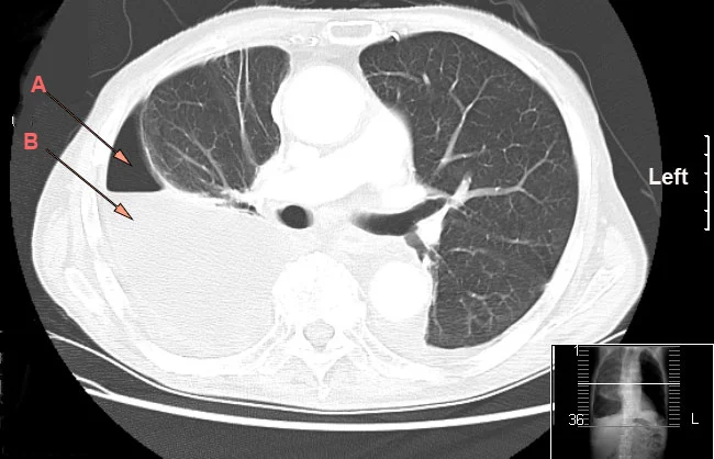
Figure 1Hydro pneumothorax
Long description
CT chest showing a large unilocular hydro-pneumothorax with associated collapse of the right lung and iatrogenic air from a previous chest drain (arrows A air, B fluid).

By the end of this page you should be able to:
- Define pneumothorax and classify it into spontaneous, traumatic, iatrogenic and tension types, distinguishing primary from secondary spontaneous pneumothorax.
- Explain why tall, thin young male smokers develop pneumothorax, and link commonly associated conditions such as COPD, cystic fibrosis, Marfan syndrome, lymphangioleiomyomatosis, Birt-Hogg-Dube syndrome and catamenial pneumothorax.
- Recognise the presentations and signs, including the emergency features of tension pneumothorax and mediastinal shift.
- Interpret chest X-ray, ultrasound and CT findings, and state the size categories used in current practice.
- Apply the current symptom-led management pathway, including observation, needle aspiration, ambulatory devices, chest drains and surgery, and know the indications, needle site and drug choices for tension decompression.
- Advise patients about smoking cessation, flying, diving, pregnancy, high-risk occupations and low-resource practice.
DefinitionPneumothorax
A pneumothorax is air in the pleural space, the potential space between the visceral and parietal pleura, making the intrapleural pressure rise from negative to atmospheric and letting part or all of the lung collapse.
The standard terminology separates pneumothorax into spontaneous and non-spontaneous forms. [6] A spontaneous pneumothorax occurs without trauma; it is primary when no clinically apparent lung disease is present and secondary when it complicates known lung disease such as COPD, interstitial lung disease or cystic fibrosis. [2][6] A traumatic pneumothorax follows blunt or penetrating chest injury, including barotrauma from mechanical ventilation. Iatrogenic pneumothorax follows a procedure such as CT-guided lung biopsy, radiofrequency ablation, cardiac device implantation or central venous catheterisation. [6] Tension pneumothorax is a life-threatening state in which air enters the pleural space through a one-way valve and cannot escape, raising intrathoracic pressure, shifting the mediastinum and impairing venous return. [4]
Clinically it is also useful to distinguish an open (sucking) pneumothorax through a chest wall defect from a closed pneumothorax, and to note pneumothorax ex vacuo, in which a non-expandable lung follows pleural pathology such as malignancy or fibrosis. [6]
Spontaneous pneumothorax is common worldwide, and it brings the greatest burden to patients with underlying lung disease. [2] A British study from 2018 reported spontaneous pneumothorax admissions of 14.1 per 100,000 people of 15 years or older, with a male to female ratio of 2.7, up from 9.1 per 100,000 in 1968. [2] A French study reported an annual rate of 22.7 cases per 100,000 people of 14 years or older. [2] Primary spontaneous pneumothorax is estimated to have an annual incidence of 18 to 28 per 100,000 males and 1 to 6 per 100,000 females. [3] Sources disagree slightly on the exact incidence, which partly reflects different case definitions and admission-based versus population-based counting; the honest summary is that it is most common in healthy young men and far less common in women. [2][3]
Mortality from spontaneous pneumothorax itself is low in primary disease but markedly higher in secondary pneumothorax because of the underlying lung disease. [2] Traumatic pneumothorax is far commoner overall, with an estimated four million cases globally. [6]
Primary spontaneous pneumothorax results from rupture of subpleural blebs or bullae, usually at the lung apex. [3][6] The changes happen in people with otherwise clinically normal lungs, though CT imaging often shows emphysema-like changes and many cases are familial. [2] Risk factors for primary disease include:
- Male sex, tall stature and low body weight, most often between 15 and 34 years of age. [3]
- Cigarette smoking, the most important modifiable risk factor, which promotes inflammation and bleb formation. [3]
- Family history and familial forms, which account for about 10% of primary pneumothoraces. [6]
- Genetic syndromes including Birt-Hogg-Dube syndrome (FLCN gene), lymphangioleiomyomatosis (TSC1 and TSC2 genes) and Marfan syndrome (FBN1 gene). [6]
- Catamenial pneumothorax, a secondary form in which thoracic endometrial deposits rupture around menstruation; this is the likely diagnosis in most women whose suspected primary pneumothorax shows a catamenial pattern, and MRI may find endometrial deposits in up to 83% of suspected cases. [6]
Secondary spontaneous pneumothorax complicates established lung disease: COPD, cystic fibrosis, interstitial lung disease, lung cancer, necrotising or cavitating infection (including tuberculosis and Pneumocystis jirovecii pneumonia in immunocompromised patients), and bullous disease. [6][8] It predominantly affects older patients. Trauma, barotrauma from positive-pressure ventilation and procedures such as CT-guided biopsy, ablation, pacemaker or central venous catheter placement account for the rest. [6] Iatrogenic pneumothorax rates include pneumothorax in about 26% of CT-guided lung biopsies, reported up to 61% in some series, 16 to 52% after lung ablation, and under 1% after pacemaker or central venous catheter placement. [6]
Air normally sits in the alveoli. When a subpleural bleb or bulla ruptures, alveolar air escapes into the pleural space. Because the pleura tries to maintain a negative pressure, even a small leak shifts the pressure toward atmospheric, the elastic lung recoils inward and the chest wall springs outward, producing partial or complete collapse. [3] In primary disease the defect often heals spontaneously, which is the rationale for conservative management. [3] In secondary disease the underlying parenchymal damage impairs healing, so persistent air leak and recurrence are more likely. [2][3]
Tension physiology develops when the rupture acts as a one-way valve: air enters during inspiration but cannot leave during expiration. Pressure rises steadily, pushing the mediastinum to the opposite side, kinking the great veins and cutting venous return, so cardiac output falls. This is an immediately lethal mechanism and is the reason tension pneumothorax is treated before imaging. [4]
The body slowly reabsorbs pleural air because nitrogen diffuses down its gradient from pleura to capillary blood. High-flow oxygen lowers the partial pressure of nitrogen in alveolar gas and steepens that gradient, speeding reabsorption threefold to fourfold in small human studies using oxygen at 16 L per minute. [2] A Korean retrospective study found oxygen at 2 to 4 L per minute gave a higher resolution rate than room air (4.27 versus 2.06% of the pneumothorax per day). [2] Even so, the optimal flow and duration are not standardised. [2]
The classic presentation is sudden, sharp, one-sided chest pain and breathlessness. Pain is pleuritic and may radiate to the shoulder or neck. Breathlessness ranges from trivial to severe, and patients often describe a click or pop at onset. Many primary pneumothoraces happen at rest; precipitants include coughing, lifting, sneezing or playing a wind instrument, but many occur with no warning. [6] A mild, small pneumothorax can be painless and found by chance. Secondary pneumothoraces often present with more marked breathlessness because reserve is limited, and older smokers may be mislabelled as a COPD exacerbation.
Constitutional symptoms such as fever and weight loss are not features of uncomplicated pneumothorax; if present, search for infection, catamenial disease or malignancy.
Look for the features considered in any acute breathlessness: respiratory rate, oxygen saturation, distress, cyanosis and the ability to speak in full sentences. The affected side shows:
- reduced or absent chest expansion;
- hyper-resonance to percussion;
- reduced or absent breath sounds and voice sounds;
- tachycardia and tachypnoea.
A small pneumothorax may have no physical signs at all, so a normal examination never excludes it. [5]
Tension pneumothorax is a clinical diagnosis. Suspect it when a patient is hypotensive and tachycardic, acutely breathless and hypoxic, with hyper-resonance and absent breath sounds on one side, tracheal deviation away from the affected side, distended neck veins, and signs of obstructive shock. Do not wait for a chest X-ray when these are present. [4] Fatal deterioration can occur within minutes; if suspected, decompress immediately and insert a drain afterward.
A pneumothorax enters the differential of almost any acute pleuritic chest pain or breathlessness. The main rivals to exclude are pulmonary embolism, myocardial infarction, pericarditis, pneumonia, exacerbation of asthma or COPD, acute bronchospasm, chest wall pain, rib fracture and pleural effusion. Clues that separate them include the sudden onset, unilateral signs and immediate hypoxia of a pneumothorax. In ventilated patients, a sudden deterioration with high airway pressure and hypoxia should prompt assessment for tension pneumothorax and for tube displacement or blockage.
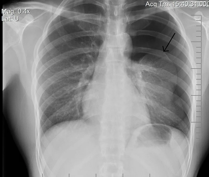
Figure 2Expiration-left-side-pneumo
Long description
Chest x-ray of a left-sided spontaneous pneumothorax with an arrow indicating the edge of the collapsed lung.

CXR is the first-line investigation. In an erect film, a pneumothorax shows as a sharp visceral pleural line with no lung markings peripheral to it, and the lung falls toward the hilum. Expiratory views can increase sensitivity. Standard radiography measures the distance from the lung edge to the chest wall; in current British Thoracic Society guidance, a pneumothorax is "large" when that rim-to-wall distance is greater than 2 cm. [3][6] Other guidelines and centres use percentage estimates, so local definitions should be checked. [3] CXR also shows associated features such as surgical emphysema, rib fractures and underlying lung disease, and it monitors resolution.
Point-of-care ultrasound is a radiation-free, bedside test. In pneumothorax it shows absence of lung sliding, because air between the pleural layers abolishes movement. [5] On M-mode the normal "seashore sign" is replaced by a "barcode" or "stratosphere sign" in pneumothorax. [5] Other useful findings are the lung point (diagnostic when seen), absence of B-lines at the affected site, and no lung pulse. [5] POCUS is particularly useful in pregnancy, in the emergency department and in intensive care, but a single negative scan does not exclude a very small pneumothorax, and it should not delay decompression in suspected tension.
CT chest is used when the diagnosis is uncertain, when a chest drain is being considered in complex or loculated disease, when a small pneumothorax is suspected, and when underlying lung disease needs definition. It is the investigation of choice for suspected familial disease, for example cystic lung disease due to lymphangioleiomyomatosis in women or a Birt-Hogg-Dube syndrome pattern. [6] In catamenial pneumothorax, MRI of the thorax may identify endometrial deposits in up to 83% of cases, especially if performed during menstruation. [6]
Arterial blood gases may show hypoxaemia. Bloods are directed at the clinical picture rather than at pneumothorax itself. In suspected tension or trauma, imaging of the neck and chest and assessment for associated injuries are needed, but never at the expense of decompression.
There are no biochemical diagnostic criteria. The diagnosis is radiological, confirmed on CXR, CT or ultrasound. In practice, patients are stratified by two questions that the 2023 British Thoracic Society guideline makes decisive: [1][6]
- Is the patient clinically stable, without respiratory distress or hemodynamic compromise?
- Is there underlying lung disease, high-risk age or smoking history, bilateral disease or haemopneumothorax?
High-risk characteristics for decompensation include tension physiology, age over 50 years with more than 20 pack-years of smoking, known underlying lung disease, significant hypoxaemia, bilateral pneumothorax and haemopneumothorax. [6] Size alone is no longer the trigger for intervention, though it guides where a drain can safely be inserted: a very small rim may need image-guided rather than bedside drainage. [2]
Ensure airway, breathing and circulation. Give oxygen to correct hypoxaemia. Analgesia matters because pain restricts ventilation; oral non-steroidal anti-inflammatory drugs or paracetamol are usually sufficient. [3] If tension pneumothorax is suspected, decompress immediately without waiting for an X-ray.
Red flagTension pneumothorax is a clinical emergency
Sudden chest pain, breathlessness, hypoxia, hypotension, tachycardia, hyper-resonant hemithorax, absent breath sounds and tracheal deviation require immediate needle decompression, then a chest drain. Do not delay for imaging.
Needle decompression converts tension to a simple pneumothorax. [4] The current Advanced Trauma Life Support (ATLS) guidance recommends a 5 cm needle for small adults or an 8 cm needle for larger adults, inserted at the 4th or 5th intercostal space in the anterior midaxillary line. The European Trauma Course (ETC) recommends a 14 or 16 gauge extra-long cannula at the 2nd intercostal space in the midclavicular line. [4] A 2025 meta-analysis concluded that, on radiological data, needle failure to reach the pleural space is common (32.84%), that each extra centimetre of needle length reduces failure by about 7.76%, and that a 7 cm needle is appropriate for right-sided tension at either the 5th intercostal space along the midaxillary line or the 2nd intercostal space along the midclavicular line; for left-sided cases the 2nd midclavicular line is preferred because of cardiac injury risk. [4] Needle decompression must always be followed by insertion of an intercostal chest drain as definitive treatment. [4]
Management of a pneumothorax in adults
- Pneumothorax confirmed or strongly suspectedAssess stability, size, risk factors and whether it is primary, secondary, traumatic, iatrogenic or tension
- Question: Tension features present?
Yes
- Immediate needle decompressionATLS: 5 cm (small adults) or 8 cm (larger adults) at 4th or 5th intercostal space anterior midaxillary line; or ETC 14-16G at 2nd intercostal space midclavicular line
- Insert intercostal chest drainDefinitive treatment after decompression
No
- Question: Clinically unstable or high-risk?
Yes (hypoxia, bilateral, underlying lung disease, haemopneumothorax, age over 50 with significant smoking)
- Chest drainUsually required for secondary spontaneous and traumatic pneumothorax; small-bore 12-14 Fr reduces pain
No
- Question: Symptom burden?
Minimal or none
- Conservative careObservation, oxygen to correct hypoxaemia, analgesia; monitor 2-4 h before discharge for primary disease and 24 h for secondary
Symptomatic
- Needle aspiration or chest drainNeedle aspiration gives quicker symptom relief; may need repeating in up to half of cases. Chest drain if aspiration fails or quicker relief with fewer procedures is prioritised. Ambulatory Heimlich valve allows shorter stay
- If persistent air leak or recurrence riskRefer for surgical opinion: VATS with bullectomy and pleurodesis per indications below
Small, stable, minimally symptomatic primary pneumothoraces are suitable for conservative management, in which the air is reabsorbed while the patient is observed, and an Australian retrospective cohort found this gave a much shorter hospital stay (0.6 versus 6.5 days) with similar one-year recurrence (11% versus 10%) and fewer complications than invasive treatment. [1][2] A 2020 randomised trial in primary spontaneous pneumothorax showed similar lung re-expansion within 8 weeks for conservative and interventional care (94.4% versus 98.5%). [2] The 2023 British Thoracic Society guideline keeps this symptom-led approach and adds that ambulatory care with a one-way valve device is recommended for primary pneumothorax with adequate support services, while chest drain insertion is usually needed for secondary pneumothorax and for trauma. [1]
Needle aspiration removes air with a 16 to 18 gauge cannula; stop and convert to a chest drain if more than 2.5 L is aspirated, since persistent air leak is likely. [2] A Cochrane systematic review found needle aspiration shorter hospitalisation and fewer adverse events than chest drain, with a similar one-year success rate, even though immediate success is lower. [2] Chest drain insertion, most often with a 12 to 14 French small-bore tube, is the most common procedure in secondary and traumatic disease; drains larger than 28 French confer no additional flow benefit. [2] After the drain is in, a digital system can help quantify air leak; clamping practice varies and its value remains debated. [2]
Persistent air leak is defined as air leak continuing for more than 2 to 5 days after drain insertion, depending on the source. [2][6] It should be reviewed for surgery when it persists despite 5 to 7 days of chest tube drainage. [2]
Surgery is recommended by the 2023 British Thoracic Society guideline for: [2]
- first pneumothorax presenting with tension, or first secondary pneumothorax with significant physiological compromise;
- second ipsilateral pneumothorax;
- first contralateral pneumothorax;
- synchronous bilateral spontaneous pneumothorax;
- persistent air leak (despite 5 to 7 days of drainage) or failure of lung re-expansion;
- spontaneous haemothorax;
- professions at risk such as pilots or divers, even after a single episode;
- pregnancy with continuous air leak.
Video-assisted thoracoscopic surgery and thoracotomy both resect bullae and carry out pleurodesis by abrasion, partial pleurectomy or talc poudrage. VATS has lower morbidity and a shorter stay, with reported recurrence of 31 per 1,000 patients compared with 15 per 1,000 after open thoracotomy; surgery halves recurrence compared with a drain alone. [2][6] Chemical pleurodesis, usually with graded talc, also reduces recurrence (risk ratio 0.56) and talc poudrage recurrence after drainage or thoracoscopy ranges from 5.6% to 24.1%. [2] Autologous blood patch pleurodesis (50 to 100 mL of the patient's own blood instilled through the drain) is an option when surgery is unsafe; reported success of autologous blood ranged from 82% at 1 to 2 mL per kg to 27% at 0.5 mL per kg and 9% with saline alone, with fever in 10 to 13% and infection in 9%. [2]
Secondary pneumothorax patients are usually older with multiple comorbidities and have higher postoperative recurrence (9.3 to 12.5%), morbidity (20.6%) and mortality (4.1% in older patients) than primary disease patients, so selection is critical. [2]
Most traumatic and iatrogenic pneumothoraces are treated with a chest drain, and large-bore drainage is favoured for major trauma because of the risk of large air leak. [6] Small, minimally symptomatic cases can be observed, particularly after iatrogenic injury. [6] All suspected high-pressure or penetrating injuries need immediate assessment for tension physiology and associated haemothorax.
- Tension pneumothorax, the immediately life-threatening conversion described above.
- Persistent air leak and failure of lung re-expansion. [2]
- Infection of the pleural space, and procedure-related infection after blood patch pleurodesis. [2]
- Surgical emphysema, sometimes dramatic but usually benign.
- Recurrence, which is common, especially in secondary disease. [2][6]
- Risks of drainage: pain, haemothorax, infection and, rarely, organ injury; small-bore tubes reduce pain. [2]
About 32% of primary pneumothoraces recur, and secondary pneumothoraces recur in 13 to 39%. [2][6] Uncertainty between series is real and reflects different case mixes. Smoking is the strongest risk factor for recurrence: quitting reduces recurrence fourfold. [6] Poorer surgical outcomes occur in older secondary-disease patients and in interstitial lung disease. [2]
Smoking cessation should be offered to every patient, because it is the only modifiable factor with a large effect. [6] There is no general population screening; instead, investigate for familial disease in younger patients, with recurrent, bilateral or unexplained pneumothoraces, or in women, where cystic lung disease or a catamenial cause must be excluded. [6] Family history and examination for skin lesions, oral fibrofolliculomas or other features of hereditary syndromes complete the assessment. [6]
Pregnancy. Small asymptomatic pneumothoraces can be observed. If symptoms arise, needle aspiration or a chest drain is used, and surgical evaluation is needed for persistent air leak because labour may worsen it. [1]
Children and younger patients. Familial forms are more likely; genetic testing should be considered. [6]
Older adults and secondary disease. These patients have the least reserve and the highest procedural risk; they warrant early chest drainage and careful selection for surgery. [2][6]
High-risk occupations. Pilots, commercial divers, military personnel and others whose collapse would endanger others or whose environment prevents treatment are usually referred for definitive surgical prevention after a single episode. [6]
Immunocompromised patients. Pneumocystis jirovecii pneumonia can be complicated by pneumothorax or other pulmonary barotrauma, particularly in advanced disease and during mechanical ventilation. [8]
Current advice is to avoid commercial air travel for 7 to 14 days after full radiographic resolution of a pneumothorax, after which gas expansion at cabin altitude is very unlikely to cause problems. [6][7] Patients can usually fly once the pneumothorax has fully resolved and the above delay has passed. [6] Emerging evidence suggests that selected stable, normoxic patients with small traumatic pneumothoraces may tolerate earlier travel, but this does not override current guidelines. [7]
Scuba diving is generally considered a lifelong contraindication after a spontaneous pneumothorax unless a definite recurrence-prevention procedure, usually VATS pleurectomy or pleurodesis, has been performed and the patient has been assessed by a diving medicine specialist. [6] The same caution applies to high-altitude and hyperbaric activity. [6]
Low-resource practice is where this condition is most dangerous, because a tension pneumothorax kills quickly and cannot wait. Priorities are:
- Teach clinical recognition of tension pneumothorax and decompress immediately with the widest-bore needle or cannula available, then insert a chest drain when one can be placed. [4]
- Chest X-ray confirms most cases, but do not delay drainage for imaging when high-risk features exist. [1]
- Use an intercostal drain where available; a one-way valve or flutter valve can allow ambulatory care when follow-up is reliable. [1]
- If thoracic surgery, graded talc and pleurodesis are unavailable, smoking cessation and patient education still reduce recurrence risk, and early referral pathways to a surgical centre should be established. [1]
- Ultrasound skills for absent lung sliding are cheap to learn and equipment is increasingly available; they allow diagnosis at the bedside. [3][5]
- Counsel every smoker; this has a larger effect than any medicine in this disease. [6]
- A pneumothorax is air in the pleural space; types are primary spontaneous, secondary spontaneous, traumatic, iatrogenic, tension and catamenial.
- Primary spontaneous pneumothorax affects tall, thin young male smokers; secondary pneumothorax complicates COPD, cystic fibrosis, interstitial lung disease, infection and cancer.
- Presentation is sudden unilateral chest pain and breathlessness, with no reliable physical signs when it is small.
- Tension pneumothorax is a clinical emergency: hypoxia, hypotension, tracheal deviation, hyper-resonance and absent breath sounds demand immediate decompression.
- CXR shows a visceral pleural line without markings beyond it; POCUS shows absent lung sliding and a barcode sign on M-mode; CT defines uncertain or complex cases.
- Modern guidance is symptom-led: small, stable, minimally symptomatic primary pneumothoraces may be observed; needle aspiration, Heimlich valve or chest drain treat symptomatic or high-risk cases.
- Small-bore (12 to 14 French) drains reduce pain; drains wider than 28 French add no flow benefit; stop needle aspiration if more than 2.5 L is withdrawn.
- VATS with pleurodesis reduces recurrence (about 31 per 1,000 versus 15 per 1,000 after open thoracotomy) and is indicated after second ipsilateral or first contralateral pneumothorax, tension or bilateral disease, persistent air leak, risky occupations and pregnancy.
- Pneumothorax recurs in about a third; smoking cessation cuts recurrence fourfold.
- Patients should avoid flying for 7 to 14 days after full radiographic resolution; scuba diving is generally contraindicated lifelong unless definitive recurrence prevention surgery has been done.
- In low-resource settings, clinical recognition, immediate decompression of tension and bedside ultrasound matter most.
A 22-year-old man, 1.88 m and 60 kg, who smokes 10 cigarettes daily, presents with sudden right-sided chest pain and mild breathlessness. Oxygen saturation is 96% on air and he is comfortable at rest. Chest X-ray shows a 1.5 cm rim of air. What is appropriate first-line management?
Show answerHide answer
Conservative management with observation, oxygen to correct any hypoxaemia, analgesia and clear safety-net advice. He is stable with a small primary spontaneous pneumothorax, so the 2023 symptom-led British Thoracic Society approach does not require invasive drainage. He should be monitored for 2 to 4 hours before discharge and given advice about smoking cessation.
How do you recognise a tension pneumothorax and what is the correct immediate action?
Show answerHide answer
Clinical diagnosis: sudden chest pain and breathlessness, hypoxia, hypotension, tachycardia, tracheal deviation away from the affected side, hyper-resonance and absent breath sounds. The immediate action is needle decompression, followed by insertion of a chest drain. ATLS recommends 5 cm for small adults or 8 cm for larger adults at the 4th or 5th intercostal space anterior midaxillary line (ETC: 14-16 gauge at the 2nd intercostal space midclavicular line). Do not wait for a chest X-ray.
What do absent lung sliding and a barcode (stratosphere) sign on ultrasound mean, and when can they mislead?
Show answerHide answer
They suggest air between the pleural layers, consistent with pneumothorax: no lung sliding and, on M-mode, a barcode rather than a seashore sign. They can mislead in apnoea, one-lung ventilation, pleural adhesions, severe lung collapse or where ventilation is reduced, so ultrasound is interpreted in clinical context and a lung point is more specific when seen.
A patient comes to clinic two weeks after a pneumothorax that has fully resolved on X-ray. He asks if he may fly and take up scuba diving. What do you advise?
Show answerHide answer
Flying is now usually allowed, because the standard advice is to avoid air travel for 7 to 14 days after full radiographic resolution. Scuba diving should not be undertaken after a spontaneous pneumothorax unless a definitive recurrence-prevention procedure such as VATS pleurectomy or pleurodesis has been performed and he has been cleared by diving medicine; a pneumothorax at depth can cause fatal barotrauma.
In a secondary pneumothorax from COPD, when should a chest drain be inserted without trying needle aspiration first, and why?
Show answerHide answer
Most symptomatic secondary spontaneous pneumothoraces are treated directly with a chest drain, because these patients have poor pulmonary reserve, high risk of persistent air leak and rapid decompensation. Needle aspiration has lower immediate success in this group and a drain provides quicker, continuous relief.
- Chronic obstructive pulmonary disease
- Cystic fibrosis
- Interstitial lung disease
- Pleural effusion
- Lung cancer
- Hypoxaemia and respiratory failure
- Bronchiectasis
- Asthma
Pleural effusion
How fluid accumulates in the pleural space, how transudates differ from exudates, and how to investigate, drain and treat effusions by cause.Chronic obstructive pulmonary disease (COPD)
Chronic lung disease with persistent airflow obstruction: clinical features, diagnosis, staging, management and complications.Hypoxaemia and respiratory failure
How hypoxaemia develops, how to read the blood gas and A-a gradient, and how to give oxygen and ventilatory support safely.
2 other topics link here.
- Subedi A, Banjade P, Joshi S, Sharma M, Surani S. Updates on British Thoracic Society Statement on Pleural Disease and Procedures 2023. The Open Respiratory Medicine Journal 2023;17:e18743064286775. Accessed .
- Cheng HS, Wong C, Chiu PH, Tong CW, Miu PLF. Management of spontaneous pneumothorax: a mini-review on its latest evidence. Journal of Thoracic Disease 2024;16(7):4756-4763. Accessed .
- K Vyas Y, Mansuriya J, Patel KA, Jani YB, Anushika. Conservative management of spontaneous pneumothorax: A review of evidence and guidelines. Bioinformation 2025;21(3):471-484. Accessed .
- Ahmad SJS, Degiannis JR, Head M, et al. Meta-analysis of the optimal needle length and decompression site for tension pneumothorax and consensus recommendations on current ATLS and ETC guidelines. World Journal of Emergency Surgery 2025;20:39. Accessed .
- Anto AM, Javed N, Nallapareddy RR, Jani S, Khaja M. Point-of-care-ultrasound in cardiac arrest: a useful tool for resuscitation. Frontiers in Medicine 2026;13:1795346. Accessed .
- Iqbal B, Hallifax R, Rahman NM. Pneumothorax: An update on clinical spectrum, diagnosis and management. Clinical Medicine (London) 2025;25(3):100327. Accessed .
- Patrick AT, Patrick AB. When Is It Safe to Fly? Early Air Travel After Small Traumatic Pneumothorax. Respirology Case Reports 2026;14(2):e70516. Accessed .
- Zhao B, Wang X, Jin Z, et al. Clinical impact and risk prediction of pulmonary barotrauma in non-HIV Pneumocystis jirovecii pneumonia: a 15-year single-center retrospective study. Frontiers in Medicine 2026;13:1761288. Accessed .
Figure credits
Educational content for medical students and doctors. It is not a substitute for clinical judgement or local guidelines.