Chronic obstructive pulmonary disease (COPD)
Chronic lung disease with persistent airflow obstruction: clinical features, diagnosis, staging, management and complications.
- Chronic obstructive pulmonary disease (COPD) is a common, preventable and treatable lung disease in which the airflow limitation is persistent and usually progressive, and is caused by a combination of small airway disease (obstructive bronchiolitis) and destruction of the lung parenchyma (emphysema). [1]
- It is diagnosed clinically and confirmed by spirometry showing a post-bronchodilator ratio of forced expiratory volume in one second to forced vital capacity (FEV1/FVC) under 0.70. [1][2]
- Smoking causes most cases in high-income countries, but biomass fuel, occupational dusts and alpha-1 antitrypsin deficiency (AATD) are major causes worldwide, including in people who have never smoked. [1]
- Stable management is built on long-acting bronchodilators (LAMA plus LABA), adding inhaled corticosteroids (ICS) when eosinophils are high or exacerbations persist, and on smoking cessation, pulmonary rehabilitation and vaccination. [1][3]
- An acute exacerbation is managed with controlled oxygen to a saturation of 88 to 92%, bronchodilators, five days of systemic corticosteroid and antibiotics when indicated, and non-invasive ventilation (NIV) for acute hypercapnic respiratory failure. [1][4]
- Prognosis is best judged by multidimensional tools such as the BODE index, rather than by lung function alone. [1][6]
By the end of this page you should be able to:
- Define COPD and state the spirometric criterion that separates it from asthma and other causes of airflow obstruction.
- Explain how smoking and biomass exposure lead to chronic bronchitis and emphysema, from the earliest pathology to the gas-exchange abnormalities.
- Recognise the clinical phenotypes of COPD and explain both what the "pink puffer" and "blue bloater" labels capture and where they mislead.
- Plan a sensible investigation pathway and interpret spirometry, the GOLD grades, the ABE groups and key blood tests.
- Stage stable COPD and choose initial and escalating pharmacotherapy by group, exacerbation history and blood eosinophil count, including roflumilast, azithromycin and dupilumab.
- Manage a patient through an acute exacerbation, including oxygen, corticosteroids, antibiotics and NIV criteria.
- Apply the criteria for long-term oxygen therapy and other interventions, recognise complications and prognosis, and adapt care in low-resource settings.
DefinitionChronic obstructive pulmonary disease (COPD)
COPD is a common, preventable and treatable disease characterised by persistent and progressive airflow limitation caused by airway or alveolar abnormalities, usually from significant exposure to noxious particles or gases. The airflow limitation results from a combination of small airway disease (obstructive bronchiolitis) and destruction of the lung parenchyma (emphysema). [1]
The Global Initiative for Chronic Obstructive Lung Disease (GOLD) defines COPD spirometrically as a post-bronchodilator ratio of forced expiratory volume in one second to forced vital capacity (FEV1/FVC) of under 0.70. [1][2] The 2025 GOLD report, summarised in an open-access review, also considers whether to use pre- or post-bronchodilator measurements: pre-bronchodilator spirometry above 0.7 is used to rule out COPD, while a ratio under 0.7 requires post-bronchodilator confirmation. [2] Fixed-ratio diagnosis keeps the test simple worldwide; the lower limit of normal may be preferred in patients over 70 to avoid over-diagnosis from normal ageing. [1][2]
COPD is a systemic disease as well as a lung disease. Extrapulmonary manifestations such as skeletal muscle wasting, cardiovascular disease, metabolic syndrome, osteoporosis, depression, anxiety and anaemia each worsen prognosis independently of lung function. [1]
COPD usually presents in adults with a relevant inhalational exposure. The onset is insidious and patients often blame early breathlessness on ageing or deconditioning, which is why only a minority of affected people in the community receive a formal diagnosis. [1] The disease spans a wide spectrum, from someone with no symptoms but spirometric obstruction, to a patient with severe respiratory failure who needs mechanical ventilation. [1]
Because the syndrome is heterogeneous, management is no longer driven mainly by a single FEV1 value. Spirometric severity (GOLD grade 1 to 4) is now reported alongside the ABE group, which is derived from symptom burden and exacerbation history and which drives drug choice. [1]
- Chronic bronchitis is a clinical syndrome: a chronic productive cough on most days for at least 3 months in each of 2 consecutive years. [1]
- Emphysema is a structural abnormality: permanent enlargement and destruction of the airspaces distal to the terminal bronchiole without obvious fibrosis. [1]
Most patients have both to varying degrees. The relative contribution of small-airway disease and emphysema varies between individuals and changes the clinical picture, the imaging and the response to treatment such as lung volume reduction. [1]
Severity of airflow limitation uses the post-bronchodilator FEV1 percent predicted:
- Grade 1 (mild): FEV1 at least 80% predicted
- Grade 2 (moderate): FEV1 50 to 79% predicted
- Grade 3 (severe): FEV1 30 to 49% predicted
- Grade 4 (very severe): FEV1 under 30% predicted [1][8]
The modern assessment separates spirometric grade from symptom and exacerbation profile:
| Group | Exacerbations per year | Symptom burden | Initial management |
|---|---|---|---|
| A | 0 or 1 moderate, no hospitalisation | CAT under 10, mMRC 0-1 | a single bronchodilator |
| B | 0 or 1 moderate, no hospitalisation | CAT 10 or above, mMRC 2 or above | LABA plus LAMA |
| E | 2 or more moderate, or 1 or more requiring hospitalisation (GOLD 2023-2025); GOLD 2026 now classifies 1 or more moderate or severe exacerbation in the previous 12 months as Group E | any | LABA plus LAMA, with ICS added if blood eosinophils are 300 cells per microlitre or more |
The cut-offs come from a peer-reviewed pharmacotherapy review, which summarises the 2023 GOLD strategy: group E carries at least 2 exacerbations a year, or at least 1 with hospitalisation, and ICS is brought in for eosinophil counts of 300 cells per microlitre or more. [3] Note that this is the ABE scheme: earlier GOLD versions used four groups (A to D). GOLD 2026 lowers the group E threshold to one moderate or severe exacerbation in the preceding 12 months, so some patients who would have been group B in 2023 to 2025 (one moderate exacerbation) are group E under GOLD 2026. [9]
The COPD Assessment Test (CAT) and the modified Medical Research Council (mMRC) dyspnoea scale grade symptom burden. A CAT under 10 with an mMRC of 0 to 1 places a patient in group A; higher scores point to group B or E. [3]
The teaching labels "pink puffer" (emphysema-predominant) and "blue bloater" (chronic bronchitis-predominant) are a contrast that still helps students link mechanism to bedside findings, but they are an oversimplification and neither predicts the patient reliably. [1]
- Emphysema-predominant ("pink puffer"). Marked air-trapping and hyperinflation produce a thin, barrel-chested patient who works hard to breathe: pursed-lip breathing, tripod posture, use of accessory muscles, diminished breath sounds and a long expiration, with relatively little cough or sputum. Because destruction of the parenchyma also destroys capillary beds, these patients tend to be relatively pink (adequately oxygenated until late) and not oedematous, but they ventilate inefficiently and are often dyspnoeic at low workloads. [1]
- Chronic bronchitis-predominant ("blue bloater"). Copious mucus and airway narrowing from inflammation produce a productive cough, frequent infections, rhonchi and wheeze. Gas exchange is poor early, giving cyanosis and secondary polycythaemia ("blue"), while oedema, raised jugular venous pressure and right-heart strain ("bloating") follow from chronic hypoxaemia and hypercapnia. [1]
| Feature | Emphysema-predominant ("pink puffer") | Chronic bronchitis-predominant ("blue bloater") |
|---|---|---|
| Main process | Destruction of the lung parenchyma with air-trapping and hyperinflation | Mucus hypersecretion and inflammatory narrowing of the airways |
| Build and breathing | Thin, barrel-chested; pursed-lip breathing, tripod posture, accessory muscle use | Productive cough with frequent infections; rhonchi and wheeze |
| Cough and sputum | Relatively little | Copious |
| Oxygenation | Relatively pink, adequately oxygenated until late | Early poor gas exchange: cyanosis and secondary polycythaemia |
| Right-heart features | Not typically oedematous | Oedema and raised jugular venous pressure from chronic hypoxaemia and hypercapnia |
| Breath sounds | Diminished, with a long expiration[1] | Rhonchi and wheeze[1] |
The contrast should be used as a link between mechanism and appearance, not as a classification. Many patients sit between the two, most do not fit either label, and the largest problem with applying the labels is that they imply a fixed, separate diagnosis when in practice small-airway disease and emphysema coexist and evolve. [1]
COPD is the third leading cause of morbidity and mortality worldwide. Global Burden of Disease estimates from 2010 put 384 million prevalent cases at roughly 11.7% of the global population. [1] Prevalence rises with age and is highest in older adults, and the global burden is projected to rise over the coming decades because of continued tobacco use and ageing populations. [1]
Rates in women have risen sharply and now equal or exceed those in men in many countries, reflecting changing smoking patterns and greater susceptibility to tobacco toxins. [1]
COPD arises from accumulated gene-environment interactions over a lifetime. [1]
- Tobacco smoking is the leading cause in high-income countries, accounting for 70 to 80% of cases; risk relates to pack-years and age of starting, and smoke from pipes, cigars, hookahs and cannabis also confers independent risk. [1]
- Biomass fuel combustion is the leading global cause, especially in resource-limited countries: wood, crop residues and animal dung burned in enclosed, poorly ventilated spaces. [1]
- Occupational exposures such as coal dust, crystalline silica, cadmium, isocyanates and organic dusts cause COPD even in lifelong non-smokers, with mining, farming, construction and textile work carrying the highest risk. [1]
- Respiratory infections, particularly severe childhood lower respiratory tract infection, recurrent pneumonia and prior pulmonary tuberculosis, impair lung development and raise lifetime susceptibility. [1]
- Alpha-1 antitrypsin deficiency (AATD) is the most common Mendelian risk factor: the Pi*ZZ genotype of SERPINA1 produces a severe protease-antiprotease imbalance, panacinar emphysema with basilar predominance and, because misfolded protein is retained in hepatocytes, liver disease. Screen for it at least once in every patient with COPD, and more urgently when emphysema begins under the age of 45, when there is little smoking history, when CT shows lower-lobe predominant emphysema, or when liver disease is unexplained. [1]
- Abnormal lung development (prematurity, low birth weight, intrauterine growth restriction, childhood asthma) lowers peak lung function and raises lifelong risk. [1]
- Asthma and airway hyperresponsiveness increase the risk of fixed airflow obstruction, and asthma-COPD overlap is a recognised, management-relevant phenotype. [1]
- Accelerated lung ageing through epigenetic and telomere mechanisms contributes independently of smoke or exposures. [1]
Importantly, 25 to 45% of people with COPD have never smoked, which is why a smoking history alone neither makes nor excludes the diagnosis. [1]
COPD is driven by an amplified and dysregulated inflammatory response to inhaled noxious stimuli in susceptible people. Cigarette smoke and other irritants activate innate and adaptive immunity, recruiting neutrophils, macrophages, CD8-positive T lymphocytes and dendritic cells into airways and parenchyma. These cells release proteases (neutrophil elastase, matrix metalloproteinases), reactive oxygen species and cytokines (interleukin-8, tumour necrosis factor-alpha, interleukin-6, interleukin-1 beta) that drive tissue injury and remodelling. [1]
In emphysema the balance between proteases and antiproteases shifts toward excess neutrophil elastase, which overwhelms alpha-1 antitrypsin and tissue inhibitors of metalloproteinases, destroying alveolar walls distal to the terminal bronchiole. Loss of elastic recoil causes dynamic airway collapse in expiration, gas trapping, hyperinflation and a flattened, mechanically disadvantaged diaphragm. Centrilobular emphysema preferentially destroys respiratory bronchioles in the upper lobes and is strongly smoking-related; panacinar emphysema, which uniformly destroys the whole acinus with lower-lobe predominance, is characteristic of AATD. [1]
Obstructive bronchiolitis is fibrosis, infiltration and remodelling of airways under 2 mm in diameter. Goblet-cell metaplasia and hypertrophy of the submucosal glands (Reid index above 0.4 in central airways) raise mucus output and cripple mucociliary clearance, while smooth-muscle hypertrophy, wall thickening and intraluminal mucus plugs narrow the lumen. Small-airway disease is the dominant contributor to airflow obstruction in mild to moderate COPD. [1]
Destruction of alveolar-capillary units and ventilation-perfusion mismatch cause the hypoxaemia, which starts on exertion and progresses to rest in advanced disease. Hyperinflation increases functional residual capacity and physiological dead space, worsening carbon dioxide elimination in severe disease. Chronic hypoxaemia drives hypoxic pulmonary vasoconstriction, pulmonary hypertension, right ventricular hypertrophy and finally cor pulmonale. [1]
Acute exacerbations are triggered by respiratory viruses (rhinovirus, influenza, respiratory syncytial virus, SARS-CoV-2; 70 to 80% of events), bacteria (Haemophilus influenzae, Streptococcus pneumoniae, Moraxella catarrhalis, and Pseudomonas aeruginosa in severe disease) or environmental pollutants. They produce an acute-on-chronic intensification of inflammation, more mucus, dynamic hyperinflation and worsening gas exchange, and in the worst cases acute-on-chronic respiratory failure. [1]
COPD presents in adults with a relevant exposure, and the onset is insidious. [1]
Cardinal symptoms:
- Dyspnoea: progressive, at first on exertion and advancing to dyspnoea at rest; grade it with the mMRC scale. [1]
- Chronic cough: often the first symptom; may be productive or dry. [1]
- Sputum: mucoid at baseline, purulent during exacerbations. [1]
- Wheeze and chest tightness: in a variable subset, more during exacerbations or in asthma-COPD overlap. [1]
- Systemic features: fatigue, unintentional weight loss and anorexia in advanced disease, each linked to worse prognosis. [1]
A complete history covers tobacco pack-years, occupational and environmental exposure, biomass fuel use, prior respiratory infection and tuberculosis, family history of emphysema or liver disease, comorbidities, inhalers and adherence, exacerbation history (frequency, severity, previous intubation), functional limits and screening for depression and anxiety. Screen every patient for AATD at least once. [1]
Signs are often absent in mild disease and appear as disease advances. [1]
- General: tachypnoea, accessory muscle use (scalenes, sternocleidomastoids), pursed-lip breathing; cachexia and muscle wasting in advanced disease; central cyanosis only when resting saturation is critically low. [1]
- Chest: barrel chest (raised anterior-posterior diameter from chronic hyperinflation); hyper-resonance to percussion with shortened cardiac and hepatic dullness; distant or diminished breath sounds; prolonged expiration (expiratory:inspiratory ratio greater than 1:3) with wheeze or rhonchi; Hoover's sign (paradoxical inward movement of the lower rib cage on inspiration) in severe hyperinflation. [1]
- Cardiovascular and extremities: raised jugular venous pressure and peripheral pitting oedema in cor pulmonale; loud pulmonic component of the second heart sound and right ventricular heave in pulmonary hypertension. Clubbing is not a typical finding in COPD, and its presence should prompt investigation for bronchiectasis, interstitial lung disease or cancer. [1]
- During an exacerbation: worsening dyspnoea at rest, sputum increasing in volume and purulence, worsening wheeze, use of all accessory muscles, paradoxical chest-wall movement, altered mentation suggesting hypercapnic encephalopathy, and haemodynamic instability. [1]
COPD must be separated from other obstructive and restrictive conditions whose presentations overlap:
| Condition | How it mimics COPD | The separating feature |
|---|---|---|
| Asthma | Wheeze, breathlessness, variable obstruction | Asthma shows marked variability and reversibility, often begins earlier in life; COPD is progressive, with largely fixed obstruction after mid-life. Patients with features of both still need ICS, because bronchodilator-only treatment is dangerous in asthma. [1] |
| Asthma-COPD overlap | Persistent obstruction in a person with asthma features | Mixed spirometric and clinical features; manage as asthma with ICS plus bronchodilators. [1] |
| Congestive heart failure | Dyspnoea, wheeze ("cardiac asthma"), cough | Basal crackles, raised jugular venous pressure, cardiomegaly, raised natriuretic peptides; improve with diuresis. [1] |
| Bronchiectasis | Cough and sputum | Wet cough, repeated infections, purulent sputum; confirmed by CT with bronchial dilatation. [1] |
| Interstitial lung disease | Breathlessness and cough | Restrictive pattern (not obstructive), reduced transfer factor, characteristic CT; fine crackles. [1] |
| Tuberculosis | Chronic cough, weight loss, haemoptysis | Epidemiological risk, fever, night sweats, upper-lobe infiltrates, positive microbiology or interferon-gamma release assay. [1] |
| Lung cancer | Weight loss and haemoptysis | Focal mass on CT, usually in a smoker; clubbing may be present. [1] |
Evaluation should be pursued in anyone with symptoms or relevant risk factors. [1] The goal is to confirm obstruction, define severity and phenotype, identify treatable traits and guide management.
Spirometry after a short-acting bronchodilator is the gold standard. A post-bronchodilator FEV1/FVC under 0.70 confirms persistent airflow limitation consistent with COPD. [1] The degree of limitation is then graded 1 to 4 by FEV1 percent predicted. [1]
Adding lung volumes and transfer factor (DLCO, how well the lungs move carbon monoxide into the blood) gives clinically useful detail. Air-trapping shows as a raised residual volume and raised RV/TLC ratio; hyperinflation as a raised total lung capacity. A reduced DLCO points to emphysematous destruction and independently predicts exercise limitation, resting and exertional hypoxaemia and mortality. A normal or preserved DLCO in an obstructed patient points toward asthma-COPD overlap or a predominantly airway disease. [1]
- Complete blood count: look for polycythaemia from chronic hypoxaemia, anaemia of chronic disease, and leukocytosis during infective exacerbations. [1]
- Alpha-1 antitrypsin level and phenotype: serum level plus Pi phenotyping (or SERPINA1 genotyping) should be measured at least once in all patients with COPD. A level under 11 micromol per litre (under about 57 mg per dL) indicates significant deficiency. [1]
- Arterial blood gas: indicated when FEV1 is under 50% predicted, saturation is under 95% on room air, clinical signs of respiratory failure are present, or when considering long-term oxygen or NIV. [1]
- Peripheral blood eosinophil count: an eosinophil count of 300 cells per microlitre or more predicts response to ICS; below 100 cells per microlitre, ICS benefit is unlikely and the risk of pneumonia may outweigh it. [1]
- B-type natriuretic peptide (BNP or NT-proBNP) and ECG: assess for comorbid heart failure and right heart strain, particularly with disproportionate dyspnoea or peripheral oedema. [1]
Figure 1Barrowchest
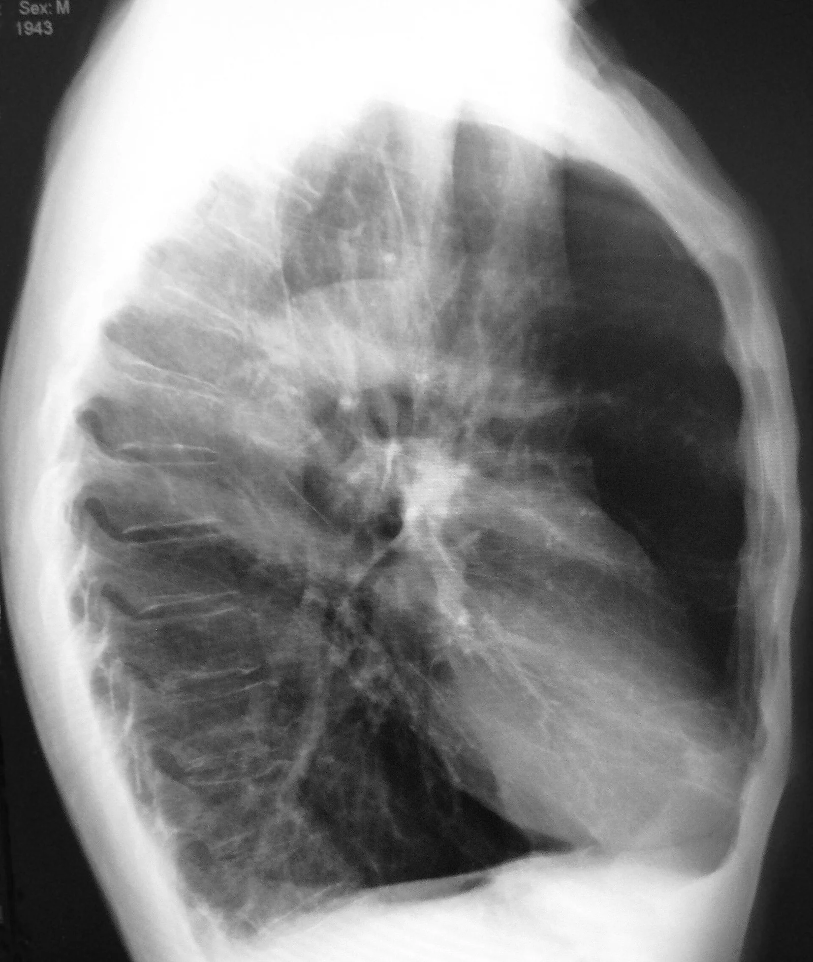
Long description
A lateral chest X-ray of a person with emphysema, showing a barrel chest and a flat diaphragm.

Figure 2Emphysema2008
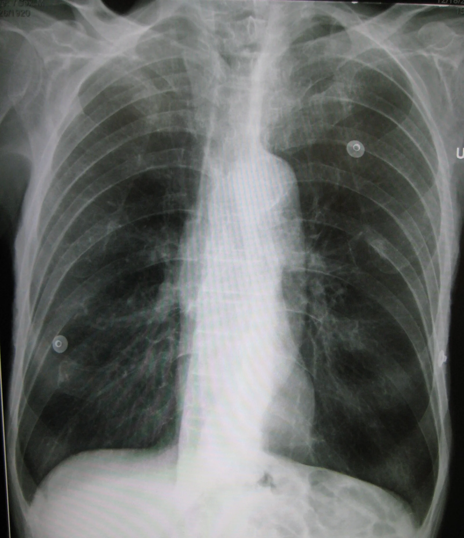
Long description
A chest X-ray of a patient with severe emphysema.

- Chest radiograph (PA and lateral) is the initial study. Classic findings are hyperinflation (flattened diaphragms, increased retrosternal airspace, horizontal ribs), attenuated peripheral markings and bullae. Bronchial wall thickening and increased bronchovascular markings suggest chronic bronchitis. It has low sensitivity for early disease but remains valuable for pneumonia, pneumothorax, effusion, pulmonary hypertension and malignancy. [1]
- High-resolution CT is not required for routine diagnosis, but it characterises emphysema phenotype, distribution and severity, bronchiectasis, air-trapping on expiratory images, bullous disease and pulmonary hypertension. It is essential for planning surgical or bronchoscopic lung volume reduction and for lung-cancer screening. [1]
The 6-minute walk test measures submaximal exercise capacity and is a validated predictor of mortality. A distance under 350 metres is associated with a markedly increased risk of death, and a change of 26 to 54 metres is the minimal clinically important difference used to judge treatment response. [1] Six-minute walk distance contributes to the BODE and ADO (age, dyspnoea, airflow obstruction) prognostic indices. [1]
The GOLD report's exacerbation typing is clinically useful:
- Type 1 (severe): all three cardinal symptoms (increased dyspnoea, increased sputum volume, increased sputum purulence)
- Type 2 (moderate): two of three
- Type 3 (mild): one cardinal symptom plus one or more supporting features, such as recent upper-respiratory infection, fever, increased wheeze or cough, or a raised heart or respiratory rate. [1]
Moderate to severe exacerbations merit arterial blood gas, chest imaging, ECG, full blood count, basic metabolic panel and sputum culture if productive. Warning signs of impending respiratory failure are: use of all accessory muscles, paradoxical breathing, saturation under 88%, rising PaCO2, pH under 7.35, falling mental status or haemodynamic instability. [1]
Diagnosis requires a compatible history and exposure plus a post-bronchodilator FEV1/FVC under 0.70. [1][2] Severity of obstruction (grade 1 to 4) and the ABE group are reported separately because they carry different implications for treatment. [1][3]
Goals are symptom relief, prevention of exacerbations and hospitalisations, slowing progression and reducing mortality. All patients need non-pharmacological measures; pharmacotherapy is individualised and escalated stepwise. [1]
- Smoking cessation is the single most effective intervention to slow FEV1 decline and cut COPD mortality, and it benefits even patients with very severe disease. Every smoker should receive pharmacotherapy (varenicline is first-line, bupropion or nicotine-replacement therapy as alternatives) plus behavioural counselling at every visit. [1][7]
- Avoidance of further exposures: occupational dust, fumes, indoor biomass combustion and outdoor air pollution. [1]
- Pulmonary rehabilitation is indicated when the mMRC is 2 or above, or the CAT 10 or above. Supervised programmes of exercise training, education and psychosocial support reduce dyspnoea, improve exercise capacity, cut exacerbation-related hospitalisation and improve quality of life. They do not change FEV1, but starting within 4 weeks after discharge reduces 30-day readmission. [1]
- Long-term oxygen therapy (LTOT): see the dedicated section below.
- Nocturnal NIV in stable disease: long-term nocturnal NIV is considered when chronic stable hypercapnia persists (PaCO2 52 mm Hg or more), especially after an exacerbation that caused acute hypercapnia; evidence supports reductions in rehospitalisation and mortality. [1][4]
- Nutrition: support for low-weight patients; low BMI is itself an independent predictor of increased mortality in COPD. [1]
Inhaled bronchodilators are the foundation of treatment. The initial agent and the escalation pathway depend on group, symptom burden, exacerbation history, eosinophil count, inhaler device capability and patient preference, with inhaler technique checked at every encounter. [1]
Short-acting bronchodilators (used as rescue by all groups):
- SABAs (salbutamol, levalbuterol): rapid-onset rescue bronchodilation. [1]
- SAMAs (ipratropium): additive bronchodilation when a SABA alone is insufficient or poorly tolerated. [1]
- SABA plus SAMA combination: more bronchodilation than either alone, and standard acute-exacerbation nebulisation. [1]
Long-acting bronchodilators (preferred maintenance):
- LAMAs (tiotropium, umeclidinium, aclidinium, glycopyrronium): 24-hour bronchodilation by M3 muscarinic blockade; they reduce dynamic hyperinflation and exacerbation frequency. Tiotropium was superior to salmeterol for exacerbation prevention in POET-COPD. [1][3]
- LABAs (salmeterol, formoterol, indacaterol, olodaterol, vilanterol): 12 to 24 hour bronchodilation via beta-2 agonism. [1]
- LABA plus LAMA dual bronchodilation (single-inhaler combinations such as umeclidinium/vilanterol, indacaterol/glycopyrronium, tiotropium/olodaterol): the preferred maintenance regimen for most symptomatic patients, with superior bronchodilation and exacerbation reduction compared with monotherapy. [1][3]
Inhaled corticosteroids: ICS monotherapy is not recommended in COPD. [1] ICS combined with a LABA adds benefit over either alone in reducing exacerbations and improving FEV1 and quality of life, but the preferred ICS-containing option today is triple therapy (ICS/LABA/LAMA), which outperforms ICS/LABA. [1][3] The evidence supports ICS when:
- blood eosinophil count 300 cells per microlitre or more (high probability of benefit),
- co-existing asthma or asthma-COPD overlap, or
- persistent exacerbations despite optimised dual bronchodilation. [1][3]
ICS (particularly fluticasone propionate) increase pneumonia risk; they should be used cautiously or avoided with recurrent pneumonia, nontuberculous mycobacterial disease, eosinophil counts under 100 cells per microlitre, or active candida infection. [1]
Triple therapy (ICS/LABA/LAMA) is indicated for symptomatic patients on dual bronchodilation who continue to exacerbate, especially with 300 eosinophils per microlitre or more. [1]
Dupilumab (anti-IL-4 receptor alpha, blocking both IL-4 and IL-13) was approved in 2024 for COPD with type 2 inflammation (blood eosinophils 300 cells per microlitre or more). BOREAS and NOTUS showed that 300 mg subcutaneously every 2 weeks reduced exacerbations and improved FEV1 in COPD with type 2 inflammation. [1][2] It is the first biologic approved for COPD.
Roflumilast, a phosphodiesterase-4 inhibitor given as 500 micrograms (0.5 mg) orally once daily, is an add-on for severe COPD (FEV1 under 50% predicted) with chronic bronchitis and frequent exacerbations despite optimised bronchodilation. It reduces exacerbations by roughly 15 to 20%. Principal adverse effects are diarrhoea, nausea, significant weight loss and insomnia; it is contraindicated with BMI under 18.5 kg per m squared and in severe hepatic impairment. [1]
Azithromycin maintenance (250 mg daily, or 500 mg three times weekly) reduces exacerbation frequency in selected patients: frequent exacerbators on optimised therapy who are ex-smokers or light smokers (benefit is weaker in active smokers, and long-term macrolides are restricted to ex-smokers or light smokers because of that lower efficacy). Careful pre-treatment assessment is mandatory: baseline ECG (QTc over 450 ms is a relative contra-indication), audiometry, sputum culture to exclude nontuberculous mycobacteria, and liver function tests, with annual audiometry thereafter. It is not an absolute contra-indication in patients who still smoke, although evidence of benefit is weaker in active smokers: a review of the MACRO trial found azithromycin mainly effective in ex-smokers with no clear preventive effect in active smokers, and GOLD restricts long-term macrolides to ex-smokers or light smokers because of lower efficacy. Continue to caution about infection, hearing, QTc and resistance rather than excluding current smokers outright. [1][10]
Methylxanthines (theophylline) are third-line because of a narrow therapeutic window and interactions; therapeutic levels are 5 to 15 micrograms per mL, with toxicity above 20 micrograms per mL. [1]
- Lung volume reduction surgery benefits upper-lobe predominant emphysema with low post-rehabilitation exercise capacity, improving survival and function in NETT-selected patients. It is contraindicated with FEV1 under 20% predicted, homogeneous emphysema, or DLCO under 20% predicted. [1]
- Bronchoscopic lung volume reduction with endobronchial valves (Zephyr) achieves sustained lobar volume loss in heterogeneous emphysema with intact interlobar fissures. LIBERATE showed improvements in FEV1, 6-minute walk distance and quality of life at 12 months; pneumothorax occurs in roughly a quarter of patients and is usually managed conservatively. [1]
- Bullectomy for giant bullae. [1]
- Lung transplantation is considered for GOLD 3 to 4 disease with a BODE index of 7 to 10, PaCO2 over 50 mm Hg, FEV1 under 20% predicted, rapidly progressive disease, or emphysema unsuitable for volume reduction. Bilateral sequential transplantation is preferred. [1]
The outline below follows an interprofessional evidence-based algorithm for applying NIV in acute exacerbation of COPD. [4]
Approach to an acute exacerbation of COPD
- Assess severityMental state, dyspnoea at rest, sputum volume and purulence, accessory muscle use, paradoxical breathing; oxygen saturation, arterial blood gas, chest radiograph, ECG, full blood count
- Controlled oxygenTarget saturation 88 to 92% with a Venturi mask to deliver precise inspired oxygen; avoid uncontrolled high-flow oxygen, which worsens ventilation-perfusion mismatch and can suppress the hypoxic drive
- BronchodilatorsNebulised salbutamol plus ipratropium every 20 minutes initially, then every 4 to 6 hours around the clock
- Systemic corticosteroidPrednisone 40 mg once daily for 5 days (or methylprednisolone 40 to 80 mg intravenously every 8 to 12 hours), started early. A five-day course was non-inferior to fourteen days with fewer adverse effects (REDUCE).
- AntibioticsIf sputum purulence accompanies increased dyspnoea or volume (type 1 or 2 exacerbation): first-line azithromycin, doxycycline or amoxicillin-clavulanate for 5 to 7 days. Escalate to levofloxacin or moxifloxacin in severe disease, recent hospitalisation or recent antibiotics; add pseudomonal cover where prior Pseudomonas or bronchiectasis exists.
- Question: Acute hypercapnic respiratory failure?
No: pH 7.35 or more
- Continue on wardDeep vein thrombosis prophylaxis, glucose control, early mobilisation; transition early to oral therapy and inhaled bronchodilators; plan 30-day follow-up
Yes: NIV criteria
- Non-invasive ventilationStrongly indicated with pH under 7.35 and PaCO2 over 45 mm Hg despite medical therapy; also with persistent hypoxaemia. Starter settings differ by source: many interprofessional algorithms use inspiratory positive airway pressure 8 to 12 cm H2O (10 most common) with expiratory positive airway pressure 3 to 5 cm H2O (5 most common), whereas the StatPearls summary uses 10 to 14 and 4 to 6 cm H2O; inspired oxygen titrated to 88 to 92%. Titration ceilings also differ by source: up to 20 to 24 cm H2O (StatPearls) or 20 to 30 cm H2O (interprofessional algorithm), raising the inspiratory pressure by 2 cm H2O at a time. Review arterial blood gases every 30 to 60 minutes.
- Question: Improves within 1 to 2 hours?
Yes
- Continue NIV, then weanWean after at least 24 hours of stable success (pH 7.35 or more, normalised respiratory rate, resolving cause), with an arterial CO2 fall of at least 10% from baseline.
No
- Invasive ventilationIf NIV fails, if the airway cannot be protected, or with haemodynamic collapse or arrest. Use lung-protective ventilation with permissive hypercapnia and prolonged expiratory time (I:E 1:3 to 1:4) to avoid dynamic hyperinflation.
- Discharge and follow-upWritten exacerbation action plan; 30-day follow-up; re-assess oxygen needs at 1 to 4 weeks and again at 12 to 16 weeks
NIV criteria and settings come from published guideline-based recommendations and an evidence-based interprofessional NIV algorithm. [4] Briefly, NIV is most likely to help when, despite standard therapy, the pH is 7.35 or less, PaCO2 is over 45 mm Hg and respiratory rate is over 20 to 24 breaths per minute; severe predictors of failure include a falling pH under 7.25 and a Glasgow Coma Scale (GCS) under 11. [4] Absolute contraindications include arrest, severe distress and unstable arrhythmia. [4]
| Element | Acute exacerbation | Stable chronic disease |
|---|---|---|
| Oxygen | Controlled oxygen to a saturation of 88 to 92% with a Venturi mask[4] | Long-term oxygen only for severe resting hypoxaemia, for at least 15 hours a day[5] |
| Bronchodilators | Nebulised salbutamol plus ipratropium, repeated early[1] | LAMA or LABA plus LAMA maintenance, with a short-acting rescue inhaler[1] |
| Corticosteroids | Prednisone 40 mg daily for 5 days, or intravenous methylprednisolone if oral is not possible[4] | Inhaled corticosteroid only with eosinophils 300 or more, asthma overlap or persistent exacerbations[1] |
| Ventilatory support | Non-invasive ventilation when pH is 7.35 or less and PaCO2 is over 45 mm Hg; invasive ventilation if it fails[4] | Nocturnal NIV for persistent hypercapnia (PaCO2 52 mm Hg or more)[1] |
| Other | Antibiotics when indicated; look for the trigger[1] | Smoking cessation, rehabilitation, vaccination, review of inhaler technique[1] |
The American Thoracic Society guideline defines eligibility: [5]
- Severe hypoxaemia: PaO2 55 mm Hg or less (7.3 kPa), or saturation 88% or less; or PaO2 56 to 59 mm Hg (7.5 to 7.9 kPa) with saturation 89% plus one of: peripheral oedema, haematocrit 55% or more, or P pulmonale on ECG.
- LTOT for at least 15 hours a day reduces 2-year mortality by about 55% and 5-year mortality by about 59% in severe resting hypoxaemia (NOTT and MRC trials). [5]
- Moderate hypoxaemia (saturation 89 to 93%) does not benefit from routine LTOT. [5]
Chronicity matters: in NOTT the criteria had to be met on at least two occasions more than a week apart over a 3-week stable period, and in the Medical Research Council (MRC) trial at least 3 weeks apart. [5] After a COPD exacerbation many patients' oxygen needs fall: the ATS guideline notes that perhaps half of those started on home oxygen at discharge may no longer need it, so reassess at 1 to 4 weeks and 12 to 16 weeks, and thereafter at least every 6 months. [5]
Review at least within 30 days of an exacerbation, with annual spirometric review as needed. Step therapy up when control is poor (confirm diagnosis, technique, adherence and triggers first) and step down to the minimum effective dose once good control has been maintained for 2 to 3 months. Provide a written exacerbation plan and check inhaler technique at every visit. [1]
- Cor pulmonale and pulmonary hypertension from chronic hypoxaemia. [1]
- Pneumothorax, particularly in bullous emphysema. [1]
- Polycythaemia (erythrocytosis) from chronic hypoxaemia, raising thrombotic risk. [1]
- Respiratory failure, type 2 in advanced disease, sometimes an acute-on-chronic presentation. [1]
- Hospital-acquired infections and osteoporosis under long-term systemic corticosteroids. [1]
- Cardiovascular events are increased during and after exacerbations and are a leading cause of death in mild to moderate disease. [1][2]
- Malnutrition and cachexia, with low BMI an independent predictor of mortality. [1]
Prognosis is variable and depends on severity, exacerbation history, comorbidities, functional status and adherence. [1] Lung function alone underestimates risk, so multidimensional tools are used:
- BODE index (body mass index, airflow obstruction by FEV1, dyspnoea by mMRC, exercise capacity by 6-minute walk distance), scoring 0 to 10, predicts 4-year all-cause mortality and outperforms FEV1 alone. [1][6] In published BODE scoring schemes points are assigned 0 to 3 for FEV1 percent predicted (0 for 65% or more, 1 for 50 to 64%, 2 for 36 to 49%, 3 for 35% or less), mMRC dyspnoea grade (0 for grades 0-1, 1 for grade 2, 2 for grade 3, 3 for grade 4), 6-minute walk distance (0 for 350 m or more, down to 3 for under 150 m), and 0 or 1 for BMI (0 if over 21 kg per m squared, 1 if 21 or less). [6]
- Lung transplant evaluation is triggered by a BODE of 7 to 10, PaCO2 over 50 mm Hg or FEV1 under 20% predicted, among other criteria. [1]
- A 6-minute walk distance under 350 m independently flags an increased risk of death. [1]
Because there is no population screening for COPD, prevention rests on exposure control: avoiding tobacco initiation and passive smoke, clean cooking and ventilation to reduce biomass exposure, and occupational dust control. [1] Early case-finding in anyone over 40 with symptoms or relevant exposures, and confirmation by spirometry, allows intervention before fixed obstruction and repeated exacerbations fix the course. [1]
Vaccination is a core part of every patient's care:
- Annual influenza vaccination (inactivated or recombinant; live attenuated vaccines are contraindicated when immunocompromised). [1]
- Pneumococcal vaccination: per current ACIP guidance, a single dose of a pneumococcal conjugate vaccine (PCV15, PCV20 or PCV21) for adults 50 or older and for adults 19 to 49 with risk conditions such as chronic lung disease; if PCV15 is used, follow with PPSV23. Age thresholds, products and sequencing vary by region and year, so the official local schedule should be used. [1][11]
- COVID-19 vaccination per current CDC and ACIP guidance. [1]
- RSV vaccination: age and product recommendations vary by region and year. Current ACIP guidance gives a single dose to all adults 75 or older and to adults 50 to 74 (earlier 60 to 74) at increased risk of severe disease; eligibility has recently widened toward age 50 in the United States. GOLD 2026 names vaccination as a core disease-modifying strategy but leaves the age and product to local guidance. [9][11]
- Tdap or Td per the standard adult schedule. [1]
- Older adults: spirometry may be harder to interpret, smoking and COPD features are commoner, and late-onset asthma can mimic the disease; recognising that fixed, progressive obstruction still requires the COPD pathway matters. [1]
- Alpha-1 antitrypsin deficiency: see aetiology above; management is according to usual COPD guidelines plus, where available and when severe deficiency with significant lung impairment is present, augmentation therapy, with smoking avoidance paramount. [1]
- Patients with overlap (asthma-COPD): maintain ICS and use bronchodilators, rather than bronchodilator-only treatment, because that is unsafe in the asthma component. [1]
Most of the global COPD burden falls in low- and middle-income countries, where biomass smoke, dusty work and late diagnosis dominate and where spirometry, CT, bronchodilator combinations, oxygen and specialist review may all be scarce. [1] Practical points:
- A clinical diagnosis from history and peak-flow or, where available, a single spirometry is better than no diagnosis; use the mMRC and CAT to phenotype and guide treatment.
- Treat tobacco dependence with very low cost tools first: behavioural counselling, with pharmacotherapy where affordable; varenicline is dosed as 0.5 mg once daily for 3 days, then 0.5 mg twice daily for 4 days, then 1 mg twice daily for 12 weeks, with an added 12-week course for those who quit. [1][7]
- Make sure inhalers can be used properly, with demonstration and, where commercial spacers are not available, bottle spacers with metered-dose inhalers; dry-powder inhalers reduce maintenance but need good inspiratory flow.
- Reserve oral corticosteroids (prednisone 40 mg daily for 5 days) and cheap antibiotics for the exacerbations that meet the criteria (purulent sputum with increased dyspnoea or volume).
- Oxygen is often the most expensive item. Begin LTOT only for severe resting hypoxaemia; re-assess needs and, where patients improve, discontinue; titrated, low-flow nasal oxygen prevents hospital-acquired hypercapnia better than high-flow masks.
- Pulmonary rehabilitation can be delivered with minimal equipment (walking, lower-limb strengthening, education) by trained physiotherapists or nurses.
- Think of tuberculosis in anyone with chronic cough, weight loss or haemoptysis, and test before assuming COPD. [1]
- COPD is a preventable, treatable disease defined by persistent, usually progressive airflow limitation (post-bronchodilator FEV1/FVC under 0.70) caused by small-airway disease and emphysema, with systemic effects. [1][2]
- Tobacco smoking causes 70 to 80% of cases in high-income countries, but 25 to 45% of cases occur in never-smokers, mostly from biomass fuel, occupational dust and AATD. [1]
- The label COPD covers two overlapping pathologies: chronic bronchitis (productive cough on most days for 3 months in 2 consecutive years) and emphysema (destruction of airspaces distal to terminal bronchioles). [1]
- "Pink puffer" and "blue bloater" are teaching contrasts, not diagnostic classes; most patients fall between them. [1]
- Clinical diagnosis is insidious and most cases are missed; screen anyone with symptoms or exposure, and confirm with spirometry. [1]
- Severity (GOLD grade 1 to 4 by FEV1) and treatment group (A, B, E by symptoms and exacerbations) are reported separately; groups use CAT (10 or more) and mMRC (2 or more) cut-offs. [1][3]
- Blood eosinophils guide ICS use: strongly favour ICS at 300 cells per microlitre or more; do not use ICS with under 100 cells per microlitre or recurrent pneumonia. [1][3]
- Stable maintenance is LABA plus LAMA for groups B and E, with early single bronchodilator for group A; escalate to ICS/LABA/LAMA (triple therapy) when exacerbations persist; add roflumilast, azithromycin or dupilumab with specific indications. [1][3]
- Acute exacerbations: controlled oxygen 88 to 92%, nebulised SABA plus SAMA, prednisone 40 mg daily for 5 days, antibiotics for purulent sputum with increased dyspnoea or volume, and NIV for pH 7.35 or less with PaCO2 over 45 mm Hg. [1][4]
- LTOT 15 or more hours a day for severe resting hypoxaemia (PaO2 55 mm Hg or less, or 56 to 59 mm Hg with complications) reduces 2-year mortality by 55% and 5-year mortality by 59%; it is not beneficial for moderate hypoxaemia. [5]
- Complications include cor pulmonale, pneumothorax, polycythaemia, respiratory failure and cardiovascular events. [1]
- Prognosis is best estimated by multidimensional indices such as BODE (0 to 10), which outperforms FEV1 alone and feeds transplant referral at 7 to 10. [1][6]
- Every patient needs smoking cessation support, pulmonary rehabilitation when symptomatic, and full vaccination (influenza, pneumococcal, COVID, RSV, Tdap), plus AATD screening. [1]
Answer each one in your head first, then open the answer.
A 62-year-old man has smoked 40 pack-years, a chronic productive cough every winter for 4 years, and progressive exertional breathlessness. Spirometry shows FEV1/FVC 0.62 after salbutamol with FEV1 58% predicted. How do you confirm and classify the disease?
Show answerHide answer
Answer: He has COPD. The diagnosis is confirmed by a post-bronchodilator FEV1/FVC under 0.70, which separates persistent obstruction from the reversible limitation of asthma. The FEV1 of 58% predicted puts him in GOLD grade 2 (moderate). Grouping follows his symptoms and exacerbations: with significant symptoms (CAT 10 or more or mMRC 2 or more) but one or fewer exacerbations a year he is group B, for which the initial maintenance is LABA plus LAMA. [1][3]
Explain why an emphysema-predominant patient is dyspnoeic at low workloads even though arterial oxygen can be normal at rest.
Show answerHide answer
Answer: Destruction of alveolar walls loses the elastic recoil that normally holds small airways open during expiration, so they collapse and trap gas; hyperinflation flattens the diaphragm into a mechanically disadvantageous position and increases the elastic work of breathing. Gas exchange is relatively preserved until late because ventilation and perfusion fall together in destroyed units, which is why these patients may stay pink. Additional work of breathing at modest loads produces dyspnoea out of proportion to resting desaturation. [1]
A patient with severe COPD on maximised inhaled therapy exacerbates twice a year. Blood eosinophils are 420 cells per microlitre. What escalation would you choose and why?
Show answerHide answer
Answer: His eosinophil count of 420 is above 300 per microlitre, so inhaled corticosteroid benefit is likely: move from LABA plus LAMA to single-inhaler triple therapy (ICS/LABA/LAMA), the preferred ICS-containing regimen. If exacerbations continue despite triple therapy he becomes a candidate for dupilumab (300 mg subcutaneously every 2 weeks), approved for type 2 COPD at 300 eosinophils per microlitre or more, with roflumilast considered if the phenotype is severe obstruction with chronic bronchitis. [1][2]
A patient arrives at the emergency department with a severe exacerbation of COPD, saturating 86% on room air, drowsy and using all accessory muscles. What are the immediate priorities up to the decision about ventilation?
Show answerHide answer
Answer: Give controlled oxygen with a Venturi mask targeting 88 to 92%, and nebulised salbutamol plus ipratropium every 20 minutes initially. Start a systemic corticosteroid (prednisone 40 mg once daily for 5 days, or methylprednisolone 40 to 80 mg intravenously every 8 to 12 hours) and give antibiotics if purulence accompanies increased dyspnoea or sputum volume. Send arterial blood gases; non-invasive ventilation is strongly indicated with pH under 7.35 and PaCO2 over 45 mm Hg despite medical therapy. Escalate to invasive ventilation if NIV fails, the airway cannot be protected, or there is haemodynamic collapse. [1][4]
What are the entry criteria and expected benefit for long-term oxygen therapy in COPD?
Show answerHide answer
Answer: LTOT is indicated for severe resting hypoxaemia: PaO2 under 55 mm Hg (or saturation 88% or less), or PaO2 56 to 59 mm Hg (saturation 89%) plus peripheral oedema, haematocrit 55% or more, or P pulmonale. Given for at least 15 hours a day it reduced 2-year mortality by about 55% (NOTT) and 5-year mortality by about 59% (MRC). Moderate hypoxaemia (saturation 89 to 93%) does not benefit from routine LTOT. Needs should be re-assessed after exacerbations because some patients recover. [5]
A BODE index is 9 in a patient with GOLD 3 obstruction. What does this imply, and what management should be considered?
Show answerHide answer
Answer: BODE combines BMI, FEV1, mMRC dyspnoea and 6-minute walk distance, scoring 0 to 10; it predicts 4-year all-cause mortality better than FEV1 alone. A score of 9 falls in the highest quartile (7 to 10), at which point, together with criteria such as PaCO2 over 50 mm Hg or FEV1 under 20% predicted, referral for evaluation for lung transplantation is considered. [1][6]
- Asthma and asthma-COPD overlap
- Acute respiratory distress syndrome and mechanical ventilation
- Cor pulmonale and pulmonary hypertension
- Alpha-1 antitrypsin deficiency
- Pneumonia
- Lung cancer and screening
- Smoking cessation
Pulmonary function tests
How lung volumes, airflow, gas transfer and blood gases are measured and what their patterns mean.Asthma
A chronic inflammatory airway disease causing wheeze and breathlessness, with reversible obstruction; modern stepwise management and acute care.Bronchiectasis
Permanent dilatation of the bronchi: causes, the cycle of inflammation and infection, diagnosis, management, complications, and low-resource care.Hypoxaemia and respiratory failure
How hypoxaemia develops, how to read the blood gas and A-a gradient, and how to give oxygen and ventilatory support safely.Pulmonary hypertension and cor pulmonale
How high pulmonary pressures arise, are classified, diagnosed and treated, and what they mean for the right heart and cor pulmonale.
8 other topics link here.
- Kanchustambham V, Brown BD. Chronic Obstructive Pulmonary Disease (COPD) - StatPearls. StatPearls [Internet]. Treasure Island (FL): StatPearls Publishing; 2026 Jan-.. Accessed .
- Halpin DMG, Singh D. What's new in the 2025 GOLD report. J Bras Pneumol 2025;51(1):e20240412. doi:10.36416/1806-3756/e20240412. Accessed .
- Duan R, Li B, Yang T. Pharmacological therapy for stable chronic obstructive pulmonary disease. Chronic Dis Transl Med 2023;9:82-89. doi:10.1002/cdt3.65. Accessed .
- Farmer MJS, Callahan CD, Hughes AM, Riska KL, Hill NS. Applying Noninvasive Ventilation in Treatment of Acute Exacerbation of COPD Using Evidence-Based Interprofessional Clinical Practice. Chest 2024;165(6):1469-1480. doi:10.1016/j.chest.2024.02.040. Accessed .
- Jacobs SS, Krishnan JA, Lederer DJ, et al. Home Oxygen Therapy for Adults with Chronic Lung Disease. An Official American Thoracic Society Clinical Practice Guideline. Am J Respir Crit Care Med 2020;202(10):e121-e141. doi:10.1164/rccm.202009-3608ST. Accessed .
- Saadhya M, et al. BODE index: A predictor of hospitalization and severity in chronic obstructive pulmonary disease patients. Journal article. (PMC12569876). Accessed .
- Marlex Pharmaceuticals, Inc. Varenicline (varenicline tartrate tablet) prescribing information. DailyMed drug label. Accessed .
- Guder G, Brenner S, Angermann CE, et al. GOLD or lower limit of normal definition? a comparison with expert-based diagnosis of chronic obstructive pulmonary disease. Respir Res 2012;13:13. doi:10.1186/1465-9921-13-13. Accessed .
- Cazzola M, Bajpai J, Calzetta L, Matera MG, Rogliani P. GOLD 2026: Transforming COPD Management with Early Intervention, Multi-dimensional Assessment, and Personalized Care. Drugs 2026;86(5):581-597. doi:10.1007/s40265-026-02303-3. Accessed .
- Pomares X. Macrolides: Indications and Contraindications. Barc Respir Netw Rev 2023. (Br n Rev 2023;9(1)). Accessed .
- Centers for Disease Control and Prevention. Adult immunization schedule and clinical guidance: RSV and pneumococcal vaccination (ACIP/CDC, 2024-2025). CDC web guidance. (RSV: adults 75+ and at-risk 50-74/60-74; pneumococcal: one dose PCV for adults 50+ and 19-49 at risk). Accessed .
Figure credits
Educational content for medical students and doctors. It is not a substitute for clinical judgement or local guidelines.