Lung cancer
Lung cancer from first suspicion to treatment by stage: risks, types, presentation, staging, molecular tests, and care in low-resource settings.
- Lung cancer is the commonest cause of cancer death worldwide, with about 2.48 million new cases and 1.81 million deaths in 2022. [5]
- Tobacco smoking is the dominant risk factor in industrialised countries, accounting for more than 80% of cases in both men and women; in never-smokers the disease has different risk factors and is commoner in women, especially in Asia. [4]
- The main groups are non-small-cell lung cancer (NSCLC: adenocarcinoma, squamous cell, large cell) and small-cell lung cancer (SCLC); carcinoid tumours are a separate group. [1]
- Most people present with cough, breathlessness, chest pain or haemoptysis, often after the cancer has spread; the commonest distant sites are liver, adrenal gland, brain and bone. [1]
- Curative treatment depends on stage: surgery or SABR for early disease, chemoradiotherapy and durvalumab for locally advanced disease, and systemic treatment chosen by PD-L1 level and by driver mutations such as EGFR and ALK for metastatic disease. [1][3]
By the end of this page you should be able to:
- Explain why lung cancer is so common and identify the main modifiable and non-modifiable risk factors.
- Describe the NSCLC and SCLC groups, where each type usually grows, and the typical associations of each.
- Recognise the pulmonary, invasive and paraneoplastic features of lung cancer and state the commonest metastatic sites.
- Choose and interpret the first-line and confirmatory investigations, including CT, PET-CT, EBUS and brain MRI.
- Apply the 9th edition TNM staging changes and explain why brain MRI is part of staging.
- Outline treatment by stage, including surgery, SBRT, chemoradiation, durvalumab, targeted therapy and immunotherapy.
- Summarise screening, prognosis and the practical priorities in a low-resource setting.
DefinitionLung cancer
Lung cancer is a malignant tumour that arises from the epithelium of the bronchi, bronchioles or alveoli. It includes non-small-cell lung cancer (NSCLC) and small-cell lung cancer (SCLC), which differ in growth rate, spread and treatment. NSCLC is subdivided further into adenocarcinoma, squamous cell carcinoma, large cell carcinoma and other less common types.
The two behave differently, so the rest of this page treats them separately once the classification has been set out.
| Type | Where it usually arises | Typical associations and behaviour |
|---|---|---|
| Adenocarcinoma | Peripheral lung, near the pleura | The commonest type in never-smokers and in women; often forms a solitary peripheral nodule; grows along alveolar walls (lepidic growth) before becoming invasive |
| Squamous cell carcinoma | Central, in a main bronchus | Strongly linked to smoking; tends to cause early bronchial obstruction, cavitation and post-obstructive infection |
| Large cell carcinoma | Often peripheral | An NSCLC that lacks the features of adenocarcinoma or squamous carcinoma; aggressive |
| Small-cell lung cancer (SCLC) | Central hilar mass, mediastinal nodes | Almost always smoking-related; rapid growth; early spread to nodes and distant sites; responds initially to chemotherapy |
| Carcinoid (neuroendocrine tumour) | Often a central endobronchial lesion | Slow-growing; may cause recurrent infections or wheeze from airway obstruction; atypical carcinoid is more aggressive |
| Pan (superior sulcus/Pancoast) tumour | Apex of the lung | A type of NSCLC that invades the brachial plexus and sympathetic chain at the apex, producing Pancoast syndrome and sometimes Horner syndrome |
SCLC is staged and treated differently from NSCLC, so it has its own later section. Adenocarcinoma is now the dominant type in many countries because squamous cancer is falling as smoking prevalence falls, while adenocarcinoma in never-smokers is rising. [4]
Lung cancer is the most frequently diagnosed cancer worldwide and the leading cause of cancer death. About 2.48 million new cases and 1.81 million deaths were recorded in 2022. [5] It is uncommon in young adults and rises steeply with age.
- Smoking prevalence still drives the pattern. In industrialised countries such as the United Kingdom and the United States, smoking accounts for more than 80% of lung cancer cases in both men and women. [4]
- In countries with low female smoking rates, most lung cancer in women occurs in never-smokers. In China, smoking accounts for 57.5% of male lung cancer but only 12.5% of female lung cancer, and more than 80% of female lung cancers occur in women who have never smoked. [4]
- Never-smoker lung cancer has its own epidemiology. Globally about two-thirds of never-smoker lung cancers occur in women, with higher prevalence in Asian populations. [4]
- Stage at diagnosis still matters most for outcome. Population-level data show that most patients are diagnosed after the cancer has spread beyond the lung. [5]
Smoking is the single most important cause. Risk rises with the number of cigarettes smoked per day, the duration of smoking and the depth of inhalation, and falls gradually after cessation. There is no safe level of tobacco exposure. [4] Passive (secondhand) smoke raises risk too, with a dose-response relationship based on duration, intensity and cumulative exposure. [4]
Radon is a radioactive gas that seeps from the ground into buildings. It is the second leading cause of lung cancer in many countries and the leading cause in never-smokers in some regions. Prolonged exposure damages airway DNA; the risk is much higher in people who also smoke. [4]
Inhaled asbestos fibres cause chronic inflammation and scarring of lung and pleural tissue and markedly raise lung cancer risk, especially in smokers. Other occupational carcinogens include silica, diesel exhaust, arsenic, chromium, cadmium, nickel and benzene. [4]
Long-term exposure to fine particulate matter (PM2.5) and to nitrogen oxides increases the risk of lung cancer and of chronic lung disease. The effect is smaller than that of smoking but affects very large populations, particularly in cities with poor air quality. [4]
- Previous chest disease. Chronic obstructive pulmonary disease and pulmonary fibrosis increase risk even after allowing for smoking.
- Radiation. Previous radiotherapy to the chest and high doses of ionising radiation raise risk.
- Family history. A first-degree relative with lung cancer approximately doubles risk in many studies.
- Genetic susceptibility. Rare inherited cancer syndromes and common low-penetrance variants contribute, although lung cancer is not generally considered a hereditary disease.
- Infection. Prior tuberculosis and recurrent pneumonia are associated with higher risk in some studies. [4]
Clinical pearlExam point
Lung cancer in a never-smoker is not a rarity. In China, more than 80% of lung cancers in women occur in women who have never smoked. Never ask only "how much did you smoke?" Always ask about radon, occupation, air pollution and family history as well. [4]
Lung cancer usually begins with repeated carcinogen damage to the bronchial epithelium, followed by a sequence of visible precancerous lesions: basal cell hyperplasia, squamous metaplasia, dysplasia, carcinoma in situ, and finally invasive cancer. [4] The whole process explains why cancer tends to arise in damaged, inflamed airways and why field cancerisation makes second cancers more likely.
Mechanistically, the tumour causes disease in four main ways.
- Local airway obstruction. A central tumour narrows or blocks a bronchus, causing post-obstructive pneumonia, collapse or air trapping distal to it. This explains wheeze, stridor and recurrent infection.
- Local invasion. Tumours can invade the chest wall, mediastinum, pleura, pericardium, recurrent laryngeal nerve, phrenic nerve, superior vena cava and the paravertebral sympathetic chain. Each produces a recognisable syndrome (see below).
- Metastatic spread. The commonest distant sites are liver, adrenal gland, brain and bone (the "LABBs"), which is why the initial CT includes the liver and adrenals and why brain MRI is built into staging. [1][5]
- Biologically active secretions. Tumour cells or their inflammatory by-products release hormones and cytokines that produce paraneoplastic syndromes, sometimes before the primary tumour is obvious.
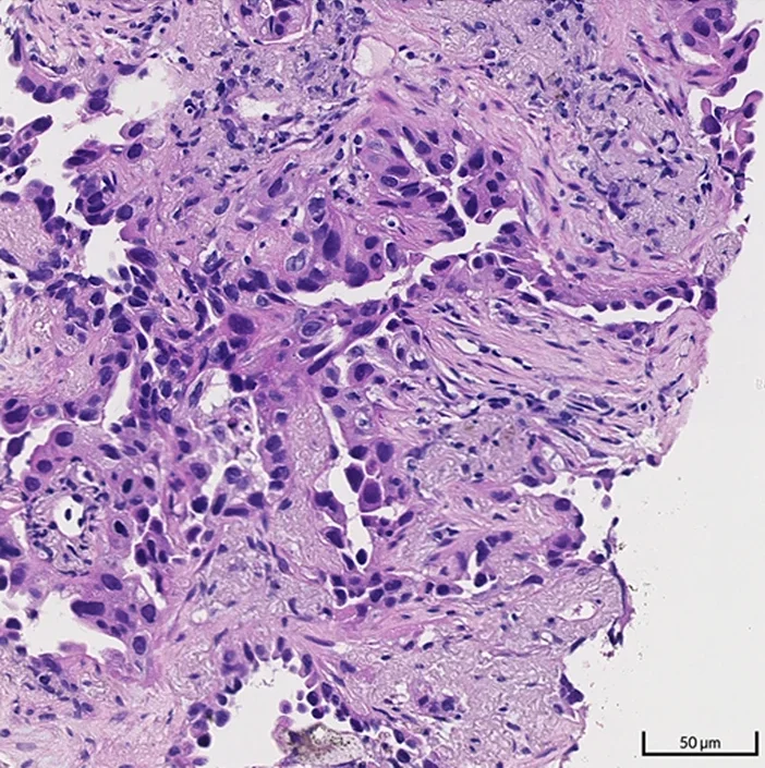
Figure 1Histopathology of lung adenocarcinoma with acinar pattern
Long description
Histopathology of lung adenocarcinoma with acinar pattern, H&E stain.

Central cancers usually present as an irregular mass in or around a main bronchus, sometimes with extensive necrosis and cavitation (especially squamous cell carcinoma). Peripheral cancers are often firm grey-white nodules with a pleural tag. SCLC typically presents as a bulky perihilar mass with early lymph node involvement.
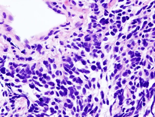
Figure 2Lung small cell carcinoma (1) by core needle biopsy
Long description
Small cell carcinoma of the lung, CT-guided core biopsy, H&E

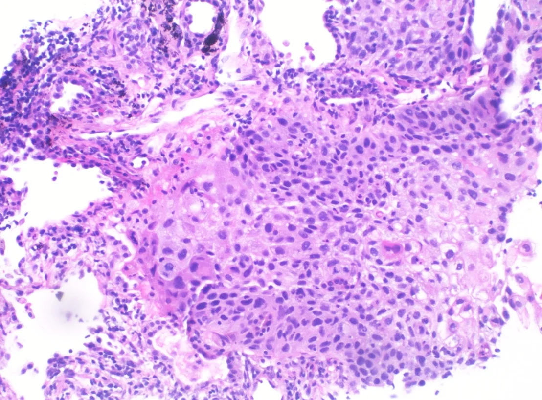
Figure 3Histopathology of squamous-cell carcinoma of the lung
Long description
Histopathology of poorly differentiated squamous-cell carcinoma of the lung from a lung biopsy, with H&E stain and a negative GATA-3 stain used to exclude metastasis from skin.

Adenocarcinoma shows gland formation and mucin production. Squamous carcinoma shows keratin pearls and intercellular bridges. Large cell carcinoma lacks these features. SCLC consists of small cells with scant cytoplasm, fine chromatin ("salt-and-pepper") and absent or inconspicuous nucleoli; they express neuroendocrine markers such as chromogranin and synaptophysin. [1] Carcinoid tumours also express neuroendocrine markers but are architecturally well differentiated.
Lung cancer is protean. It may be found by accident on a chest film taken for another reason, it may present with a single symptom, or it may declare itself with metastatic disease.
- Cough, often new or changed, is the commonest symptom. [1]
- Haemoptysis (coughing up blood) is a red flag at any age in a smoker. [1]
- Breathlessness from obstruction, effusion, anaemia or loss of functional lung.
- Chest or shoulder pain, pleuritic or dull; shoulder pain suggests apical involvement.
- Weight loss and anorexia, often with malaise and fatigue.
- Hoarse voice from recurrent laryngeal nerve palsy.
- Bone pain from skeletal metastasis.
- Headache, confusion or fits from brain metastasis.
- Swelling of the face and neck (facial plethora, dilated neck veins) from superior vena cava obstruction.
Figure 4Superior.vena.cava.syndrome.aak

Long description
Superior vena cava syndrome with dilated superficial veins

- Reduced air entry, dullness and bronchial breathing over a post-obstructive lobe.
- Stridor or wheeze from central obstruction.
- Clubbing and hypertrophic osteoarthropathy (see paraneoplastic section).
- Cervical or supraclavicular lymphadenopathy.
- Pancoast features: shoulder/medial arm pain, weakness of the small hand muscles and, sometimes, Horner syndrome (ptosis, miosis and anhidrosis) from sympathetic chain invasion. [1]
- Horner syndrome also occurs with metastatic disease elsewhere, but a new Horner syndrome with shoulder pain points to an apical tumour.
- Superior vena cava syndrome: facial and arm swelling, plethora, distended neck and chest wall veins, breathlessness, headache. This is an oncological emergency.
- Pleural effusion: dull percussion note, reduced breath sounds, tracheal deviation away from the effusion.
Paraneoplastic syndromes are effects of the tumour that are not explained by its mass or metastases. They are more often seen with SCLC but occur in NSCLC too.
- SIADH (syndrome of inappropriate antidiuresis): hyponatraemia with concentrated urine, usually small-cell disease. [1]
- Hypercalcaemia from PTHrP (parathyroid hormone-related peptide): thirst, polyuria, confusion and constipation, typically squamous cell carcinoma. [1]
- Cushing syndrome: weight gain, hypertension, hypokalaemia and hyperglycaemia from ectopic ACTH secretion, most often SCLC.
- Lambert-Eaton myasthenic syndrome: proximal muscle weakness that improves with repeated effort, reduced or absent reflexes, often with autonomic features; associated with SCLC.
- Hypertrophic osteoarthropathy: clubbing plus painful symmetrical periostitis of the long bones ("Pierre Marie-Bamberger syndrome").
- Other, rarer syndromes include cerebellar degeneration, limbic encephalitis and dermatomyositis.
| Organ system | Syndrome | Features | Usual tumour type |
|---|---|---|---|
| Endocrine | SIADH | Hyponatraemia with concentrated urine[1] | Small cell |
| Endocrine | Hypercalcaemia from PTHrP | Thirst, polyuria, confusion, constipation[1] | Squamous cell |
| Endocrine | Cushing syndrome (ectopic ACTH) | Weight gain, hypertension, hypokalaemia, hyperglycaemia | Small cell |
| Neurological | Lambert-Eaton myasthenic syndrome | Proximal weakness that improves with repeated effort, absent reflexes, autonomic features | Small cell |
| Neurological | Cerebellar degeneration, limbic encephalitis | Rarer; may precede the cancer diagnosis | Mainly small cell |
| Musculoskeletal | Hypertrophic osteoarthropathy | Clubbing with painful symmetrical periostitis of the long bones | Mainly non-small-cell |
| Skin | Dermatomyositis | Rarer | Various |
When these syndromes appear, the underlying tumour should be sought even if the primary is not yet visible.
Red flagDo not ignore
- Haemoptysis at any age, especially in a smoker or former smoker.
- A new persistent cough that does not settle.
- Finger clubbing with weight loss.
- Horner syndrome or shoulder pain suggesting an apical tumour.
- Superior vena cava syndrome: urgent investigation.
- Unexplained hyponatraemia or hypercalcaemia in an adult smoker.
The differential includes both non-malignant conditions that mimic lung cancer and other cancers that can spread to the lung.
| Condition | Feature that separates it from lung cancer |
|---|---|
| Chronic obstructive pulmonary disease (COPD) | Chronic, progressive dyspnoea; no progressive lesion or weight loss; spirometry shows obstruction |
| Pneumonia or tuberculosis | Fever, productive cough, response to antibiotics or anti-TB treatment; imaging improves on treatment |
| Bronchiectasis | Chronic productive cough, dilated airways on CT, no mass |
| Pulmonary fibrosis | Progressive dry cough and breathlessness, basal reticulation on CT, not a discrete mass |
| Benign pulmonary nodule | Stable on serial imaging (no growth over 2 years), calcification in benign patterns |
| Metastasis to the lung from another primary | Known cancer elsewhere; multiple nodules; PET-CT and histology identify source |
| Lymphoma or thymoma | Mediastinal mass with specific histology and markers |
| Pulmonary embolism | Pleuritic chest pain, breathlessness, normal chest radiograph, raised D-dimer; CT pulmonary angiography diagnostic |
The table is a teaching aid. In practice the diagnosis is made by biopsy, and the clinical picture guides how quickly it is pursued.
The aims are: to confirm the diagnosis and type; to stage the disease; and to identify any driver mutation. The tests should be chosen so that one investigation gives as much diagnostic and staging information as possible. [1]
Figure 5Where a lung mass shows on a lateral chest view
Long description
A side-on drawing of the chest with the front on the left and the back on the right. The sternum is at the front, the spine at the back, the heart shadow in the lower front, the hilum in the middle and the diaphragm below. Orange letters A, B and C mark the retrosternal space, the hilum and the retrocardiac zone over the lower spine, the places where a mass is best seen on a lateral view or can hide on a frontal one.
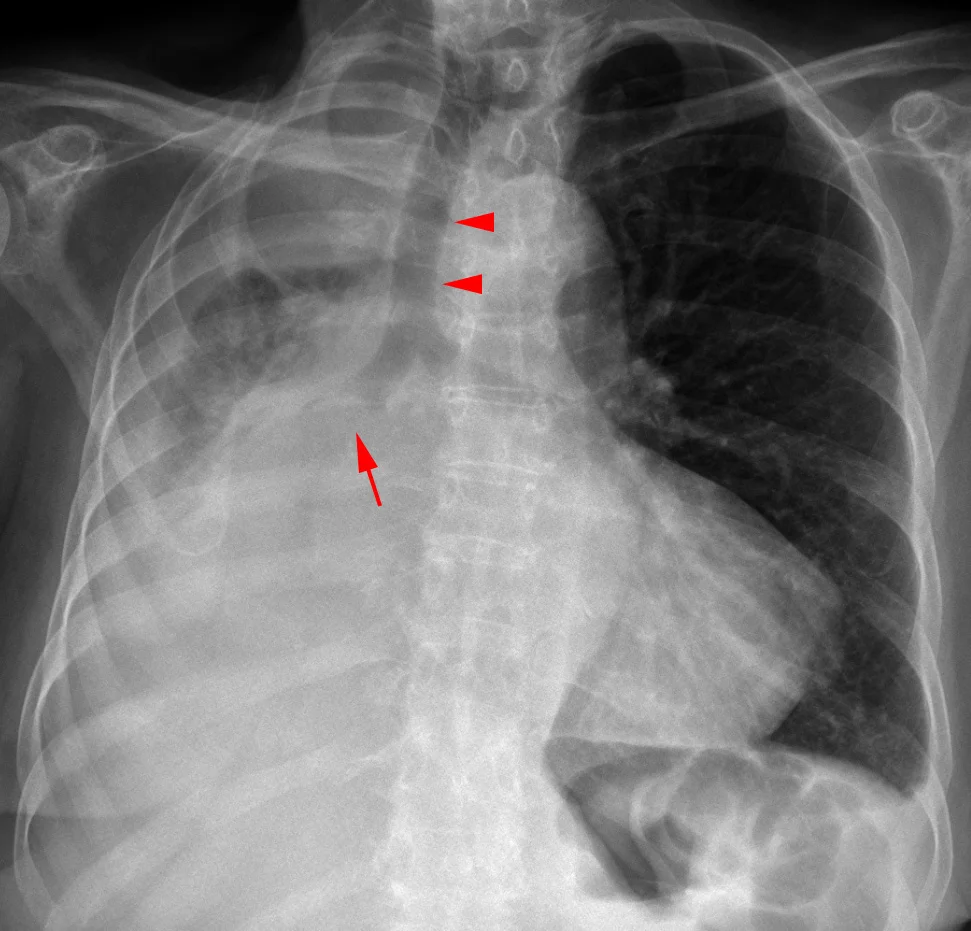
Figure 6Mediastinalshift bei BC
Long description
A chest X-ray described as showing mediastinal shift to the right from subtotal atelectasis of the right lung due to a bronchial carcinoma, with a Pfeil/arrow.

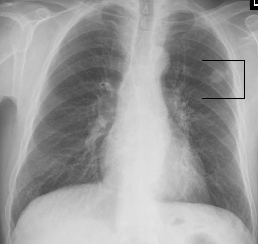
Figure 7Thorax pa peripheres Bronchialcarcinom li OF markiert
Long description
A conventional posteroanterior (PA) chest X-ray showing a rounded density in the left lung identified as a peripheral bronchial carcinoma, with a marker around the lesion.

- Contrast-enhanced CT of the chest, with the liver, adrenals and lower neck included, is the first imaging test. [1]
- PET-CT should be offered to everyone who may be treated with curative intent, and it must be available with rapid access. [1]
- MRI of the chest is not used routinely to stage the primary tumour, but is used when assessing chest wall or apical (superior sulcus) tumours. [1]
- Brain imaging is part of staging for curative-intent treatment: no dedicated brain imaging for stage 1 disease without neurological symptoms; contrast-enhanced brain CT for stage 2; contrast-enhanced brain MRI for stage 3. With neurological symptoms, CT followed by MRI, or MRI first, is used. [1]
- Bone assessment starts with X-ray for focal bone pain; if negative, bone scintigraphy or MRI. Bone scintigraphy is avoided when PET-CT has not shown bone metastases. [1]
- Peripheral lesions: image-guided biopsy. [1]
- Central lesions: flexible bronchoscopy if nodal staging does not influence treatment. [1]
- Intrathoracic nodes: offer PET-CT first for nodes under 10 mm maximum short axis on CT; for nodes 10 mm or more, PET-CT followed by EBUS-TBNA or EUS-FNA, or both. [1]
- EBUS-TBNA (endobronchial ultrasound-guided transbronchial needle aspiration) samples paratracheal and peri-bronchial nodes and lesions. [1]
- Surgical mediastinal staging is considered if EBUS/EUS sampling is negative but clinical suspicion of nodal disease remains high. [1]
- Bigger or metastatic lesions may be biopsied first if the nodal result would change the plan. [1]
Tissue must be adequate for diagnosis, subtyping and molecular marker testing. [1][3] If a lesion is near an airway, a core or cell block may be enough; otherwise a CT-guided biopsy or surgical sample is required. Ask the pathologist to reserve material for predictive testing rather than using it all for diagnosis.
First investigations for suspected lung cancer after an abnormal chest film
- Contrast-enhanced chest CT (include liver, adrenals, lower neck)Characterise the primary; assess nodal and distant disease
- Question: Is curative treatment still possible?
Yes or maybe
- PET-CT if not already done
- Question: Nodes under 10 mm short axis on CT?
Yes
- PET-CT firstBiopsy only if PET-positive or change in plan
No (10 mm or more)
- PET-CT then EBUS-TBNA (and/or EUS-FNA)Surgical mediastinal staging if sampling negative but suspicion high
- Brain imaging by stageNo dedicated brain imaging for stage 1 without symptoms; brain CT for stage 2; brain MRI for stage 3
- Biopsy (peripheral: image-guided; central: bronchoscopic)Reserve tissue for molecular testing
No (advanced disease)
- Least invasive biopsy of the easiest metastatic siteTissue for type and molecular markers
- Brain MRI if symptomatic, or to plan radiotherapy
- Symptom control and palliative assessment
There are no laboratory criteria that make the diagnosis; it is a diagnosis of histology plus imaging. In practice, a confident diagnosis requires a tissue biopsy with immunohistochemistry to distinguish adenocarcinoma, squamous carcinoma and SCLC, together with a CT or PET-CT showing a compatible lesion. [1][3]
The current international staging system is the 9th edition of the TNM classification (IASLC/UICC), based on a database of tens of thousands of lung cancer cases. [2] Three points matter to a student:
- The T and M-N categories keep the 8th edition structure; the 9th edition refines rather than replaces it. [2]
- N2 is now split into N2a and N2b: N2a is metastasis in a single ipsilateral mediastinal or subcarinal node station; N2b is metastasis in multiple ipsilateral stations. [2]
- M1c is now split into M1c1 and M1c2: M1c1 is multiple extrathoracic metastases in one organ or organ system; M1c2 is multiple metastases in multiple organ systems. [2]
The T categories by tumour size remain those established in the 8th edition, and the M1a/M1b distinctions also remain familiar. The staging tables in the UICC 9th edition should be consulted for the exact stage groups from IIb to IVb, which were revised in this edition. [2]
Pleural and pericardial effusion with positive cytology counts as M1a. N3 disease (contralateral mediastinal nodes, scalene or supraclavicular nodes) makes the cancer at least stage IIIB/IIIC in most groupings.
Management should always be discussed in a thoracic oncology multidisciplinary team.
- Stop smoking as soon as lung cancer is suspected, with support; do not delay surgery to allow quitting. [1]
- Supportive care: nutrition, breathlessness management, exercise and pulmonary rehabilitation.
- Palliative care should be introduced early for anyone with symptoms, not reserved for the end of life (see below).
- Stage 1-2 (selected T1-T2b, N0, M0): offer lobectomy (open or thoracoscopic) if the patient is fit. [1]
- If lobectomy is contraindicated or declined: sublobar (wedge or segmental) resection or SABR for T1a-T2b, N0, M0. [1]
- T3 tumours with chest wall involvement: complete (en bloc) resection. [1]
- Stage 3A (N2) operable disease: consider chemoradiotherapy plus surgery only in experienced centres; schedule surgery 3-5 weeks after chemoradiotherapy. [1]
- Radical radiotherapy doses when conventional fractionation: 55 Gy in 20 fractions over 4 weeks, or 60-66 Gy in 30-33 fractions over 6-6.5 weeks. [1]
- Lymph node sampling or en bloc resection of hilar and mediastinal nodes at surgery. [1]
For fit patients with resectable disease at high risk of recurrence (tumours over 4 cm or node-positive), systemic treatment is now standard around surgery. [1]
- Neoadjuvant nivolumab with chemotherapy (NICE technology appraisal). [1]
- Neoadjuvant then adjuvant durvalumab with platinum chemotherapy (no EGFR or ALK driver). [1]
- Neoadjuvant then adjuvant pembrolizumab with platinum chemotherapy (high-risk NSCLC). [1]
- Adjuvant cisplatin-based chemotherapy for node-positive (T1a-4, N1-2, M0) and selected T2b-4, N0 tumours over 4 cm. [1]
- Adjuvant osimertinib for 3 years for completely resected stage 1b-3a NSCLC with EGFR exon 19 deletions or exon 21 (L858R) mutations. [1]
- Adjuvant alectinib for completely resected stage 1b (tumours 4 cm or more) to 3a ALK-positive NSCLC. [1]
- Adjuvant immunotherapy (atezolizumab or pembrolizumab, chosen by tumour PD-L1 level, driver mutations and recurrence risk) after platinum-based chemotherapy in some patients. [1]
- Consider chemoradiotherapy if the patient cannot or will not have surgery. [1]
- Durvalumab after chemoradiotherapy is recommended where PD-L1 is 1% or more and the disease has not progressed. [1]
Treatment is now chosen by histology (squamous vs non-squamous) and by biomarker status, not by histology alone. [1]
- High PD-L1 expression, no driver mutation: immunotherapy alone is often considered; the exact threshold and agent follow current local guidance. [1]
- Lower PD-L1 expression, no driver mutation: a PD-1/PD-L1 inhibitor combined with platinum-doublet chemotherapy is the usual approach. [1]
- EGFR (exon 19 deletion or L858R): a third-generation EGFR tyrosine kinase inhibitor such as osimertinib is the preferable first-line option, especially with brain metastases. [3]
- ALK: alectinib, or lorlatinib, as first-line. [3]
- Other drivers (ROS1, BRAF V600, MET exon 14 skipping, RET, KRAS G12C, NTRK, HER2): match with the corresponding licensed agent. [1][3]
- Brain metastases often respond to modern targeted drugs (especially EGFR and ALK tyrosine kinase inhibitors); whole-brain or stereotactic radiotherapy is used when systemic control is inadequate or for symptoms.
Every patient with advanced non-squamous NSCLC should have a defined minimum panel: EGFR, ALK, ROS1, BRAF V600, NTRK, and MET exon 14 skipping/amplification, RET rearrangement, KRAS G12C and HER2. [3]
- Testing is avoided in confident squamous cancer as routine, except for young patients (under 50 years), never-smokers (fewer than 100 cigarettes in a lifetime) or former light smokers (under 15 pack-years, all kinds of tobacco), and long-term ex-smokers who stopped more than 15 years ago; in regions with high EGFR prevalence in squamous tumours it may be tested regardless of smoking history. [3]
- NGS (next-generation sequencing) multiplex panels are preferred where available; RNA-based NGS is preferred for fusions. [3]
- Squamous/aspirate material is often adequate; liquid biopsy (circulating tumour DNA) is an acceptable alternative when tissue is insufficient or a repeat biopsy is unsafe. [3]
- Add PD-L1 tumour proportion score (TPS) on tissue, because it selects immunotherapy.
- Repeat testing at progression (especially for EGFR T790M) is standard. [3]
SCLC is almost always smoking-related and is treated as a systemic disease.
- Definitions (usual clinical practice): limited stage means disease confined to one hemithorax that can be encompassed in a single radiotherapy field (roughly T1-4, N0-3, M0); extensive stage means spread beyond that (M1a/b including cerebral metastases). [1]
- Limited stage: cisplatin-based chemotherapy (4-6 cycles) with concurrent twice-daily thoracic radiotherapy started during the first or second cycle, or sequential radiotherapy if concurrent treatment is not tolerated. [1]
- Prophylactic cranial irradiation (PCI) 25 Gy in 10 fractions is offered in limited stage if the disease has not progressed after first-line treatment. [1]
- Durvalumab is now recommended after chemoradiotherapy in limited-stage disease if the cancer has not progressed. [1]
- Extensive stage: platinum-etoposide chemotherapy (up to 6 cycles), plus durvalumab or atezolizumab in patients with performance status 0-1. [1]
- PCI may be considered in extensive stage after a response to first-line treatment. [1]
- Thoracic radiotherapy in extensive stage is considered after a good response in the chest and at distant sites. [1]
- After relapse: re-challenge with platinum-etoposide if the relapse is late; otherwise an anthracycline-containing regimen or topotecan; palliative radiotherapy for symptoms. [1]
- Tarlatamab, a DLL3-directed bispecific T-cell engaging antibody, has been appraised for relapsed extensive-stage disease that has progressed after two or more lines of treatment; it was not recommended for routine use in the UK (NICE technology appraisal). [1]
- Treat symptoms early: opioids for cancer pain and breathlessness, dexamethasone for cerebral oedema or SVC obstruction, nerve blocks and radiotherapy for focal pain. [1]
- Drainage and, when appropriate, pleurodesis for symptomatic malignant pleural effusions. [1]
- Airway stenting or laser therapy for central obstruction causing life-threatening dyspnoea. [1]
- Palliative radiotherapy relieves haemoptysis, pain, cough and SVC obstruction; single-fraction radiotherapy is used for painful bone metastases. [1]
- Palliative care clinicians, social care and psychological support should be involved from the point of diagnosis when symptoms are present. [1]
There is no single agreed follow-up schedule. After curative treatment, review and imaging are scheduled by the multidisciplinary team and CT is usually reserved for new symptoms or reassessment after radical treatment. [1] Recurrence of SCLC in particular should prompt rapid reassessment for further systemic therapy or palliation.
Every patient with a suspected or confirmed lung cancer should be referred to a chest physician and discussed in a lung cancer multidisciplinary team. [1] Refer urgently for:
- suspected lung cancer on imaging or with a new persistent cough or haemoptysis;
- superior vena cava syndrome or spinal cord compression;
- Pancoast tumour with neurological signs;
- fistula, major bleeding, or febrile neutropenia during chemotherapy.
- Airway obstruction leading to post-obstructive pneumonia, lobe collapse, or lobar air trapping.
- Pleural effusion (often malignant) and pericardial effusion.
- Superior vena cava syndrome, a cardiothoracic emergency.
- Spinal cord compression from vertebral metastasis.
- Metastatic disease to liver, adrenal, brain, bone and elsewhere.
- Paraneoplastic syndromes (SIADH, hypercalcaemia, Cushing syndrome, Lambert-Eaton syndrome, hypertrophic osteoarthropathy).
- Treatment toxicity: cytopenias, neuropathy, pneumonitis (with radiotherapy or immune checkpoint inhibitors), skin and gut toxicity, infection.
- Second primary cancers, especially in continuing smokers.
Prognosis depends on stage, performance status, histology, biomarker profile and comorbidity. Five-year survival is high for a resected stage I tumour and poor once the disease has spread to distant sites; modern targeted therapies and immunotherapy have improved outcomes for metastatic disease but have not cured most patients. [5] Accurate staging at the outset is therefore the most important way to choose between a curative and a palliative plan.
- Smoking cessation is the single most effective measure; it reduces lung cancer risk even when stopped in middle age. [1]
- Avoid asbestos, silica and other occupational carcinogens, with dust control, protective equipment and legislative standards. [4]
- Reduce radon exposure by testing buildings and remediating high readings. [4]
- Air quality policies matter for never-smokers and for the whole population. [4]
- Diet and exercise are not protective specifically, but support overall health.
The standard evidence-based screening test is low-dose CT (LDCT) of the chest.
- The US Preventive Services Task Force recommends annual LDCT for adults aged 50-80 years with at least a 20 pack-year smoking history who currently smoke or quit within the past 15 years. [4]
- Trials in this group showed a mortality benefit from LDCT screening, but with false positives, overdiagnosis and the harms of invasive follow-up.
- Screening never-smokers (including high-risk Asian women) remains controversial because the benefit is unproven and overdiagnosis is a real concern. [4]
- Screening programmes must include smoking cessation support; otherwise a negative scan can be misread as a licence to smoke.
- In low-resource settings, screening is usually not implemented, and the priority is early diagnosis of symptomatic disease and tobacco control.
- Pregnancy. Lung cancer in pregnancy is rare and management is multidisciplinary; chemotherapy is generally avoided in the first trimester.
- Older adults. Age alone should not exclude curative treatment; use comprehensive geriatric assessment to balance benefit and toxicity.
- Immunocompromised patients are more likely to have infection mimicking cancer; insist on tissue diagnosis when imaging is atypical.
- Never-smokers and light smokers. Think of EGFR, ALK and other drivers; molecular testing is especially important. [3]
When advanced imaging, bronchoscopy and modern drugs are unavailable, the following sequence is the best available.
- Chest X-ray followed by ultrasound where a mass or effusion is suspected; look for a hilar mass, collapse or effusion. [1]
- Sputum cytology can confirm cancer when bronchoscopy is unavailable and the lesion is central. [1]
- ** CT scan of chest, abdomen and pelvis** when available, is the single most useful staging step; include the liver and adrenals if the protocol allows. [1]
- Obtain tissue by the simplest safe route (bronchoscopy if available, otherwise image-guided needle biopsy, pleural fluid cytology or lymph node biopsy). Save material for later molecular testing if resources ever become available.
- Clinically stage the patient with examination, brain imaging only for focal neurological signs or headache, and focal bone X-rays for bone pain. Do not chase distant metastases if the result would not change management.
- Surgery (lobectomy with mediastinal node sampling) is the curative option where theatre and pathology are available and disease is stage 1-2.
- Short-course palliative radiotherapy and oral morphine are inexpensive, effective and widely available; prioritise symptom relief.
- Document outcomes in a simple audit register so that local pathways can be improved.
- Lung cancer is the leading cause of cancer death worldwide, with about 2.48 million new cases and 1.81 million deaths in 2022. [5]
- Smoking causes more than 80% of cases in industrialised countries; in countries with low female smoking, most lung cancer in women occurs in never-smokers. [4]
- The main types are adenocarcinoma, squamous, large cell and small cell; SCLC is almost always smoking-related and behaves as a systemic disease. [1]
- Risk factors besides smoking include radon, asbestos and other occupational exposures, air pollution, previous chest disease, radiation and family history. [4]
- Typical presentation is cough, haemoptysis, breathlessness, chest pain and weight loss; local invasion produces Pancoast/Horner, SVC syndrome and hoarse voice.
- Paraneoplastic syndromes include SIADH, PTHrP hypercalcaemia, Cushing syndrome, Lambert-Eaton syndrome and hypertrophic osteoarthropathy.
- The commonest metastatic sites are liver, adrenal, brain and bone (the LABBs).
- Diagnosis is tissue plus imaging; the core staging sequence is contrast CT, PET-CT, EBUS and brain MRI by stage. [1]
- The 9th edition TNM refines the 8th edition by splitting N2 into N2a/N2b and M1c into M1c1/M1c2. [2]
- Early disease is treated with surgery or SBRT; stage 3 with chemoradiotherapy plus durvalumab; metastatic disease by PD-L1 level and by driver mutation. [1][3]
- Molecular testing (EGFR, ALK, ROS1, BRAF, MET, RET, KRAS G12C, NTRK, HER2) is standard in advanced non-squamous NSCLC. [3]
- SCLC limited stage is treated with platinum chemotherapy, concurrent radiotherapy and PCI; durvalumab improves outcomes; extensive stage adds durvalumab or atezolizumab. [1]
- Screening with annual low-dose CT is recommended for people aged 50-80 with at least 20 pack-years who smoke or quit within 15 years. [4]
- In low-resource settings, prioritise tissue diagnosis, sputum cytology, CT staging, surgery for early disease and cheap symptom control.
Answer each one in your head first, then open the answer.
A 62-year-old smoker has a new cough, haemoptysis and weight loss over 3 months. Chest X-ray shows a hilar mass. What should the next investigation be, and what must the sample be adequate for?
Name the commonest distant metastatic sites of lung cancer and give the mnemonic.
Show answerHide answer
Liver, adrenal gland, brain and bone - the "LABBs". They are the reason staging includes the liver and adrenals on chest CT and brain MRI when radical treatment is planned. [1]
A patient with stage 3 NSCLC being treated with curative intent has no neurological symptoms. What brain imaging is appropriate?
Show answerHide answer
Contrast-enhanced brain MRI. CT of the head is used if clinical features suggest intracranial disease, and dedicated brain imaging may be omitted in stage 1 disease without neurological symptoms. [1]
Explain the difference between N2a and N2b in the 9th edition TNM and why it matters.
Show answerHide answer
N2a is metastasis in a single ipsilateral mediastinal or subcarinal node station; N2b is metastasis in multiple ipsilateral stations. The split reflects a real survival difference and helps choose between surgery plus chemoradiotherapy and definitive chemoradiation. [2]
A 58-year-old woman with stage IIIB EGFR-mutant NSCLC (exon 19 deletion) is fit. After concurrent chemoradiotherapy with no progression, what systemic treatment is recommended and on what molecular basis?
Show answerHide answer
Durvalumab is recommended after chemoradiation when PD-L1 is 1% or more. In addition, for EGFR-mutant disease osimertinib is the standard targeted agent in advanced disease; the exact sequence in stage III is decided by the multidisciplinary team and by whether the EGFR mutation was already known. [1][3]
Which patients should receive prophylactic cranial irradiation in limited-stage SCLC, at what dose, and what is the new additional systemic option?
Show answerHide answer
PCI 25 Gy in 10 fractions is offered when the disease has not progressed after first-line treatment in patients with performance status 0-2. Durvalumab is now recommended after chemoradiotherapy in limited-stage SCLC if the disease has not progressed. [1]
Who is eligible for LDCT screening, and what is the controversy in never-smokers?
Show answerHide answer
Annual LDCT for adults aged 50-80 years with a smoking history of 20 pack-years or more who currently smoke or quit within 15 years. Screening never-smokers is controversial because the benefit is unproven and overdiagnosis is a concern. [4]
Lung cancer interlocks with chronic obstructive pulmonary disease, tuberculosis, malignant pleural effusion, superior vena cava syndrome and metastatic brain disease. Prerequisites: none noted. Related topics will be linked by the discipline once verified registry entries are published.
Solitary pulmonary nodule
A rounded opacity up to 3 cm surrounded by aerated lung: how to tell benign from malignant and what to do next.Pleural effusion
How fluid accumulates in the pleural space, how transudates differ from exudates, and how to investigate, drain and treat effusions by cause.Pneumoconioses
Inhaled workplace dusts that scar the lungs: what silicosis, coal disease, asbestosis, beryllium disease, and byssinosis are, and how they differ.Chronic obstructive pulmonary disease (COPD)
Chronic lung disease with persistent airflow obstruction: clinical features, diagnosis, staging, management and complications.
4 other topics link here.
- National Institute for Health and Care Excellence (NICE). Lung cancer: diagnosis and management. NICE guideline NG122, published 28 March 2019, last updated 08 March 2024. Accessed .
- Brierley JD, Van Eycken LJ, Giuliani ME, Rous BA, Huang SH, Asamura H, Gospodarowicz MK, Lee AW, O'Sullivan B. The 9th Edition of the UICC TNM Classification of Malignant Tumours: Updates and Rationale for Change. International Journal of Cancer 2026;159(7):1588-1601. doi:10.1002/ijc.70561. Accessed .
- Lee SH, Menis J, Kim TM, Kim HR, Zhou C, Kurniawati SA, Prabhash K, Hayashi H, Lee DD, Imasa MS, Teh YL, Yang JC, Reungwetwattana T, Sriuranpong V, Wu CE, Ang Y, Sabando M, Thiagarajan M, Mizugaki H, Noronha V, Yulianti M, Zhang L, Smyth E, Yoshino T, Park JO, Pentheroudakis G, Park S, Peters S, Ahn JB, Popat S. Pan-Asian adapted ESMO Clinical Practice Guidelines for the diagnosis, treatment and follow-up of patients with oncogene-addicted metastatic non-small-cell lung cancer. ESMO Open 2024;9(12):103996. doi:10.1016/j.esmoop.2024.103996. Accessed .
- Ding Y, Tao H, Zhu C, Li H, Zhang Y, Du Y, Du L, Gong W, Wang L. Lung cancer in never-smoking females: epidemiology, risk factors and screening. Frontiers in Public Health 2026;14:1834538. doi:10.3389/fpubh.2026.1834538. Accessed .
- Zhai J, Zheng H, Xue L. Global disparities in lifetime risk of lung cancer: links to socioeconomic development, smoking, and fine particulate matter from GLOBOCAN 2022. Frontiers in Public Health 2026;14:1842037. doi:10.3389/fpubh.2026.1842037. Accessed .
Figure credits
- Figure 1: Chen CL, Chen CC, Yu WH, Chen SH, Chang YC, Hsu TI, via Wikimedia Commons, CC BY 4.0, CC BY 4.0.
- Figure 2: No machine-readable author provided. KGH assumed (based on copyright claims)., via Wikimedia Commons, CC BY-SA 3.0, CC BY-SA 3.0.
- Figure 3: Mikael Häggström, M.D., via Wikimedia Commons, CC0, CC0.
- Figure 4: EMAHkempny, via Wikimedia Commons, CC BY-SA 4.0, CC BY-SA 4.0.
- Figure 5: Original illustration by MedGrasp, CC BY 4.0.
- Figure 6: Hellerhoff, via Wikimedia Commons, CC BY-SA 3.0, CC BY-SA 3.0.
- Figure 7: Lange123 at German Wikipedia, via Wikimedia Commons, CC BY-SA 3.0, CC BY-SA 3.0.
Educational content for medical students and doctors. It is not a substitute for clinical judgement or local guidelines.