Pneumoconioses
Inhaled workplace dusts that scar the lungs: what silicosis, coal disease, asbestosis, beryllium disease, and byssinosis are, and how they differ.
- Pneumoconioses are the diseases caused by breathing in inorganic dusts at work: silica, coal dust, asbestos, beryllium, certain metals and fibres. Most are fibrotic, progress slowly, and can worsen even after exposure has stopped.[1]
- Silicosis comes from respirable crystalline silica. It is seen in stone work, mining, ceramics, foundries and sandblasting, and develops as upper lobe nodules that can merge into progressive massive fibrosis, with swollen lymph nodes that may show eggshell calcification. Silicosis strongly increases the risk of tuberculosis and is also linked to lung cancer.[1][2]
- Coal workers' pneumoconiosis has a similar upper lobe simple nodular picture after coal dust exposure, but tends to be milder in its simple form and can also progress to progressive massive fibrosis.[1][3]
- Asbestosis is a dose-dependent UIP-type fibrosis causing lower lobe scarring after asbestos exposure, usually with 20 to 30 years of latency. It runs with pleural plaques and diffuse pleural disease, and asbestos exposure also causes lung cancer and malignant mesothelioma.[1][3]
- Chronic beryllium disease is a granulomatous disease that mimics sarcoidosis, occurring in industries such as ceramics, aerospace, nuclear engineering and lamp manufacture, and is diagnosed by the beryllium lymphocyte proliferation test.[3][5]
- Byssinosis is a cotton-dust and textile-dust airway disease, treated like occupational asthma and often worst on the first day back at work.[1]
- There is no cure. The central actions are to stop or reduce exposure, screen for tuberculosis, encourage smoking cessation, treat complications, and arrange early specialist care and compensation review.[1]
By the end of this page you should be able to:
- Define a pneumoconiosis and split the major types by dust, site of fibrosis and imaging pattern.
- Explain why inhaled dust damages the lung, and why different dusts prefer different parts of the lung.
- Recognise silicosis, coal workers' pneumoconiosis, asbestosis, berylliosis and byssinosis from exposure history and imaging.
- Plan first-line and specialist investigation, including beryllium lymphocyte proliferation testing, and name the pathological hallmarks of each.
- State the evidence for progression after exposure ends, tuberculosis risk, and lung cancer risk in silica disease.
- Explain exposure limits, surveillance programmes and workplace control, and why artisanal and informal mining is a particular problem.
- Outline management, compensation and referral pathways, and the low-resource adaptations that matter worldwide.
DefinitionPneumoconiosis
A pneumoconiosis is a lung disease caused by the inhalation and retention of inorganic dusts at work (and occasionally from the general environment). The dust is biologically active rather than inert: it sets up inflammation, fibrosis and sometimes granuloma formation. Most forms are fibrotic, most have long latency, and many progress after the person has stopped being exposed.[1]
Medically useful distinctions are these.
By fibrogenicity. Silica, asbestos and coal mine dust are fibrogenic: they scar. Some other dusts are largely inert or only weakly fibrogenic, and cause pigment overload rather than dense fibrosis. Beryllium and some metals drive a granulomatous reaction rather than ordinary fibrosis.
By the dust involved. The name of the disease usually names the dust: silicosis (crystalline silica), coal workers' pneumoconiosis (coal mine dust, which itself contains silica), asbestosis (asbestos fibres), chronic beryllium disease or berylliosis (beryllium), talcosis (talc), siderosis (iron), and others.
By severity. Many diseases exist as simple disease (small nodules, often with little impairment) and complicated disease, the most important form being progressive massive fibrosis: conglomerate masses of fibrotic nodules usually larger than 1 cm and sometimes larger than 2 cm, typically in the upper lobes, with volume loss and traction bronchiectasis around them.[3][4]
By tempo. Silicosis can be chronic (10 or more years of low to moderate exposure), accelerated (within 10 years of moderate to high exposure, often faster progressing) or acute (weeks to 5 years after a very high exposure, with features of silicoproteinosis).[3][4]
Silicosis is only the most studied example. In every case, the diagnosis requires both a compatible exposure and a compatible lung picture; exposure history alone is not enough, and CT appearances alone are not enough.
Worldwide, pneumoconiosis is not a historical curiosity. The Global Burden of Disease study identified 23,695 new cases of silicosis in 2017, 39% of the 60,055 new cases of pneumoconiosis identified that year, and over 20,000 new silicosis cases were counted. Those figures are considered a substantial underestimate, because many countries lack reporting systems and occupational lung surveillance.[2] In formal case series, chronic silicosis begins after years of exposure; by contrast, recent artificial stone silicosis cases have occurred in workers in their twenties or thirties after a few years of high exposure.[1][2]
The burden is uneven. In 2017 East Asia had the highest regional silicosis incidence, with 15,980 cases, and China and India carried the largest national case numbers. More than 23 million workers in China and more than 10 million in India are estimated to be exposed to respirable crystalline silica. South and sub-Saharan Africa have rising incidence, with artisanal and informal gold mining an important driver. European and North American incidence has fallen, but remains in people who are still exposed, and is thought to be under-diagnosed, particularly in construction.[2]
Silica stands out in numbers: in the United States an estimated 2.3 million workers are exposed to respirable crystalline silica, of whom 1.85 million are in construction and 320,000 in general industry and maritime work. Workers in insecure jobs, immigrants and small, informal businesses carry disproportionate risk because they face more intense exposures and fewer protections.[2]
Respiratory disease caused by work is broader than pneumoconiosis. Occupational exposure accounts for roughly one in six cases of COPD and asthma worldwide, and in the United Kingdom around 12,000 deaths each year are attributed to prior occupational exposure to lung disease causes, about 60% of them from cancer.[1]
Risk is exposure, dose and time, modified by setting. The occupations most strongly linked to each disease are well established.
| Disease | Main exposure | Typical workplaces and tasks | Dose-response and latency |
|---|---|---|---|
| Silicosis | Respirable crystalline silica (quartz, cristobalite, tridymite) | Mining, quarrying, stone cutting, foundry work, glass and ceramics, tunneling, construction, demolition, abrasive blasting, artificial stone fabrication, hydraulic fracturing | Chronic: 10 or more years of low to moderate exposure. Accelerated: under 10 years of moderate to high exposure. Acute: weeks to 5 years of very high exposure.[1][4] |
| Coal workers' pneumoconiosis | Coal mine dust, which itself contains variable silica | Underground coal mining, coal handling and transport | Often decades of exposure; simple upper lobe nodular disease can appear, with possible progression to progressive massive fibrosis.[1][3] |
| Asbestosis | Asbestos fibres | Asbestos mining and milling, shipbuilding and repair, insulation, pipefitting, plumbing, carpentry, construction and demolition of older buildings | 20 to 30 years typically; development and severity are dose-dependent.[1][3] |
| Berylliosis | Beryllium metal, alloy and oxide dust | Ceramics, aerospace, nuclear engineering, electronics, fluorescent lamp manufacture, alloy processing and welding | Usually chronic; exposure can be downstream and unrecognised when beryllium is a component of alloys.[3][6] |
| Byssinosis | Cotton, flax or hemp (textile) dust | Textile spinning and weaving, cotton ginning, other organic dust occupations | Short-latency, exposure-related symptoms; often worse at the start of the working week.[1] |
Other risk factors matter. Insecure and informal work raises exposure intensity.[2] Dry cutting, lack of water suppression, poor ventilation and inconsistent respiratory protection are the specific drivers of artificial stone silicosis outbreaks worldwide.[2] The value of the exposure is the product of concentration and time: long-service workers in dust also develop disease, not only those in short, very high exposures, although accelerated forms are now the ones producing young patients.
The story is the same in broad outline for most dusts, with differences that explain the different organs and lobes involved.
Where the dust lands. Particles that are roughly 1 to 5 micrometres across, with a density similar to water, reach the alveoli; larger or denser particles impact on airway walls and are cleared by the mucociliary elevator; smaller ones are exhaled. In the upright, resting lung the upper zones have poorer perfusion and aeration, so dust is retained there rather than washed away. This is the basic reason most pneumoconioses prefer the upper lobes. Asbestos fibres behave differently: they are long and narrow, with an aspect ratio (length to diameter) around 3 to 1, and they settle more in the lower lobes and pleura.[4]
What the dust does. Inhaled silica particles generate reactive free radicals on their surface, damage DNA and kill cells directly. Silica also triggers a robust respiratory burst in alveolar macrophages, releasing oxidants, proteases and pro-inflammatory cytokines such as TNF-alpha and IL-1. The cycle of macrophage death and re-ingestion of the same dust drives the severe fibrosis of silicosis. Crystalline silica, quartz in particular, is classified as a Group 1 carcinogen, acting through oxidative DNA damage and chronic inflammation.[4]
Coal dust produces similar but generally less rapidly fibrotic injury through oxidative stress, release of enzymes, lipid and protein damage, and activation of fibroblasts and collagen deposition. Contaminants in coal dust such as heavy metals, transition metals and polycyclic aromatic compounds add to the oxidative load, which helps explain why coal disease can look radiologically like silicosis in some miners.[4]
Asbestos fibres cannot be fully engulfed by macrophages; frustrated phagocytosis sustains the inflammation, and the retained fibres cause lower lobe parenchymal fibrosis and pleural disease. The cancer risks come partly from the genotoxicity of the fibres and partly from the chronic inflammation they sustain.
Beryllium works by immune sensitisation rather than direct toxicity: inhaled beryllium is taken up by antigen-presenting cells, which present it to T cells, so that sensitised T cells drive non-necrotising granuloma formation in the lung and sometimes elsewhere. This is why berylliosis looks like sarcoidosis, and why it is sometimes called chronic beryllium disease.[6]
Why disease progresses after exposure ends. The latency between first exposure and disease explains why an older worker can develop new or worsening disease years after leaving the job, and why removal from exposure is prevention rather than cure. Macrophages already laden with dust continue to release mediators; the particles themselves are insoluble. Occupational diseases are conventionally classed as short-latency (occupational asthma, hypersensitivity pneumonitis) or long-latency (silicosis, other pneumoconioses, asbestos disease, lung cancer, occupational COPD), the latter usually 10 or more years after first exposure, and mesothelioma can appear 40 years later.[1]
Figure 1Asbestosis - Fibrous pleural plaque (7468458430)
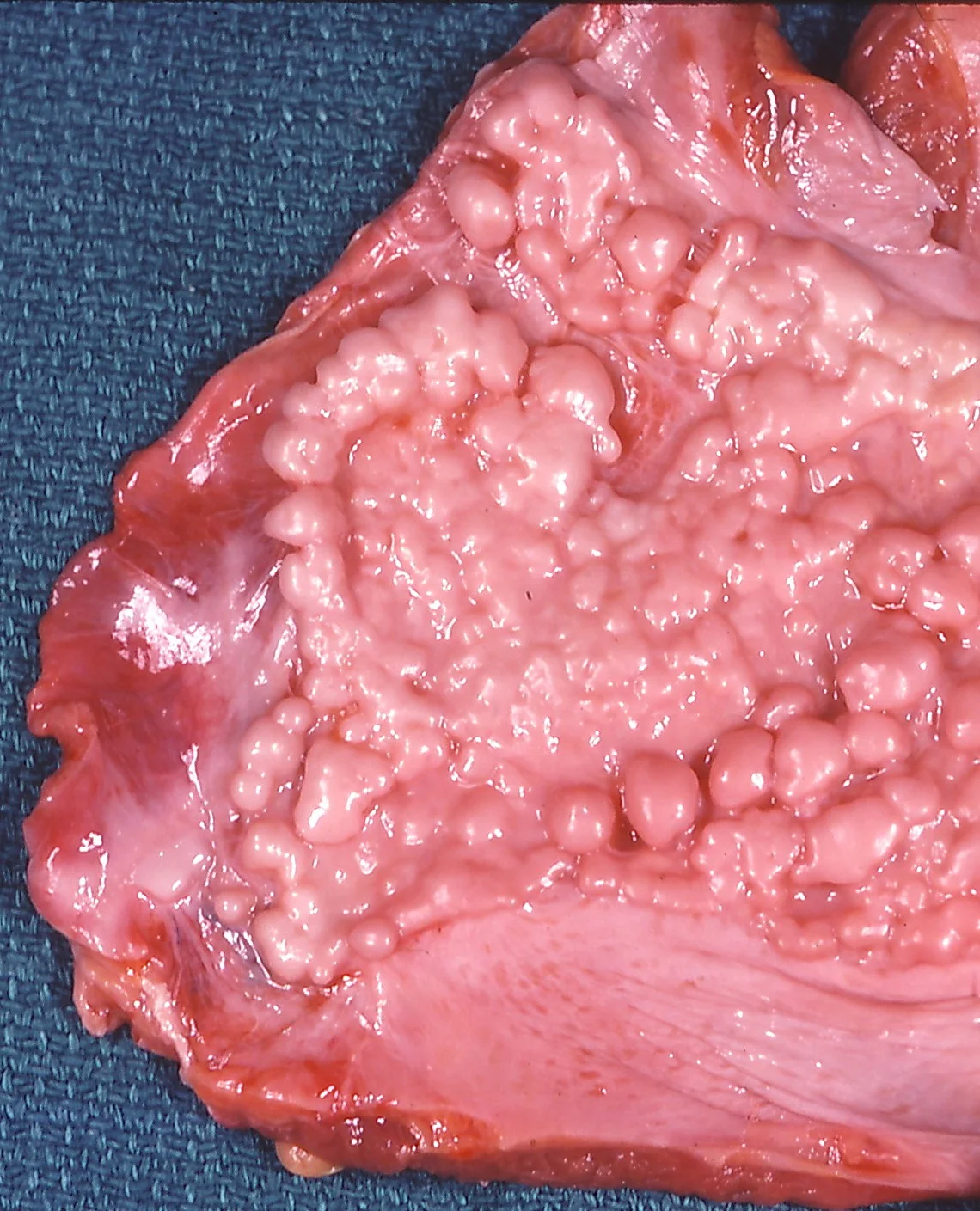
Long description
Gross photograph of a fibrous pleural plaque with a verrucous surface on the diaphragmatic pleura.

Figure 2Ferruginous body
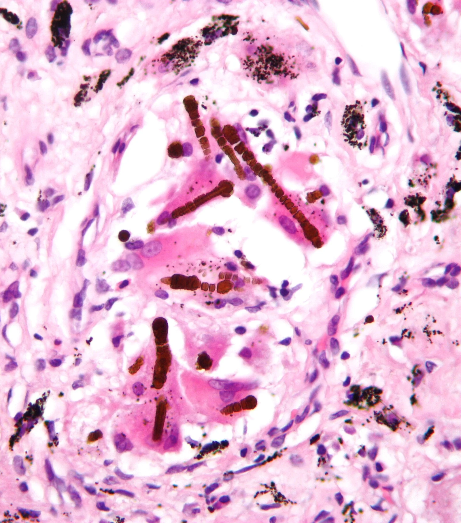
Long description
High-magnification micrograph of ferruginous bodies, also asbestos bodies, in asbestosis; the specimen is lung biopsy, H&E stain, and the bodies are described as rust-coloured (red/brown) baton-shaped segmented rods, with anthracotic pigment also visible.

Pathology confirms what imaging suggests and helps distinguish dust disease from mimics.
Silicosis. The characteristic lesion is a sharply circumscribed nodule of whorled, densely hyalinised collagen, often 1 cm across or less in simple disease. Macrophages form a mantle around young lesions. Under polarised light the nodules contain brightly birefringent particles; larger birefringent particles within them are often silicates rather than pure silica. In long-standing nodules calcification and even ossification can occur. Nodules may merge into conglomerate masses (progressive massive fibrosis) at least 1 cm, sometimes at least 2 cm, across, and may protrude through the visceral pleura. Enlarged mediastinal nodes with nodular fibrosis and peripheral (eggshell) calcification are seen in 75% of silicosis patients. Acute silicosis (silicoproteinosis) shows alveolar filling with granular eosinophilic material that is strongly positive with periodic acid-Schiff after diastase. Immunological disease can coexist: polyclonal hypergammaglobulinaemia, raised rheumatoid factor and antinuclear antibodies, systemic sclerosis and rheumatoid disease.[4][3]
Coal workers' pneumoconiosis. Centrilobular black pigmented macules, which progress to random nodules when associated with fibrosis, mainly in the upper lobes. When the disease becomes complicated it forms large, usually bilateral conglomerate masses similar to progressive massive fibrosis. Emphysematous change develops at the periphery of the macules, and complications include tuberculosis and ischaemic necrosis leading to cavitation. Coal dust from anthracite mines, which has more crystalline quartz, can produce silicosis-like pathology; bituminous and lignite coal tends to a silicatosis-like picture. Emphysema is common around macules. Tuberculous infection is an important complication.[4]
Asbestosis. Asbestosis is dose-dependent pulmonary fibrosis with lower lobe and posterior predominance, often presenting on HRCT as a usual interstitial pneumonia pattern: peripheral reticulation, traction bronchiectasis and, in severe disease, honeycombing. Pleural plaques often coexist, but asbestosis is a parenchymal diagnosis and should not be confused with pleural disease. Asbestos (ferruginous) bodies are asbestos fibres coated by an iron-rich protein, visible in tissue and BAL: a pointer to exposure, though their absence does not exclude disease.[1][3]
Berylliosis. Non-necrotising epithelioid granulomas that are histologically almost indistinguishable from sarcoidosis, found especially in the upper lung, with perilymphatic, peribronchovascular and subpleural nodules. Hilar and mediastinal node involvement is common, while upper lobe fibrosis and honeycombing are uncommon compared with sarcoidosis.[3][6]
The lung is not the only affected tissue. Beryllium disease in particular can be multi-organ: hepatic, renal, skin, ocular, cardiac and neurological involvement has now been well reported, so that extrapulmonary sarcoidosis in a beryllium-exposed worker should prompt reconsideration of the diagnosis.[6]
Silicosis. Simple silicosis is often symptom-free and found on surveillance. Chronic disease presents with cough and exertional breathlessness, with reduced pulmonary function as the nodular load increases. Complicated disease (progressive massive fibrosis) causes more severe breathlessness, upper lobe volume loss, and risk of haemoptysis in cavitating nodules. Acute silicosis is severe and rapidly progressive and behaves clinically like alveolar proteinosis. Accelerated silicosis, now seen in artificial stone work, affects young workers and carries high morbidity and mortality.[1][2]
Coal workers' pneumoconiosis. Simple disease causes little in the way of symptoms, with an upper lobe nodular picture and the possibility of progression to progressive massive fibrosis. Emphysema and chronic airflow limitation are common fellow travellers through smoking and dust.[1][3]
Asbestosis. Exertional breathlessness and dry cough after long, usually heavy past exposure, with lower lobe basal crackles and clubbing in more advanced disease. Latency is typically 20 to 30 years, and severity is dose-dependent. Asymptomatic pleural plaques are the commonest asbestos-related finding and do not usually alter lung function.[1][3]
Berylliosis. Nonspecific: dry cough, exertional dyspnoea, fatigue, fever, weight loss and arthralgia, sometimes with extrapulmonary disease. The imaging mimics sarcoidosis and the presentation is classically mistaken for it before exposure is suspected. CBD is associated with lower lung function than sarcoidosis, and a restrictive pattern is more common (41% versus 15% in one study).[3][5]
Byssinosis. An airway disease of textile workers exposed to cotton, flax or hemp dust. Symptoms follow exposure: chest tightness, cough and wheeze, traditionally worse on the first day back after a break, called Monday fever. The mechanism is an innate and airway response rather than granuloma formation, and it overlaps with occupational asthma and COPD.[1]
Two questions arise in practice: when a patient with possible dust exposure has a lung problem, what else could it be, and when a nodular or fibrotic lung disease is found, what is the dust diagnosis?
| Condition | Exposure clue | Site and pattern | What separates it |
|---|---|---|---|
| Chronic silicosis | Silica jobs: mining, stone, foundry, blasting, artificial stone | Upper lobe well-defined nodules; may calcify; can merge into PMF; eggshell calcification of nodes | Well-defined nodules and frequent calcification distinguish it from coal disease; TB is a frequent complication.[1][3][4] |
| Coal workers' pneumoconiosis | Coal mining and coal handling | Upper lobe centrilobular macules and nodules; less calcified | Nodules less well defined than in silicosis; emphysema is typical; advanced disease is PMF.[1][3] |
| Asbestosis | Asbestos exposure 20 to 30 years before | Lower lobe posterobasilar fibrosis, UIP pattern; may have pleural plaques | Dose-dependent lower lobe fibrosis with pleural disease; distinguish from idiopathic pulmonary fibrosis by exposure history.[1][3] |
| Berylliosis | Beryllium in ceramics, aerospace, nuclear, welding, alloys | Upper lobe perilymphatic nodules, nodular septal and subpleural disease; mimics sarcoidosis | BeLPT positive; non-necrotising granulomas; more extrapulmonary disease possible.[3][5][6] |
| Byssinosis | Cotton, flax or hemp dust | Airway disease (occupational asthma type) rather than parenchymal nodules | Chest tightness after exposure, Monday fever; obliterative or asthma-pattern rather than nodular.[1] |
| Tuberculosis or silicotuberculosis | Same as silicosis plus TB risk | Consolidation, cavitation, miliary or endobronchial disease | Silicosis raises TB risk markedly (relative risk 4.01 in radiological silicosis), and the two can coexist.[2][4] |
| Sarcoidosis | None (idiopathic) | Hilar nodes, upper lobe perilymphatic, non-necrotising granulomas | BeLPT and exposure history separate it from berylliosis; silicosis has better-defined calcified upper lobe nodules.[3][5] |
| Idiopathic pulmonary fibrosis | None (idiopathic) | Lower lobe UIP fibrosis without asbestos exposure or pleural plaques | Exposure history and pleural findings point toward asbestosis.[1][3] |
Figure 3Asbestosis and cryptococcosis - Pleural plaques - CT scan Case 194 (5999300496)
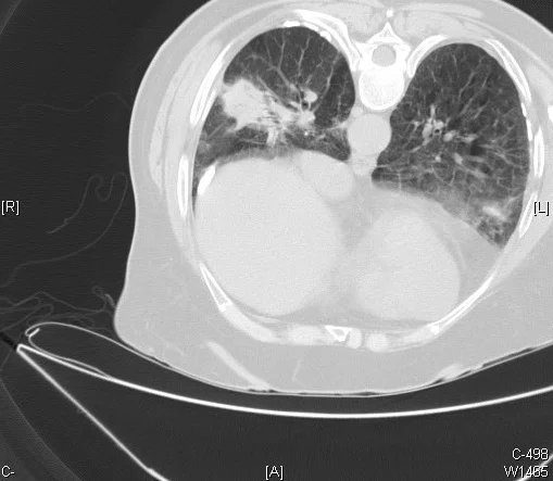
Long description
A chest CT scan showing multiple calcified pleural plaques in a patient with significant asbestos exposure.

Occupational history. The single most important investigation is a careful, job-by-job occupational history covering current and past work, dates of first and last exposure, the exact tasks, visible dust, control measures, respiratory protection, smoking and any symptoms that relate to work or time away from it. Long latency means that jobs held decades ago still matter, and a diagnosis is often missed unless this history is taken. Since pneumoconioses are compensable, it is worth asking who the employer was and whether changes to a job would be acceptable.[1][2]
Imaging. Chest radiography is the starting point and the basis of working surveillance, although it is less sensitive than CT for early lung lesions. High-resolution CT of the chest is the central diagnostic test: it defines nodularity, progressive massive fibrosis, lymph node calcification, pleural plaques, asbestosis-type basal fibrosis and beryllium-type perilymphatic disease, and helps exclude malignancy when appearances are asymmetric. Expiratory-phase imaging adds sensitivity for air trapping in airway disease. A multidisciplinary discussion that includes exposure history, imaging and pathology is recommended. The imaging patterns considered characteristic are interpretation by a radiologist familiar with occupational lung disease.[1][2][3]
Lung function. Spirometry and diffusing capacity show whether restriction, obstruction or a mixed pattern is present, and record progression. In beryllium disease the FVC fall is greater than in sarcoidosis, and a restrictive pattern is more common.[5]
Tissue and cell diagnosis. In cases where imaging and exposure do not agree, or where infection, malignancy or granulomatous disease must be excluded, bronchoalveolar lavage, transbronchial biopsy or cryobiopsy, and surgical lung biopsy all have roles. Lavage captures lymphocyte and dust profiles; biopsy shows silica or asbestos fibres, granulomas and coated asbestos (ferruginous) bodies. Polarised light microscopy reveals birefringent crystals, and specialist centres use analytic electron microscopy and mass spectrometry to identify the dust, mostly in compensation disputes. For surgical biopsies, the site should target the upper lobe when simple silicosis or CWP is suspected and a lower lobe when asbestosis is suspected, and a sample at least 3 to 5 cm long and 3 cm deep should be inflated with fixative.[4]
Beryllium testing. In any granulomatous lung disease with possible exposure, request a BeLPT on blood, and repeat borderline or negative tests. Sputum conversion or two positive tests, one positive and one borderline, or one positive BAL-cell test, support the diagnosis of chronic beryllium disease in a person with compatible granulomatous inflammation.[5][6]
Tuberculosis work-up. Because silicosis, coal workers' pneumoconiosis and asbestos exposure settings all raise tuberculosis risk (relative risk about 4.0 in radiological silicosis), screen for tuberculosis in line with local guidelines in anyone with a relevant exposure and compatible symptoms, including latent infection where policy supports it.[2][4]
A worker with cough, breathlessness or abnormal imaging and possible dust exposure
- Occupational historyList every job, dates, tasks, dust types and control measures; note smoking.
- ImagingChest radiograph then HRCT (plus expiratory views if airway disease is suspected)
- Question: What does the imaging show?
Upper lobe small nodules, possible calcification or eggshell nodes
- Silicosis or CWPConsider accelerated or acute silicosis if exposure was recent and heavy
- Screen for tuberculosis
- Assess for silica exposure and compensation
Lower lobe UIP-type fibrosis with pleural plaques
- Asbestosis likelyDocument exposure dates; exclude lung cancer and mesothelioma
- Refer for specialist review
Granulomatous nodules mimicking sarcoidosis
- Request BeLPT if any beryllium exposure is possible
- Consider silicosarcoidosis overlap
Airway disease (asthma-type)
- Occupational asthma with latency, irritant asthma or byssinosis
- Serial peak-flow records and specific challenge if needed
Undifferentiated or progressive fibrosis
- Multidisciplinary review and pathology if needed
- Consider antifibrotic therapy and transplantation referral
Pneumoconiosis severity is described rather than scored on blood tests.
- Simple versus complicated: in silicosis and CWP the simple form has small nodules; the complicated form (progressive massive fibrosis) has conglomerate fibrotic masses at least 1 cm across, often 2 cm or more, with volume loss and paracicatricial emphysema. Eggshell nodal calcification is characteristic of silicosis.
- Acute, accelerated or chronic silicosis are distinguished mainly by exposure level and latency (weeks to 5 years; under 10 years; 10 or more years).[3][4]
- Asbestosis is diagnosed on exposure plus UIP-type lower-lobe fibrosis; severity follows the burden of fibrosis and the extent of functional restriction, and severity of asbestosis is dose-dependent.[1][3]
- Berylliosis requires granulomatous inflammation plus evidence of beryllium sensitisation on BeLPT, using the criteria of two positive tests, one positive plus one borderline, or one positive BAL-cell test.[5][6]
- Byssinosis is usually diagnosed clinically on exposure and characteristic symptoms, with lung function showing variable obstruction.[1]
There is no universally applied single staging score for all pneumoconioses; the ILO radiographic classification of pneumoconiosis exists for dust surveillance, and regional practice varies. Severity and occupationally induced disability are in practice rated locally.
There is no cure for an established pneumoconiosis, because the dust is insoluble and the scarring is permanent. Management therefore targets progression, infection, cancer risk, symptoms and the workplace.
- Remove or reduce exposure. Complete cessation of exposure is ideal. If the patient is still working, the balance between health and employment must be discussed individually: outright advice to stop work is not always realistic or in the patient's best interests, but ongoing exposure drives progression, and beryllium sensitisation in particular can worsen if exposure continues.[1][6]
- Coordinate with a specialist occupational lung disease centre as soon as a case is suspected. Early specialist input improves outcomes. Where undifferentiated fibrosis is progressing, antifibrotic therapy may improve lung function, and advanced disease should be assessed for transplantation.[1]
- Treat tuberculosis. Tuberculosis should be actively screened for in silicotic patients and treated when present, and contacts managed by local policy. Tuberculosis is the commonest infectious complication of silicosis.[2][4]
- Screen for malignancy and fibrosis. Quartz (crystalline silica) is a Group 1 carcinogen, and asbestos exposure raises lung cancer and mesothelioma risk. Surveillance should follow local cancer-screening pathways; for asbestos patients the dose-response relationship means that heavier exposures carry higher risk.[2][4]
- Supportive care. Oxygen and pulmonary rehabilitation for respiratory failure, vaccination against influenza and pneumococcus according to local policy, and treatment of COPD or asthma symptoms when present. Many affected patients have considerable smoking history, and smoking cessation is strongly advised because tobacco and occupational dusts interact to raise lung cancer and respiratory failure risk.[1]
- Beryllium disease. Management follows sarcoidosis principles with corticosteroids, and escalation to methotrexate or biologics in refractory disease has been described in severe systemic cases; these should be handled in a specialist centre. Exposure cessation is essential.[5][6]
Any mobile nodular disease seen on imaging, any undifferentiated fibrosis in a dust-exposed worker, any positive BeLPT, any tuberculosis in a person with silica exposure, or persistent respiratory symptoms despite clearance of infection merits referral to an occupational lung disease service. Early referral is particularly important because statutory compensation time limits, such as the 3-year limit on starting claims in the UK, may apply.[1]
- Progressive massive fibrosis: the feared outcome of silicosis and coal workers' pneumoconiosis, producing upper lobe masses, volume loss and respiratory failure.
- Tuberculosis and silicotuberculosis: silicosis multiplies tuberculosis risk and compounds severity.[2]
- Cancer: crystalline silica is an IARC Group 1 lung carcinogen, and asbestos exposure causes lung cancer and malignant mesothelioma.[2][4]
- Caplan syndrome and autoimmune disease: rheumatoid nodules in silicotic lungs, plus connective tissue disease associations.[4]
- Pleural disease from asbestos: plaques, diffuse thickening, benign effusions and the risk of mesothelioma and lung cancer. In a minority (10 to 15%) of asbestosis cases there is no imaging evidence of pleural disease.[1][3]
- Airway disease: byssinosis and occupational asthma, COPD acceleration, bronchiectasis and emphysema; pulmonary rehabilitation is often needed.
- Emphysema and pneumothorax: emphysema around CWP macules; complicated disease can cavitate after ischaemia or tuberculosis.[4]
Pneumoconioses are chronic, slowly progressive and usually irreversible. Simple nodular silicosis or CWP may remain stable for years, but disease can progress after exposure has stopped, especially in complicated disease. Accelerated silicosis can evolve over months and leads to premature progressive massive fibrosis in young workers. In silicosis the natural course also reflects the extent of tuberculosis, autoimmune disease and cancer risk, which is why screening modifies outcomes. Asbestosis is dose-dependent and develops over decades. Berylliosis tends to be persistent and granulomatous, sometimes needing long-term immunosuppression. Recovery of saturated lung function after established fibrosis is not expected; the realistic goals are preserving remaining function, preventing infection and avoiding further dust exposure.
Prevention is by far the most effective intervention, because treatment changes little.
Control of exposure. The hierarchy of controls applies: eliminate or substitute the dust hazard where possible (for example banning or limiting high-silica artificial stone), wet cutting and water suppression, local exhaust ventilation, reduced dust-generating tasks, and well-fitted respiratory protection worn correctly. Many outbreaks have been driven by dry cutting of artificial stone. Avoiding dry sweeping and dry drilling and running dust sources under water are the simplest low-cost steps. Exposure to coal dust and asbestos should be controlled in the same way.[2][4]
Exposure limits. There is no universal protective respirable silica exposure limit. Existing occupational exposure limits for respirable crystalline silica vary between countries from 0.025 mg per cubic metre to 0.35 mg per cubic metre over an 8-hour shift. The US permissible exposure limit is 0.05 mg per cubic metre as a time-weighted average; the American Conference of Governmental Industrial Hygienists recommends 0.025 mg per cubic metre; the European directive permits 0.1 mg per cubic metre; Canada's limit is 0.025 mg per cubic metre. Most low and middle income countries have no legislated exposure limit, or have limits that are not enforced. At 0.1 mg per cubic metre exposure from age 20 to 65, the excess lifetime risk of lung cancer by age 75 is 1.1% to 1.7% above a background of 3% to 6%; at 0.05 mg per cubic metre over 45 years of work, 19 per 1000 people are at risk of lung cancer mortality and 75 per 1000 of radiographic silicosis.[2]
Surveillance and health checks. Health surveillance of exposed workers is recommended. In China, policy calls for at least 95% of dust-exposed workers to undergo health surveillance. The US introduced a silica inspection and compliance emphasis programme to support the 2016 to 2018 standard. In Brazil, workers exposed to dust must have annual chest radiography and spirometry every 2 years. In South Africa, statutory limits (0.05 to 0.10 mg per cubic metre) exist but underreporting is common, and South Africa is the only African country with a national programme for silicosis elimination. Early disease detected through surveillance tends to be milder and more treatable. In high-exposure settings such as sandstone mines in Rajasthan, 52% of workers had radiographic silicosis and 7.5% progressive massive fibrosis in one survey, with 12% of those with silicosis also having tuberculosis.[2]
Education and informal workers. Targeted programmes for artisanal and small-scale miners should include education, engineering solutions and medical screening, and should be paired with workplace regulation rather than blamed on individual behaviour.[2]
Artisanal and small-scale mining is one of the largest occupational health problems for silica disease worldwide. In Zimbabwe, 11.2% of artisanal gold miners had silicosis and 4.0% had tuberculosis. In Peru, 26% of workers studied had silicosis. Copper and cobalt miners, quarry and construction workers, ceramics and foundry workers, hydraulic fracturing workers and agricultural workers all carry risk. Workers in insecure jobs, often immigrants in small informal enterprises, typically face the highest exposure with the fewest protections.[2]
In a low-resource setting a usable approach is:
- Recognise the job history first: mining, stonecutting, ceramics, sandblasting, tunnelling, construction, demolition, textiles.
- Use simple control measures: water on the dust source, ventilation, less dusty methods, well-fitted respirators with fit testing, worker education in the local language.
- Screen with a careful occupational history, a basic chest radiograph and spirometry, and refer when facilities exist. If testing is unavailable, clinical criteria (symptoms, compatible imaging where possible) drive case finding.
- Document the cases and pass information to compensation and public health systems where they exist; silicosis remains compensable and undiagnosed cases otherwise receive no support.
- Treat tuberculosis promptly when suspected and arrange smoking cessation; oxygen and supportive care should not be delayed because advanced disease is now present.
- Do not wait for sophisticated tests before acting. The key low-cost interventions are removing the worker from further exposure where feasible, stopping dust at source, and treating tuberculosis.
Pneumoconiosis is defined by accumulated exposure over years, so disease in children or adolescents is unusual and should prompt a search for intense, unprotected workplace or take-home exposure, and for beryllium sensitisation if granulomatous disease is present. With beryllium disease, workplace surveillance programmes in dedicated beryllium facilities try to identify sensitisation before symptoms develop, and this is why systematic screening is valuable.[5][6]
For workers who are still in employment, the decision to continue working should be individualised and made with the employer and occupational health, and the patient's own livelihood considered alongside the health risks. An adverse reaction to dust is an indication to move the worker out of exposure if possible.[1]
Documenting exposure is both a clinical and a medico-legal necessity. In the UK, the Industrial Injuries Disablement Benefit (IIDB) covers prescribed occupational diseases and support is based on disability severity. Claims against an employer for negligence or breach of statutory duty must usually be started within 3 years of diagnosis because of the statute of limitations, which makes early diagnosis and early specialist input important. Systems differ by country: some have statutory schemes, others rely entirely on private litigation, and many countries have poor surveillance so compensation is rare for informal workers.[1][2]
The items that matter most in a claim are the specific substances, plant or processes, the dates and duration of exposure, the controls in use, the respiratory protection issued and whether it was adequate, any lung function or radiology records from health surveillance, and written witness statements. Artisanal and informal workers usually have none of these and frequently miss out on compensation. When a case is confirmed, early discussion with an occupational medicine or specialist lung disease service is the best way to connect the patient with the right local scheme.
- Pneumoconioses are inorganic dust diseases of the lung: fibrotic or granulomatous, with long latency, often progressing after exposure stops.
- Silicosis: respirable crystalline silica; stone work, mining, ceramics, foundry work, sandblasting, artificial stone and hydraulic fracturing; simple nodular upper lobe disease that merges into progressive massive fibrosis; eggshell calcified mediastinal nodes; tuberculosis risk multiplied and quartz an IARC Group 1 lung carcinogen.
- Coal workers' pneumoconiosis shares the upper lobe simple nodular pattern and can progress to progressive massive fibrosis; emphysema is common.
- Asbestosis: dose-dependent UIP-type lower lobe fibrosis after asbestos, with 20 to 30 years of latency; pleural plaques and diffuse thickening coexist, and asbestos exposure causes lung cancer and mesothelioma.
- Berylliosis: granulomatous, sarcoidosis mimic, workplace exposure in ceramics, aerospace, nuclear engineering and alloy work; diagnosed with beryllium sensitisation on BeLPT.
- Byssinosis: cotton, flax or hemp dust; airway disease with Monday fever.
- Imaging: chest radiograph first, then HRCT, which is the central diagnostic test; expiratory views for air trapping.
- Pathology: silicotic whorled collagen nodules; birefringent crystals; ferruginous asbestos bodies; non-necrotising granulomas in berylliosis; anthracotic pigment in CWP.
- Latency and post-exposure progression mean that retired and former workers still need assessment.
- Management: no cure; remove or reduce exposure, treat tuberculosis, control comorbid airflow disease, consider antifibrotics for progressive fibrosis, oxygen, transplantation when appropriate, and strong smoking cessation advice.
- Surveillance: exposure limits vary widely (0.025 to 0.35 mg per cubic metre silica); 0.05 mg per cubic metre in the US; water suppression, wet cutting and respiratory protection are the effective interventions.
- Compensation depends on national systems and strict time limits; exposure history must be taken in detail for both care and claims.
Answer each one in your head first, then open the answer.
A 58-year-old man who spent 25 years in an underground coal mine now has a chest radiograph showing upper lobe nodularity and bilateral conglomerate masses. What is the likely diagnosis, and what complication or late consequence must be considered?
Show answerHide answer
Complicated coal workers' pneumoconiosis with progressive massive fibrosis. Because long latency is typical, progression can continue after leaving the mine. Tuberculosis, ischaemic necrosis and cavitation, emphysema and respiratory failure also need consideration; there is no cure, and the goal is preserving function, preventing infection and stopping further dust exposure. Underlying quartz in some coal dust can produce silicosis-like pathology, so tuberculosis screening is warranted.
A 45-year-old maintenance worker in an aerospace plant has been told he has sarcoidosis after granulomatous inflammation on biopsy and mediastinal lymphadenopathy. Under what circumstance should you reconsider the diagnosis, and what test should be done?
Show answerHide answer
If there is any beryllium exposure from alloys, welding, ceramics or component manufacture, suspect chronic beryllium disease. Upper lobe perilymphatic granulomatous disease mimics sarcoidosis. Check BeLPT (blood, with BAL-cell testing if needed); two positive tests, or one positive plus one borderline, or one positive BAL test together with granulomatous inflammation, supports the diagnosis. Exposure cessation and specialist care are then essential.
In radiological silicosis, what is the relative risk of tuberculosis and why does this matter for practice?
Show answerHide answer
A pooled relative risk of 4.01 (95% confidence interval 2.88 to 5.58) for tuberculosis in people with radiological silicosis. It matters because tuberculosis case finding and treatment, and screening in high-exposure settings, are core to care of silicotic patients, tuberculosis is the commonest infectious complication of silicosis, and untreated disease progresses.
Name a workplace where silicosis has re-emerged in young workers, and state the control measure most clearly linked to it.
Show answerHide answer
Artificial (engineered) stone fabrication for domestic benchtops and kitchen worktops; dry cutting without water suppression has been a widespread practice and is strongly linked to the outbreaks. Other key measures are water on the dust source, local exhaust ventilation, wet methods and respiratory protection.
Why does a dose-response relationship matter when you are deciding whether asbestosis is the cause of a patient's UIP-type lower lobe fibrosis?
Show answerHide answer
Asbestosis is dose-dependent and usually develops after 20 to 30 years of heavy exposure, in jobs such as shipbuilding, insulation and pipefitting. Heavier exposure raises both the probability and severity of disease, and other fibrotic conditions, particularly idiopathic pulmonary fibrosis, can look identical on imaging. The exposure history is therefore critical to the diagnosis, and pleural plaques increase the probability.
In chronic beryllium disease, how does lung function compare with sarcoidosis?
Show answerHide answer
Patients with chronic beryllium disease have lower FVC (mean z score -1.58 versus -0.75) and a restrictive pattern is more common (41% versus 15%, odds ratio 3.99). This supports careful physiological monitoring and early specialist management.
- Interstitial lung disease
- Lung cancer
- Hypersensitivity pneumonitis
- Asthma and occupational asthma
- Pulmonary function tests
- Pleural effusion
- Tuberculosis
DONE would be declared after checking.
Interstitial lung disease
A restrictive interstitial lung disease: how idiopathic pulmonary fibrosis and other causes are classified, diagnosed, and managed.Hypersensitivity pneumonitis
Immune-mediated lung disease from inhaled antigens: farmer's, bird fancier's, humidifier and hot-tub lung; non-fibrotic and fibrotic forms.Lung cancer
Lung cancer from first suspicion to treatment by stage: risks, types, presentation, staging, molecular tests, and care in low-resource settings.Pleural effusion
How fluid accumulates in the pleural space, how transudates differ from exudates, and how to investigate, drain and treat effusions by cause.
6 other topics link here.
- Howlett P, Szram J, Feary J. Occupational lung disease: What the general physician needs to know. Clin Med (Lond) 2025;25(3):100305. doi:10.1016/j.clinme.2025.100305. Accessed .
- Hoy RF, Jeebhay MF, Cavalin C, et al. Current global perspectives on silicosis: convergence of old and newly emergent hazards. Respirology 2022;27(6):387-398. doi:10.1111/resp.14242. Accessed .
- Matyga AW, Chelala L, Chung JH. Occupational lung diseases: spectrum of common imaging manifestations. Korean J Radiol 2023;24(8):795-806. doi:10.3348/kjr.2023.0274. Accessed .
- Calabrese F, Montero-Fernandez MA, Kern I, et al. The role of pathologists in the diagnosis of occupational lung diseases. Virchows Arch 2024;485(2):173-195. doi:10.1007/s00428-024-03845-1. Accessed .
- Jouanjan L, Terschluse C, Zissel G, et al. Beryllium Lymphocyte Proliferation Test: differential diagnosis of sarcoidosis and chronic beryllium disease. Chest 2025;168(6):1404-1414. doi:10.1016/j.chest.2025.06.034. Accessed .
- Jouanjan L, Zissel G, Agarwal P, et al. Systemic chronic beryllium disease: beyond lung disease. Occup Environ Med 2026;83(5):e110668. doi:10.1136/oemed-2025-110668. Accessed .
Figure credits
Educational content for medical students and doctors. It is not a substitute for clinical judgement or local guidelines.