Interstitial lung disease
A restrictive interstitial lung disease: how idiopathic pulmonary fibrosis and other causes are classified, diagnosed, and managed.
- Interstitial lung disease is not one disease but a family of disorders that scar or inflame the lung interstitium, the tissue between the air sacs and blood vessels, producing a restrictive pattern on breathing tests.
- The hallmark pattern is a reduced total lung capacity and forced vital capacity with a preserved or raised forced expiratory volume in one second to forced vital capacity ratio, plus a fall in the diffusing capacity for carbon monoxide.
- Causes are grouped by origin: idiopathic interstitial pneumonias, connective-tissue-disease-related disease, drug-induced disease, occupational dusts, and granulomatous diseases.
- Idiopathic pulmonary fibrosis is the most important fibrotic type: it shows a usual interstitial pneumonia pattern on high-resolution computed tomography, has a poor prognosis, and is treated with the antifibrotic drugs nintedanib or pirfenidone, together with oxygen and symptom relief.
- Diagnosis and treatment decisions are usually ratified by multidisciplinary discussion that brings together pulmonologists, radiologists, pathologists, and rheumatologists.
By the end of this page you should be able to:
- Explain what the lung interstitium is and why disease there produces a restrictive physiology.
- Recognise the restrictive spirometric pattern and say what each abnormal test reflects.
- Classify the common causes of interstitial lung disease by category and named example.
- Describe the usual interstitial pneumonia pattern on high-resolution computed tomography and distinguish it from other patterns.
- Outline a sensible investigation pathway, including serology, bronchoalveolar lavage, biopsy, and multidisciplinary discussion.
- Plan management of idiopathic pulmonary fibrosis with drug, dose, route, duration, and monitoring.
- Define progressive pulmonary fibrosis and state the criteria and treatment recommended for it.
- Adapt care when tests or drugs are unavailable, including in low-resource settings.
DefinitionInterstitial lung disease
Interstitial lung disease is a broad term for disorders that damage the lung interstitium, the alveolar epithelium, basement membrane, and perivascular and perilymphatic tissues that sit between the air spaces and the pulmonary capillaries. The damage may be inflammatory, fibrotic, or both, and it reduces the compliance of the lung so that less air can be held at a given pressure.
The term is an umbrella, not a single diagnosis, and it encompasses a large family of conditions with very different causes, treatments, and outcomes. What unites them is that the primary problem is in the tissue of the lung rather than in the airways or pleura, and that the clinical and physiological presentation is usually restrictive.
The restrictive pattern itself reflects a stiff lung with small volumes. The forced vital capacity falls because the total volume the lungs can hold is reduced. The total lung capacity falls for the same reason. The forced expiratory volume in one second falls in proportion to the forced vital capacity, so the ratio between them is preserved, or sometimes even raised, because the lung empties relatively quickly once it is stiff. The transfer factor for carbon monoxide, usually recorded as the gas transfer for carbon monoxide, falls because the thickened or scarred alveolar-capillary membrane and the loss of ventilated surface area reduce gas transfer into the blood even before oxygen levels fall at rest.
In practical terms, a reduced total lung capacity and forced vital capacity with a preserved or raised forced expiratory volume in one second to forced vital capacity ratio, together with a reduced diffusing capacity for carbon monoxide, point toward an interstitial process. Extrinsic causes of restriction such as obesity, chest wall deformity, neuromuscular weakness, or a large pleural effusion can mimic part of this picture, but they do not usually reduce the diffusing capacity in the same way and the history points elsewhere.
Causes are best organised by origin rather than by pattern. The same patient may have more than one contributor, for example rheumatoid arthritis with a methotrexate pneumonitis, so the classification is a framework for thinking rather than a rigid pigeonhole.
| Category | Named examples |
|---|---|
| Idiopathic interstitial pneumonias | Idiopathic pulmonary fibrosis (usual interstitial pneumonia pattern); nonspecific interstitial pneumonia; cryptogenic organising pneumonia; acute interstitial pneumonia; desquamative interstitial pneumonia; lymphocytic interstitial pneumonia; pleuroparenchymal fibroelastosis. |
| Connective tissue disease | Rheumatoid arthritis, systemic sclerosis, polymyositis and dermatomyositis, Sjögren syndrome, systemic lupus erythematosus, mixed connective tissue disease. |
| Drug induced | Amiodarone, bleomycin, methotrexate, nitrofurantoin, and many cytotoxic and immunomodulatory drugs. |
| Occupational | Asbestosis, silicosis, coal worker's pneumoconiosis, chronic beryllium disease, hard metal disease, hypersensitivity to organic dusts at work. |
| Granulomatous | Sarcoidosis, chronic beryllium disease, hypersensitivity pneumonitis. |
| Other | Hypersensitivity pneumonitis from domestic or occupational antigens, radiation-induced lung injury, post-infectious or post-acute respiratory distress syndrome fibrosis, and interstitial lung abnormalities that are subclinical. |
Idiopathic pulmonary fibrosis is defined as a chronic, fibrosing interstitial pneumonia of unknown cause associated with the radiological and histological features of usual interstitial pneumonia.[1] It occurs primarily in older adults, is characterised by progressive worsening of breathlessness and lung function, and carries a poor prognosis.[1] It is the prototype of a progressive fibrotic interstitial lung disease and the condition for which most of the pharmacological evidence exists.
Nonspecific interstitial pneumonia is a more uniform, less temporally heterogeneous fibrosis that is often seen in younger patients and in connective tissue disease. Its high-resolution computed tomography pattern frequently shows ground glass with fine reticulation rather than honeycombing, and subpleural sparing is a clue. It tends to respond better to immunosuppression than idiopathic pulmonary fibrosis does.
Cryptogenic organising pneumonia is an idiopathic interstitial pneumonia in which the alveolar spaces fill with plugs of loose connective tissue. It presents subacutely with cough and breathlessness and often responds well to corticosteroids, which is one reason not to lump every interstitial lung disease together therapeutically.
Connective tissue disease deserves special attention because the lung disease may precede the joint or skin disease by months or years. High-resolution computed tomography patterns in these patients resemble the idiopathic interstitial pneumonias, including nonspecific interstitial pneumonia, usual interstitial pneumonia, organising pneumonia, and lymphocytic interstitial pneumonia.[3] Serologic markers are therefore used as screening tools: high titres of rheumatoid factor and anti-cyclic citrullinated peptide antibodies are associated with pulmonary involvement in rheumatoid disease, anti-topoisomerase I antibodies are strongly associated with interstitial lung disease in systemic sclerosis, and antisynthetase antibodies such as anti-Jo-1 and anti-MDA5 antibodies are linked to interstitial lung disease and sometimes to rapidly progressive disease in the inflammatory myopathies.[3]
Drug-induced disease should always be considered, because the list of offending agents is long and the presentation can be delayed. A current narrative review lists amiodarone, methotrexate, and nitrofurantoin among the commonly used drugs known to cause drug-induced interstitial lung disease, alongside immune checkpoint inhibitors and biologics, and lists bleomycin among the agents associated with a usual interstitial pneumonia pattern of basal reticulation and honeycombing.[4] The essential first step in suspected drug-induced disease is to withdraw the culprit agent, with tailored immunosuppression and supportive care as needed.[4]
Occupational disease begins with the exposure history. Asbestosis affects shipyard, construction, insulation, and brake mechanics. Silicosis follows silica dust in mining, quarrying, sandblasting, and tunnelling. Chronic beryllium disease occurs in electronics, aerospace, telecommunications, and nuclear industries, and typically shows upper lobe fibrosis with traction bronchiectasis and interlobular septal thickening.[5] Hypersensitivity pneumonitis from mouldy hay, bird proteins, or contaminated water systems belongs in this group when an exposure can be identified. The challenge is that occupational lung disease is frequently missed because clinicians do not ask about work, dusts, and hobbies; a focused occupational history is therefore a diagnostic test in its own right.
Granulomatous disease includes sarcoidosis, which often appears on high-resolution computed tomography as small nodules with a perilymphatic distribution, and chronic beryllium disease. Hypersensitivity pneumonitis also sits in this family. When nodules predominate, think sarcoidosis, infection, or hypersensitivity pneumonitis rather than idiopathic pulmonary fibrosis.
Idiopathic pulmonary fibrosis occurs primarily in older adults and is uncommon before middle age.[1] Connective tissue disease and hypersensitivity pneumonitis tend to present at younger ages and in women, while occupational disease mirrors the distribution of the underlying exposure. Because interstitial lung disease is a family of conditions rather than a single diagnosis, reported prevalence figures depend heavily on whether a study counts all interstitial abnormalities or only clinically apparent disease, and on access to chest imaging. The practical message is that the condition is common enough in respiratory and rheumatology clinics that a low threshold for chest imaging in an older patient with unexplained breathlessness is justified.
The central story is one of repeated micro-injury to the alveolar epithelium followed by disordered repair. In idiopathic pulmonary fibrosis, the current model is that ageing and environmental insults injure the type II pneumocyte. Instead of regenerating a normal epithelium, the repair response activates fibroblasts and myofibroblasts through signalling such as transforming growth factor beta, producing fibroblast foci that lay down collagen in a disorganised way. The fibrosis is patchy and heterogeneous, with dense scar and fibroblast foci abutting areas of relatively normal lung, and it advances from the periphery of the lobule inward.
Three consequences follow directly.
- The lung becomes stiff. Compliance falls, so for any given inspiratory effort the lung expands less. Volumes fall and breathing becomes work.
- The diffusion barrier thickens and surface area is lost, so the diffusing capacity for carbon monoxide falls early, often before resting oxygen saturation deteriorates.
- The distortion of the airways produces traction bronchiectasis, and the cystic destruction at the lung bases produces honeycombing. These are the structural signatures of advanced fibrosis.
Honeycombing on computed tomography corresponds to bronchiolar cysts that develop after collapse of fibrotic alveolar septa and dilatation of terminal airways.[1] It is defined as clustered, thick-walled cystic spaces of similar diameter, measuring between 3 and 10 mm but sometimes up to 2.5 cm, and it is an essential computed tomography criterion for the typical, definite usual interstitial pneumonia pattern when it is seen with a basal and peripheral predominance.[1] Honeycombing must be distinguished from paraseptal emphysema, whose cysts are usually larger and are not associated with surrounding fibrosis or traction bronchiectasis.[1]
Inflammatory interstitial diseases such as nonspecific interstitial pneumonia and hypersensitivity pneumonitis share the reduced compliance and volumes, but the dominant process is inflammation that may be at least partly reversible, which is why recognising the pattern matters for prognosis and treatment.
The classic presentation is of a patient in late middle age or older who develops slowly progressive breathlessness on exertion and a dry cough over months to years. A rapid diagnostic series emphasises that the main symptoms and signs of idiopathic pulmonary fibrosis and of non-idiopathic progressive pulmonary fibrosis include dyspnoea, dry cough, inspiratory crackles, and finger clubbing.[2] The same breathlessness is what brings patients to attention, and it is often attributed to ageing, deconditioning, heart failure, or airways disease before the diagnosis is made.
History. Patients describe exertional dyspnoea that worsens insidiously, a persistent dry or minimally productive cough, and fatigue. A careful history is the diagnostic backbone:
- Occupations and hobbies: mining, quarrying, shipyard, construction, farming, bird keeping, asbestos handling, exposure to mouldy or water-damaged buildings, silica dust, and solvents.
- Drug history: amiodarone, methotrexate, nitrofurantoin, bleomycin, checkpoint inhibitors, and biologics.
- Connective-tissue features: arthralgia or arthritis, Raynaud phenomenon, dry eyes and mouth, muscle weakness, skin thickening or rash, dysphagia.
- Onset and progression: an insidious course over months favours idiopathic pulmonary fibrosis, while a subacute course over weeks favours organising pneumonia or hypersensitivity pneumonitis.
- Family history: familial pulmonary fibrosis and early disease suggest genetic forms and sometimes telomere-related disease.
Examination. Fine inspiratory crackles that sound like separating velcro are strongly associated with the presence of pulmonary fibrosis and are best heard at the posterior lung bases; they persist throughout inspiration and are not cleared by coughing.[2] Advanced disease may show finger clubbing, which is common in idiopathic pulmonary fibrosis but rare in hypersensitivity pneumonitis and sarcoidosis, making it a useful pointer. Tachypnoea, accessory muscle use, reduced chest expansion, and oxygen desaturation on exertion appear as disease progresses. Cyanosis, elevated jugular venous pressure, a right ventricular heave, and an accentuated pulmonary component of the second heart sound signal pulmonary hypertension and cor pulmonale, which are late complications.
Red flags. Acute deterioration beyond the usual pace, new fever, or increasing oxygen requirement should trigger a search for an acute exacerbation, infection, pulmonary embolism, or decompensated heart failure rather than being dismissed as progression.
The key task is to separate the fibrotic interstitial lung diseases from one another and from conditions that mimic them. The table compares the common ones on features that can be obtained from history, examination, and imaging.
| Condition | Typical patient and course | Distinguishing features |
|---|---|---|
| Idiopathic pulmonary fibrosis | Older adult, male predominance, insidious over years | Basal and peripheral honeycombing and traction bronchiectasis on high-resolution computed tomography; clubbing common; progressive. |
| Nonspecific interstitial pneumonia | Younger than idiopathic pulmonary fibrosis, often female, connective tissue disease | Ground glass with fine reticulation, traction bronchiectasis, relative subpleural sparing; less honeycombing. |
| Cryptogenic organising pneumonia | Subacute over weeks, often post-infection or drug-related | Peripheral or peribronchial consolidation, often migrates; frequently corticosteroid responsive. |
| Hypersensitivity pneumonitis | Any age, exposure history to birds, moulds, dusts | Centrilobular nodules, mosaic attenuation, air trapping; three-density sign when fibrotic; may have marked lymphocytosis in bronchoalveolar lavage. |
| Connective tissue disease related | Arthralgia, Raynaud, rash, known autoimmunity | Autoantibodies present; patterns include nonspecific interstitial pneumonia or organising pneumonia; earlier onset. |
| Drug-induced | Temporal relation to culprit drug; any age | Pattern varies with drug; withdrawal often leads to stabilisation. |
| Sarcoidosis | Younger adult, black ethnic predominance in some populations | Perilymphatic nodules, upper and mid zone predominance, hilar lymphadenopathy; granulomas on biopsy. |
| Occupational lung disease | Relevant exposure history | Distribution follows the agent: basal for asbestos, upper zones for silicosis and beryllium. |
| Extrinsic restriction | Obesity, neuromuscular, chest wall | Reduced volumes but diffusing capacity usually preserved; history obvious. |
Pulmonary function testing quantifies the restrictive defect and provides a baseline for monitoring. Expect forced vital capacity and total lung capacity below the lower limit of normal, a forced expiratory volume in one second to forced vital capacity ratio that is preserved or increased, and a reduced diffusing capacity for carbon monoxide.[1] A decline in forced vital capacity of 5% or more over one year is a marker of disease progression in idiopathic pulmonary fibrosis.[1] Six-minute walk distance and resting and exertional oxygen saturation add functional context and feed into prognosis and oxygen prescription.
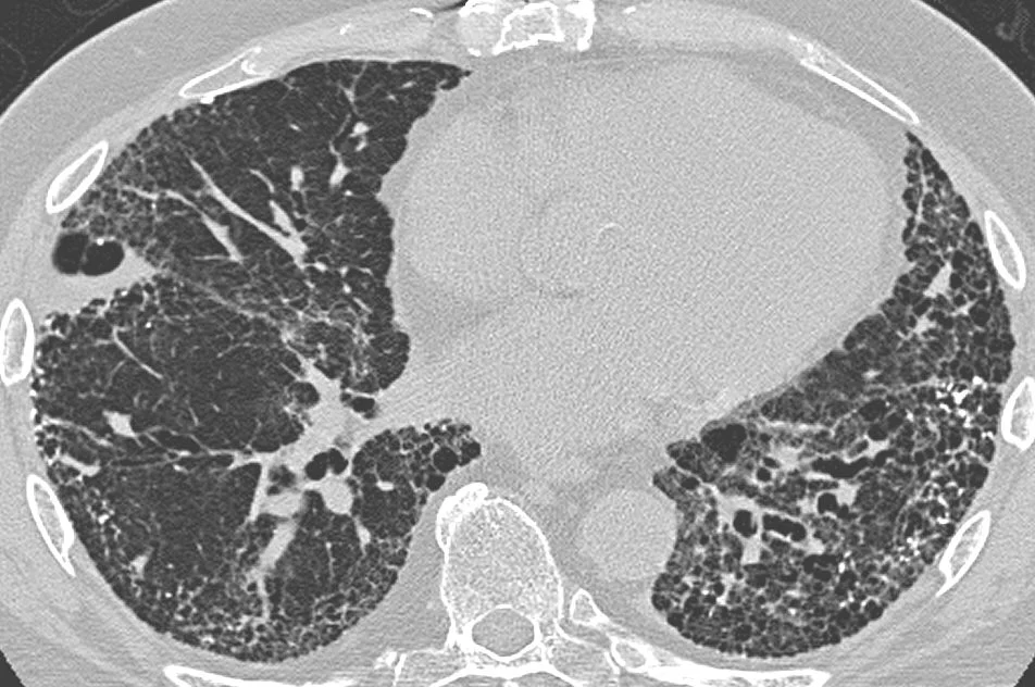
Figure 1CT scan in usual interstitial pneumonia (UIP)
Long description
A CT scan of usual interstitial pneumonia (UIP) showing interstitial thickening, architectural distortion, honeycombing and bronchiectasis.

High-resolution computed tomography of the chest is the pivotal investigation. It defines the pattern, suggests the cause, and in many cases removes the need for biopsy. Four categories are used, and the distribution and features carry diagnostic weight.[1]
| Category | Distribution and CT features | Confidence of histological UIP |
|---|---|---|
| UIP pattern | Subpleural and basal predominant, often heterogeneous. Honeycombing with or without traction bronchiectasis/bronchiolectasis, irregular septal thickening, reticulation, mild GGO, possible pulmonary ossification. | Confident (over 90%) |
| Probable UIP pattern | Subpleural and basal predominant, often heterogeneous. Reticular pattern with traction bronchiectasis/bronchiolectasis, may have mild GGO, absence of subpleural sparing. | Provisional high confidence (70-89%) |
| Indeterminate for UIP | Diffuse without subpleural predominance; peribronchovascular with subpleural sparing; suggesting NSIP; perilymphatic; suggesting sarcoidosis; upper or mid lung; suggesting fibrotic HP, CTD-ILD, or sarcoidosis; subpleural sparing. | Provisional low confidence (51-69%) |
| Alternative diagnosis | CT findings suggestive of another disease, including cysts suggesting cystic lung disease, mosaic attenuation or three-density sign suggesting hypersensitivity pneumonitis, consolidation suggesting organising pneumonia, nodules suggesting sarcoidosis, and pleural plaques suggesting asbestosis. | Low to very low confidence (under 50%) |
Honeycombing is a basal peripheral essential criterion for the typical definite pattern and is usually associated with traction bronchiectasis and a varying amount of ground glass.[1] The categories are not merely academic: they drive whether a biopsy is needed and how confidently a diagnosis of idiopathic pulmonary fibrosis can be made. Criteria reported to support a high-confidence diagnosis of idiopathic pulmonary fibrosis in the absence of a biopsy include a woman over 60 years of age, extensive reticulation of more than 30% of the lung on high-resolution computed tomography with age over 70, increased neutrophils or absence of lymphocytosis in bronchoalveolar lavage fluid, and multidisciplinary discussion producing a confident diagnosis.[1]
Nobody with interstitial lung disease should be labelled idiopathic until autoimmune disease has been considered. A panel tailored to the clinical picture typically includes antinuclear antibody, rheumatoid factor and anti-cyclic citrullinated peptide antibodies, anti-topoisomerase I, anti-threonyl-tRNA synthetase and other myositis-specific antibodies, anti-MDA5, anti-Sjögren syndrome A antibody, and screening for systemic sclerosis and inflammatory myopathy. Positive results move the diagnosis toward connective tissue disease associated interstitial lung disease, which changes treatment.[3]
Bronchoalveolar lavage does not by itself diagnose most interstitial lung diseases, but it changes probabilities. An increased proportion of neutrophils with little or no lymphocytosis supports the fibrotic, usual interstitial pneumonia end of the spectrum, while a marked lymphocytosis points toward hypersensitivity pneumonitis or organising pneumonia.[1] It is also useful to exclude infection, haemorrhage, and malignancy, and to look for eosinophilia or other clues. The strongest signal it provides is often negative: a high neutrophil count makes a confident non-biopsy diagnosis of idiopathic pulmonary fibrosis more plausible.[1]
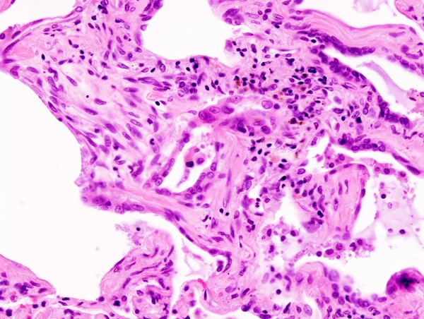
Figure 2Usual interstitial pneumonia (1)
Long description
A histopathological image of pulmonary fibrosis representing usual interstitial pneumonia (UIP), autopsy specimen, stained with hematoxylin and eosin.

Not every patient needs a lung biopsy. When the high-resolution computed tomography shows a clear usual interstitial pneumonia pattern in an appropriate clinical setting, biopsy adds little. Biopsy is considered when the imaging is indeterminate or suggests an alternative diagnosis, when the clinical picture does not fit, or when a histological diagnosis would change management. Transbronchial lung cryobiopsy has been conditionally accepted in the 2022 guideline as an alternative to surgical lung biopsy in centres with appropriate expertise, and the same careful patient selection applies as for surgical biopsy: severe physiological impairment, such as forced vital capacity under 50% predicted or diffusing capacity under 35%, moderate or severe pulmonary hypertension, uncorrectable bleeding risk, or significant hypoxaemia with arterial partial pressure of oxygen under 55 to 60 mmHg are relative contraindications because they raise the risk of complications.[1] An adequate biopsy is read against the full clinical and radiological picture in multidisciplinary discussion. When no biopsy can be obtained or is unsafe, an indeterminate pattern remains a working diagnosis that is revised as the disease declares itself.
Diagnosis in interstitial lung disease has shifted from a single-clinician label to a team decision. High-resolution computed tomography interpretation, pathology where available, clinical history, and serology are reviewed together by pulmonologists, radiologists, pathologists, and rheumatologists. Multidisciplinary discussion increases diagnostic confidence and reduces the chance that a treatable condition is called irreversible fibrosis.[1] It is also where treatment plans, including whether to use immunosuppression, antifibrotics, or transplant referral, are ratified.
Diagnostic approach to suspected interstitial lung disease
- Suspect ILDChronic dyspnoea, dry cough, fine inspiratory crackles, finger clubbing, compatible physiology
- History and examinationExposures, drugs, connective tissue features, occupation and hobbies, smoking, family history
- Pulmonary function testsRestrictive pattern: low FVC and TLC, preserved or raised FEV1/FVC, low DLCO
- High-resolution CT of the chestCategorise as UIP, probable UIP, indeterminate for UIP, or alternative diagnosis
- Question: CT pattern?
Typical UIP or probable UIP in a fitting clinical setting
- Multidisciplinary discussion confirms idiopathic pulmonary fibrosisSerology and, if obtained, bronchoalveolar lavage neutrophilia supports the diagnosis
Indeterminate or alternative diagnosis on CT
- Question: Is serology or an exposure suggestive of CTD, HP, drug, or occupation?
Yes
- Treat as that disease; biopsy only if it would change management
No
- Consider transbronchial cryobiopsy or surgical lung biopsy where safeAvoid in FVC under 50%, DLCO under 35%, severe pulmonary hypertension, or significant hypoxaemia
- Reassess with the multidisciplinary teamIntegrate history, physics, serology, imaging, bronchoalveolar lavage, and histology into one diagnosis
Management splits by underlying cause. Idiopathic pulmonary fibrosis is the model used below because it is the best studied; where other diseases differ, the differences are pointed out.
For all fibrotic interstitial lung diseases, sensible general measures include smoking cessation, avoidance of further occupational or domestic exposures, treatment of gastro-oesophageal reflux and obstructive sleep apnoea, vaccination against influenza and pneumococcal disease, pulmonary rehabilitation when breathlessness limits exercise, and nutritional support. Notably, the 2022 guideline makes conditional recommendations against antacid medication and antireflux surgery as treatments for idiopathic pulmonary fibrosis aimed at improving respiratory outcomes, because the systematic reviews informing the committee did not show a consistent benefit.[1] Gastro-oesophageal reflux should still be treated when it is symptomatic.
Idiopathic pulmonary fibrosis is treated with one of two antifibrotic drugs, nintedanib or pirfenidone. They are not interchangeable in every patient, and choice depends on comorbidities, liver function, interactions, tolerability, and cost. [7] [8] The decision to start is now made earlier rather than later, because the goal is to slow decline rather than to reverse established scar.
| Drug | Regimen | Dose reductions and monitoring |
|---|---|---|
| Nintedanib | 150 mg orally twice daily, about 12 hours apart, with food | Reduce to 100 mg twice daily for mild hepatic impairment (Child-Pugh A) or for intolerance; maintain reduced dose or discontinue if not tolerated at 100 mg twice daily. Liver function tests before starting and periodically during treatment because of hepatotoxicity; interrupted and then reintroduced at 100 mg twice daily when enzymes normalise, with escalation back to full dose possible. Common adverse effects include diarrhoea, nausea, vomiting, abdominal pain, appetite loss, weight loss, headache, and hypertension. |
| Pirfenidone | Titrate over 14 days to 801 mg three times daily with food (2403 mg per day in total): 267 mg three times daily for days 1 to 7, 534 mg three times daily for days 8 to 14, then 801 mg three times daily from day 15 onwards | Reduce to 267 mg three times daily with a strong CYP1A2 inhibitor such as fluvoxamine, and to 534 mg three times daily with a moderate inhibitor such as ciprofloxacin 750 mg twice daily. Liver function tests before starting and periodically; dose holds for hepatotoxicity or severe skin and gastrointestinal reactions. Administration with food reduces nausea and dizziness; photosensitivity and rash occur. |
| Prednisone and immunosuppression | Not first-line for idiopathic pulmonary fibrosis | Combination prednisone, azathioprine, and N-acetylcysteine increased harm in idiopathic pulmonary fibrosis, so this regimen is not used. Corticosteroids are used for acute exacerbation, organising pneumonia, and connective tissue disease associated disease. |
Nintedanib is a small-molecule tyrosine kinase inhibitor that targets fibroblast growth factor, platelet-derived growth factor, and vascular endothelial growth factor receptors, reducing the signalling that drives fibroblast proliferation and collagen production. Pirfenidone is an antifibrotic with antioxidant and anti-inflammatory properties that downregulates transforming growth factor beta signalling and collagen synthesis. Real-world practice emphasises that both reduce rather than halt disease progression, which is why patients must be counselled that the medication will not make them feel better or restore lung function, and that adherence to a daily regimen with food matters.
For connective tissue disease associated interstitial lung disease, immunosuppression remains the main therapeutic strategy, with glucocorticoids, mycophenolate, rituximab, cyclophosphamide, or tacrolimus chosen according to the underlying disease and its extrapulmonary activity.[3] Nintedanib is endorsed as an additional option for systemic sclerosis associated interstitial lung disease, and as a treatment when disease behaves progressively fibrotic despite immunosuppression, on the basis of shared fibrotic mechanisms.[3] The 2022 guideline makes a conditional recommendation for nintedanib in progressive pulmonary fibrosis and calls for more research into pirfenidone in that setting.[1]
Oxygen is prescribed for chronic hypoxaemia rather than for breathlessness alone. The American Thoracic Society home oxygen guideline recommends strong support for long-term oxygen in patients with interstitial lung disease who have severe chronic resting hypoxaemia, defined as an arterial partial pressure of oxygen at most 55 mmHg or an oxygen saturation at most 88%, or an arterial partial pressure of oxygen of 56 to 59 mmHg or saturation of 89% together with edema, a haematocrit of 55% or more, or P pulmonale on the electrocardiogram. Ambulatory oxygen is conditionally recommended for severe exertional hypoxaemia.[6] In fibrotic interstitial lung disease, resting hypoxaemia tends to appear late; exertional desaturation often comes first and should be checked with a walked test or the six-minute walk.
For breathlessness, pulmonary rehabilitation, breathing retraining, pacing, and fan therapy help many patients. Opioids in low dose and benzodiazepines are used for refractory breathlessness and anxiety in palliative care, and treatment of cough includes simple measures first, with neuromodulators reserved for stubborn cases.
Lung transplantation is the only intervention that clearly improves survival in selected patients with idiopathic pulmonary fibrosis. Patients at increased risk of mortality should be referred for transplant at diagnosis, not after functional reserve has been exhausted, because waiting lists are long.[1] Evaluation every three to six months tracks progression, with forced vital capacity and diffusing capacity used as sentinel measures.[1] Eligibility is individual and includes age, comorbidities, frailty, and psychosocial factors; single and bilateral lung transplantation are both performed, and in experienced centres survival is good. The presence of significant fibrotic honeycombing on imaging and rapid decline are signals to make or confirm the referral quickly.
An acute exacerbation of idiopathic pulmonary fibrosis is a sudden, unexplained worsening of breathlessness over days to weeks, with new widespread parenchymal abnormality on imaging and no alternative cause such as infection, heart failure, pulmonary embolism, or pneumothorax. It is a dramatic and often terminal event. Acute exacerbations may be treated with corticosteroids, but mechanical ventilation is not recommended for the majority of patients with respiratory failure.[1] Management is supportive: oxygen, treatment of reversible triggers, intravenous corticosteroids, empirical antibiotics when infection cannot be excluded, and discussion of goals of care. This is an area where honest early advance care planning, made while the patient can participate, replaces crisis decision making.
Palliative care should be introduced early, alongside rather than instead of disease-directed treatment. Patients may benefit from help with symptom management, including cough, dyspnoea, and anxiety.[1] Patient values and preferences should be explored explicitly, and decisions about pharmacological escalation, oxygen, ventilation, and transplant pursued or declined recorded and revisited as the disease changes.[1]
Patients are reviewed three to six months apart, or more often if they are deteriorating.[1] Each visit checks symptoms, oxygen saturation at rest and on exertion, forced vital capacity and diffusing capacity, high-resolution computed tomography when there is concern about progression or a change in pattern, weight and nutritional status, drug tolerance, and the patient's own priorities. Exacerbations, hospitalisations, deteriorating or newly developed pulmonary hypertension, and quality of life are all relevant but are less sensitive as single follow-up measures than the absolute change in forced vital capacity.
Progressive pulmonary fibrosis is the broader label applied when any interstitial lung disease other than idiopathic pulmonary fibrosis behaves progressively. It was introduced because antifibrotic therapy now has a role outside idiopathic pulmonary fibrosis. The diagnosis requires at least two of three criteria, occurring within the past year and with no alternative explanation: worsening respiratory symptoms, radiological progression on chest imaging, and physiological progression in the form of an absolute decline in forced vital capacity of 5% or more or an absolute decline in diffusing capacity for carbon monoxide of 10% or more within one year of follow-up.[1] Worsening symptoms alone is not enough without another criterion, and other possible causes of decline, such as infection or cardiac decompensation, must be excluded first.
Forced vital capacity is used as the sentinel physiological measure because it is associated with prognosis and is reproducible across centres.[1] The absolute rather than relative change is preferred because it forecasts poorer outcomes better in the available data.
The 2022 guideline makes a conditional recommendation for nintedanib in progressive pulmonary fibrosis and recommends further research into pirfenidone in this population.[1] In practice, when a patient with fibrotic hypersensitivity pneumonitis, connective tissue disease associated fibrosis, unclassifiable fibrotic interstitial lung disease, or another fibrotic disorder continues to worsen despite appropriate treatment of the underlying cause, nintedanib is considered and discussed. Combination with immunosuppression is not contraindicated when the underlying disease requires it, but drug interactions and hepatotoxicity are monitored.
- Respiratory failure from progressive stiffening and loss of gas exchange surface, eventually often with chronic hypoxaemia.
- Pulmonary hypertension and cor pulmonale, driven by chronic hypoxia, vascular remodelling, and destruction of the vascular bed; suspected when dyspnoea worsens out of proportion to lung function or when there are signs of right heart strain.
- Acute exacerbation as above, which is a feared and often fatal complication.
- Lung cancer risk is increased in fibrotic lung disease, partly through shared smoking exposure and partly through the scarring itself.
- Venous thromboembolism, because immobility, hypoxia, and systemic inflammation all promote clotting.
- Sleep disordered breathing and cor pulmonale overlap, where nocturnal desaturation accelerates pulmonary vascular disease.
- Treatment toxicity from antifibrotics, particularly hepatotoxicity, severe diarrhoea, and photosensitivity.
- Osteoporosis and muscle deconditioning from inactivity and, where used, corticosteroids.
Prognosis varies enormously by cause. Idiopathic pulmonary fibrosis has a poor prognosis and a relentlessly progressive course, although the rate of decline varies between individuals and the advent of antifibrotic therapy has improved survival compared with historical series.[1] Nonspecific interstitial pneumonia and hypersensitivity pneumonitis generally fare better if inflammation is controlled early. Connective tissue disease associated disease depends on the underlying disorder, with systemic sclerosis associated fibrosis behaving aggressively when progressive. The most useful prognostic anchor in the clinic is the rate of forced vital capacity decline and the extent of honeycombing on imaging; both predict mortality better than a single cross-sectional measurement. The conversation about prognosis should be honest and revisited often, and it should sit alongside active symptom control and support.
There is no screening programme for idiopathic pulmonary fibrosis, because no test is specific enough before symptoms arise. Prevention is therefore practical: avoid occupational and domestic exposures, wear respiratory protection in dusty work, stop smoking, and treat connective tissue disease early. Early identification of interstitial lung abnormalities on chest computed tomography done for other reasons is increasingly common and warrants a respiratory and rheumatology review rather than reassurance, because some progress to clinical disease.
Children. Interstitial lung disease is rare in children and the differential is different, dominated by genetic and developmental surfactant disorders, infection, and immune dysregulation. Management is in a specialist centre.
Older adults. Idiopathic pulmonary fibrosis is a disease of older adults, so polypharmacy, falls, frailty, comorbidity, and cognitive ability all complicate treatment. Drug interactions, particularly CYP1A2 inhibitors with pirfenidone, matter more than in younger patients.
Pregnancy. Connective tissue disease associated interstitial lung disease in pregnancy requires coordinated care between respiratory, rheumatology, and obstetric teams. Antifibrotic drugs are not used in pregnancy; treatment of flares relies on glucocorticoids and, where needed, agents compatible with pregnancy under specialist supervision.
Families. Familial pulmonary fibrosis should be suspected when more than one first-degree relative is affected or disease appears at a young age. Patients and families benefit from genetic counselling and, where appropriate, telomere genetics testing.
The diagnostic pathway should be slimmed to what is available without losing accuracy.
- A careful history of exposures, occupations, hobbies, and drugs provides most of the aetiological diagnosis and costs nothing.
- Chest radiography plus careful auscultation for velcro-like crackles detect most clinically significant disease; high-resolution computed tomography is ideal but a conventional computed tomography series is still far better than a plain film alone.
- Spirometry with forced vital capacity and total lung capacity where the body plethysmograph exists, and diffusing capacity where the gas analysers exist; in their absence, a peak flow and oxygen saturation are better than nothing and a six-minute walk with oximetry detects exertional desaturation.
- Where a biopsy is not available, a multidisciplinary discussion with a clinician, a radiologist, and, if possible, a remotely reviewed pathology slide remains valuable, and a provisional diagnosis with a planned review is acceptable.
- Nintedanib and pirfenidone are expensive and may be unavailable; when they are not, the emphasis falls on early identification of reversible or treatable forms such as organising pneumonia, infection, drug toxicity, and connective tissue disease, all of which respond to immunosuppression or drug withdrawal.
- Oxygen concentrators, portable cylinders, and pulmonary rehabilitation are often more accessible than antifibrotics and should be prioritised.
- Palliative care, symptom control, and advance care planning are low-cost interventions that matter most to patients as disease progresses.
- Interstitial lung disease is an umbrella diagnosis whose unifying physiology is restriction: reduced total lung capacity and forced vital capacity, preserved or raised forced expiratory volume in one second to forced vital capacity ratio, and reduced diffusing capacity for carbon monoxide.
- Causes are grouped into idiopathic interstitial pneumonias, connective tissue disease, drug-induced disease, occupational dust exposures, granulomatous diseases, and other categories.
- Idiopathic pulmonary fibrosis is a chronic, fibrosing interstitial pneumonia of older adults with a usual interstitial pneumonia pattern, progressive course, and poor prognosis.
- A typical definite usual interstitial pneumonia pattern requires basal and peripheral honeycombing, defined as clustered thick-walled cysts of 3 to 10 mm up to 2.5 cm, usually with traction bronchiectasis.
- Three to six monthly follow-up with forced vital capacity and diffusing capacity tracks progression; an absolute forced vital capacity decline of 5% or more over one year signals progression.
- Antifibrotic therapy uses nintedanib 150 mg twice daily or pirfenidone titrated to 801 mg three times daily, with food and liver function monitoring; it slows but does not reverse fibrosis.
- Progressive pulmonary fibrosis outside idiopathic pulmonary fibrosis requires worsening symptoms plus either radiological or physiological decline, and nintedanib carries a conditional recommendation.
- Long-term oxygen is indicated for severe chronic resting hypoxaemia, defined as an arterial partial pressure of oxygen at most 55 mmHg or saturation at most 88%, or 56 to 59 mmHg with end-organ evidence; ambulatory oxygen is conditionally recommended for exertional hypoxaemia.
- Transplant referral should be made early for suitable patients at increased mortality risk, and evaluation occurs every three to six months.
- Acute exacerbations may be treated with corticosteroids, but mechanical ventilation is not recommended for most patients with respiratory failure.
- Inflammatory forms such as organising pneumonia and many connective tissue disease associated diseases can respond to immunosuppression; antifibrotics are used in fibrotic and progressive disease.
- Diagnosis is increasingly made by multidisciplinary discussion integrating history, imaging, serology, lavage, and histology; no single test is sufficient on its own.
Answer each one in your head first, then open the answer.
A 72-year-old man has slowly progressive breathlessness over two years and a dry cough. High-resolution CT shows basal and peripheral honeycombing with traction bronchiectasis. What is the likely diagnosis, and what physiological pattern would you expect?
Show answerHide answer
Idiopathic pulmonary fibrosis, showing a usual interstitial pneumonia pattern. The spirometric pattern is restrictive: forced vital capacity and total lung capacity are reduced, the forced expiratory volume in one second to forced vital capacity ratio is preserved or raised, and the diffusing capacity for carbon monoxide is reduced. Finger clubbing is common in this disease and the crackles are fine inspiratory crepitations at the bases.
A patient with rheumatoid arthritis has breathlessness and high-resolution CT shows a nonspecific interstitial pneumonia pattern with fine reticulation and subpleural sparing. Which serologic and clinical clues point toward connective tissue disease associated interstitial lung disease, and how does management differ from idiopathic pulmonary fibrosis?
Show answerHide answer
High titres of rheumatoid factor and anti-cyclic citrullinated peptide antibodies support rheumatoid disease related lung involvement. Connective tissue disease associated interstitial lung disease is usually managed primarily with immunosuppression, such as glucocorticoids or mycophenolate, chosen for the systemic disease, with nintedanib added when disease behaves progressively fibrotic. Idiopathic pulmonary fibrosis is not treated with chronic immunosuppression, which caused harm in combination, but with an antifibrotic alone.
List the criteria used to diagnose progressive pulmonary fibrosis in a patient whose interstitial lung disease is not idiopathic pulmonary fibrosis.
Show answerHide answer
At least two of three criteria occurring within the past year with no alternative explanation: worsening respiratory symptoms, radiological progression, and physiological progression defined as an absolute decline in forced vital capacity of 5% or more or an absolute decline in diffusing capacity for carbon monoxide of 10% or more. The 2022 guideline gives a conditional recommendation for nintedanib in this setting.
A patient starting nintedanib asks what dose to take and how it should be taken. What do you say, and what monitoring is needed?
Show answerHide answer
Nintedanib is taken as 150 mg by mouth twice daily, about 12 hours apart, with food, swallowed whole without chewing. In mild hepatic impairment it is reduced to 100 mg twice daily, and the same reduced dose is used when 150 mg is not tolerated. Common problems are diarrhoea, nausea, and vomiting; liver function tests are checked before starting and periodically because of hepatotoxicity, with interruption and reintroduction at 100 mg twice daily once enzymes normalise.
An 80-year-old with severe fibrosis has been on home oxygen for six months and now presents with a two-week worsening and new widespread ground glass on CT. How do you approach the acute event, and what is the role of mechanical ventilation?
Show answerHide answer
First exclude infection, pulmonary embolism, heart failure, and pneumothorax. If these are excluded, this is an acute exacerbation of fibrotic interstitial lung disease: treat with supportive oxygen, empirical antibiotics when infection cannot be excluded, and corticosteroids, and arrange discussion of goals of care. Mechanical ventilation is not recommended for the majority of patients with respiratory failure, so honest advance care planning replaces late crisis decisions.
In a low-resource clinic without high-resolution CT or biopsy, what is the practical approach to a patient with probable fibrotic interstitial lung disease?
Show answerHide answer
Take a detailed exposure, drug, and family history, listen for velcro-like basal crackles and look for clubbing, obtain chest radiography and spirometry with diffusing capacity if available, and use conventional CT if possible. Focus on identifying reversible or treatable disease such as drug toxicity, infection, organising pneumonia, or connective tissue disease, because these respond to drug withdrawal or immunosuppression. Prioritise smoking cessation, pulmonary rehabilitation, oxygen, and early palliative care; refer for antifibrotic therapy where funds and access allow.
Interstitial lung disease connects closely to Pulmonary function tests, COPD, Bronchiectasis, Hypersensitivity pneumonitis, and Eosinophilic pulmonary syndromes, which are separate MedGrasp topics.
Pulmonary function tests
How lung volumes, airflow, gas transfer and blood gases are measured and what their patterns mean.Sarcoidosis
A multisystem granulomatous disease: how noncaseating granulomas form, how sarcoidosis presents by organ, and how it is diagnosed and treated.Pneumoconioses
Inhaled workplace dusts that scar the lungs: what silicosis, coal disease, asbestosis, beryllium disease, and byssinosis are, and how they differ.Hypersensitivity pneumonitis
Immune-mediated lung disease from inhaled antigens: farmer's, bird fancier's, humidifier and hot-tub lung; non-fibrotic and fibrotic forms.Pulmonary hypertension and cor pulmonale
How high pulmonary pressures arise, are classified, diagnosed and treated, and what they mean for the right heart and cor pulmonale.
6 other topics link here.
- Raghu G, Remy-Jardin M, Richeldi L, et al. Idiopathic Pulmonary Fibrosis (an Update) and Progressive Pulmonary Fibrosis in Adults: An Official ATS/ERS/JRS/ALAT Clinical Practice Guideline. Am J Respir Crit Care Med 2022;205(9):e18-e47. doi:10.1164/rccm.202202-0399ST. Accessed .
- Bermudo G, Rivera-Ortega P, Molina-Molina M, et al. The benefits of a rapid diagnostic primary care circuit for interstitial lung disease. NPJ Prim Care Respir Med 2025;35:56. doi:10.1038/s41533-025-00462-6. Accessed .
- Ozduygu G, Kaenmuang P, Guedes ABF, et al. Systemic Autoimmune Rheumatic Disease-Associated Interstitial Lung Disease: A Pulmonary Perspective on Recent Guidelines. Adv Ther 2026;43(10):4355-4379. doi:10.1007/s12325-026-03701-5. Accessed .
- Poveda JJR, Tamayo MH, Sellares J, et al. Drug-Induced Pulmonary Toxicity in the Era of Immunotherapy and Biologics: A Narrative Review of Mechanism, Diagnosis, and Management. Pulm Ther 2026;12(1):99-116. doi:10.1007/s41030-025-00338-7. Accessed .
- Feary J, Lindstrom I, Huntley CC, et al. Occupational lung disease: when should I think of it and why is it important?. Breathe (Sheff) 2023;19(2):230002. doi:10.1183/20734735.0002-2023. Accessed .
- Jacobs SS, Krishnan JA, Lederer DJ, et al. Home Oxygen Therapy for Adults with Chronic Lung Disease. An Official American Thoracic Society Clinical Practice Guideline. Am J Respir Crit Care Med 2020;202(10):e121-e141. doi:10.1164/rccm.202009-3608ST. Accessed .
- Boehringer Ingelheim Pharmaceuticals, Inc. OFEV (nintedanib) prescribing information (DailyMed label). DailyMed (FDA label), accessed 2026-10-27. Accessed .
- Genentech USA, Inc. ESBRIET (pirfenidone) prescribing information (DailyMed label). DailyMed (FDA label), accessed 2026-10-27. Accessed .
Figure credits
Educational content for medical students and doctors. It is not a substitute for clinical judgement or local guidelines.