Bronchiectasis
Permanent dilatation of the bronchi: causes, the cycle of inflammation and infection, diagnosis, management, complications, and low-resource care.
- Bronchiectasis is permanent, abnormal dilatation of the bronchi with inflamed, thickened walls. It is diagnosed by a compatible history plus the characteristic appearance on high-resolution CT.
- One underlying cause is found in many patients: post-infective damage (often tuberculosis in high-TB settings), cystic fibrosis, primary ciliary dyskinesia, immunodeficiency, allergic bronchopulmonary aspergillosis, obstruction, rheumatoid arthritis and alpha-1 antitrypsin deficiency are among the more common.
- The disease is driven by a vicious cycle: poor mucus clearance lets bacteria persist, which provokes neutrophilic inflammation, which injures the airway wall and worsens clearance, leading to further dilatation and infection.
- The classic symptom is a chronic productive cough with purulent sputum, with recurrent chest infections, intermittent haemoptysis and, in some, digital clubbing.
- Management rests on airway clearance, treating exacerbations with antibiotics, and prevention with vaccination. Long-term macrolides or inhaled antibiotics are used for patients with frequent exacerbations, and brensocatib is now an option in selected adults and adolescents aged 12 years and older.
By the end of this page you should be able to:
- Define bronchiectasis and classify it by cause and by morphology.
- Explain the vicious cycle from the initial insult to permanent airway dilatation.
- Recognise the typical history and examination findings and know when to suspect bronchiectasis.
- Interpret the key investigations, especially the HRCT signs and sputum culture.
- Outline the management of stable disease, exacerbations, haemoptysis and frequent exacerbators.
- Explain the place of surgery, bronchial artery embolisation and brensocatib.
- Adapt assessment and care to low-resource settings, including post-tuberculosis bronchiectasis.
DefinitionBronchiectasis
Permanent dilatation of the bronchi, with destruction of the bronchial wall and impaired mucociliary clearance, accompanied by a chronic cough producing sputum and recurrent respiratory infections. The diagnosis rests on this definition plus characteristic clinical symptoms; the dilatation is confirmed on imaging.
Radiologically and clinically, bronchiectasis is therefore not simply a scan finding: a slightly widened bronchus in an asymptomatic person is not the same disease. The spectrum runs from occasional infections in an otherwise well person to daily purulent sputum, frequent exacerbations and respiratory failure.
Morphological types. Cylindrical (tubular) bronchiectasis shows uniform airway dilation; varicose (varicoid) shows irregular beaded dilatation; cystic (saccular) shows balloon-like dilatation with air-fluid levels. These patterns usually coexist and often reflect how long the airway has been damaged rather than the cause.
Anatomical distribution gives clues to aetiology. Diffuse disease is typical of cystic fibrosis, primary ciliary dyskinesia and immunodeficiency. Focal disease suggests obstruction (foreign body, tumour, stricture) or prior localised infection. Upper-lobe predominance is characteristic of post-tuberculosis and post-NTM disease; lower-lobe disease is more typical of other infection-driven and post-aspiration bronchiectasis.
Clinically significant bronchiectasis is further described by the number of lobes involved, the frequency and severity of exacerbations, chronic infection with particular organisms (especially Pseudomonas aeruginosa), and lung function. Many clinicians now label individual treatable traits such as airway infection, breathlessness, eosinophilic inflammation and comorbid disease.
Bronchiectasis is the third most common chronic respiratory disease worldwide, after asthma and chronic obstructive pulmonary disease.[1] Recent estimates suggest roughly 1 in 200 adults have bronchiectasis, although increasing rates of diagnosis mean this is likely an underestimate.[1] The rates of hospitalisation and mortality from bronchiectasis are rising by approximately 4 percent per year.[1]
The burden is not evenly distributed. The prevalence in the United States has been estimated at 139 to 213 cases per 100,000 adults, while in the United Kingdom it has been estimated at 566 per 100,000 women and 485 per 100,000 men, with the highest reported estimates in China (about 1200 per 100,000 people above 40 years of age).[3] In parts of Africa and South Asia the aetiological mix is dominated by post-infectious disease, often post-tuberculosis, and patients present younger and more often male than in the high-income registries.[3]
Bronchiectasis is highly heterogeneous: a single label covers elderly never-smokers with a single basal segment dilated after a pneumonia, children with primary ciliary dyskinesia, and young adults with cystic fibrosis. This heterogeneity should be kept in mind when reading any single guideline.
Aetiological testing is part of the management, because treating the underlying cause changes outcomes. Bronchiectasis is described as idiopathic in roughly 20 to 40 percent of cases, although some of that reflects incomplete testing rather than true unknown cause.[1]
The major causes and risk factors are summarised below.
| Cause | Typical clues | Comment |
|---|---|---|
| Post-infective (severe pneumonia, pertussis, measles, tuberculosis) | Clear before-and-after history; upper-lobe, focal or widespread scarring depending on the infection | Probably the most common identifiable cause worldwide |
| Cystic fibrosis | Onset in childhood, pancreatic insufficiency, malabsorption, sinus disease, male infertility | Test with sweat chloride and CFTR analysis |
| Primary ciliary dyskinesia | Chronic rhinosinusitis, otitis media, situs inversus, neonatal respiratory distress, infertility | Responsible for up to 10 percent of cases, often beginning in childhood |
| Immunodeficiency | Recurrent or severe infections from early life; common variable immunodeficiency most common in adults | Low immunoglobulins; replacement reduces further damage |
| Allergic bronchopulmonary aspergillosis (ABPA) | Asthma or cystic fibrosis, very thick and pluggy sputum, eosinophilia | A high proportion of asthmatics with positive Aspergillus skin tests are affected |
| Tuberculosis | Prior TB (especially smear-positive or cavitary disease), haemoptysis, upper-lobe bronchiectasis | Incidence of bronchiectasis after TB ranges from 16 to 65 percent in reported studies |
| Obstruction (tumour, foreign body, lymphadenopathy, strictures) | Focal disease, recurrent distal infection, non-resolving atelectasis | Missed if not imaged beyond the bronchiectatic segment |
| Rheumatoid arthritis (and other connective tissue diseases) | Known inflammatory disease, dry eyes and mouth, joint deformity | About 5 to 10 percent of patients with rheumatoid arthritis, inflammatory bowel disease and other connective tissue diseases have bronchiectasis |
| Alpha-1 antitrypsin deficiency | Basal emphysema, early-onset disease, family history, liver disease in childhood | An uncommon, treatable cause of lower-lobe disease |
| Non-tuberculous mycobacterial infection | Tall, thin female patients, non-productive cough, weight loss, poor response to standard antibiotics | Mycobacterium avium complex is the most common organism; classically tall, thin patients |
| Gastro-oesophageal reflux and aspiration | Nocturnal cough, regurgitation, risk factors for aspiration | Under-recognised treatable trait |
| Young syndrome, Kartagener features | Infertility, chronic sinus disease | Rare or hereditary |
Immunodeficiency deserves specific mention. Primary immunodeficiency, especially common variable immunodeficiency, often presents only with recurrent infections; secondary immunodeficiency from lymphoma, myeloma, drugs or HIV also causes bronchiectasis. Serum immunoglobulins should be measured in every new patient, because immunoglobulin replacement reduces further lung damage.
Cystic fibrosis. In any young adult or adolescent with bronchiectasis, especially with pancreatic symptoms, chronic sinusitis or infertility, cystic fibrosis and its milder variants must be considered. The sweat chloride test is the standard biochemical screen: a value of 60 mmol/L or greater is indicative of cystic fibrosis, 40 to 59 mmol/L is intermediate and 39 mmol/L or less makes cystic fibrosis unlikely in a person above 6 months of age.[6] Genetic testing and a CF care centre referral are needed when results are abnormal or intermediate.
Alpha-1 antitrypsin deficiency. Patients present with early emphysema, often basal and panlobular, in addition to bronchiectasis. A low serum alpha-1 antitrypsin level leads to phenotyping and genotyping (for example, the PiZZ genotype). Augmentation therapy exists but is only available in some health systems.
Primary ciliary dyskinesia. Nasal nitric oxide is low, and the diagnosis is made by high-speed videomicroscopy, electron microscopy, or genetics, because PCD is genetically heterogeneous. Associated findings include chronic rhinosinusitis, otitis media, situs inversus (Kartagener syndrome), neonatal respiratory distress and male or female infertility. The defect lies in ciliary ultrastructure and function, so mucus transport fails from early life.
ABPA. ABPA produces bronchiectasis through an allergic response to Aspergillus fumigatus, usually in asthma or cystic fibrosis. It is thought to be the cause of bronchiectasis in roughly 10 percent of patients, with geographical variation.[1] Diagnostic tests include total IgE, Aspergillus-specific IgE and IgG, a blood count showing eosinophilia, sputum culture and CT showing central bronchiectasis (often with high-attenuation mucus plugs). In classic Rosenberg-Patterson criteria the total IgE exceeds 1000 IU/mL.[4] Some criteria (including the ISHAM working group) accept lower total IgE values when every other criterion is met, and a positive type I skin test can substitute when specific IgE is unavailable.[4] Cystic-fibrosis-specific ABPA uses its own criteria with a total IgE greater than 1000 IU/mL or, if lower, a doubling from baseline.
The pathophysiology can be understood as a sequence that becomes self-sustaining.
- An initial insult reduces clearance or defence. Infection, obstruction, impaired immunity or a congenital ciliary defect damages the mucociliary escalator or overwhelms local defences.
- Mucus retention and bacterial colonisation follow. Bacteria that would normally be cleared persist in dilated, secretion-filled airways.
- Chronic inflammation is triggered. Neutrophils dominate; they release elastases, matrix metalloproteinases, reactive oxygen species and neutrophil extracellular traps, which perpetuate inflammation and damage the bronchial wall and elastic tissue.
- Structural damage causes irreversibility. Destruction of elastic and muscular support leads to permanent dilatation, while scarring and narrowing of smaller airways trap further secretions.
- The cycle restarts. Worse clearance permits more infection, which drives more inflammation, which drives more dilatation.
This is Cole's vicious cycle, now more often described as a vicious vortex, because each component acts on all the others rather than in a simple line.[1][2] The trigger, or "entry point", varies with the underlying cause: in cystic fibrosis it is abnormal ion transport and mucus; in immunodeficiency it is failed clearance of normal organisms; in obstruction it is stagnant secretions behind a narrowed airway.[2]
Neutrophil serine proteases are central to the damage, especially neutrophil elastase, which slows ciliary beat frequency, stimulates mucus secretion and correlates with disease severity, dyspnoea, radiological extent, lung function (FEV1) and mortality.[2] Non-tuberculous mycobacteria and Pseudomonas aeruginosa thrive in damaged airways and in turn amplify inflammation.
Once dilated, the bronchus cannot return to normal diameter with treatment; management aims to arrest the cycle, not to reverse structural damage.
The symptom complex reflects the cycle of retained secretions and infection.
- Chronic productive cough, usually daily, with mucoid or purulent sputum. Sputum volume is a measure of disease activity, and three or more of the classic signs (increased cough, increased sputum volume or change in consistency, increased purulence, breathlessness or reduced exercise tolerance, fatigue or malaise, haemoptysis) developing over 48 hours mark an exacerbation.[5]
- Recurrent chest infections or "winter chest" that needs antibiotics repeatedly.
- Pleuritic chest pain or localised ache.
- Haemoptysis, often small-volume and related to an exacerbation, but sometimes the leading complaint.
- Breathlessness on exertion, often with wheeze in those with coexisting asthma or COPD.
- Fatigue, weight loss, anorexia and a general sense of ill health in severe or infective disease.
- Rhinosinusitis and nasal polyps, especially in PCD and cystic fibrosis.
Patients often describe years of a "smoker's cough" without smoking, or a label of asthma or COPD that never responded to treatment. Diagnostic delay is common: about a quarter of patients wait more than ten years for the diagnosis of bronchiectasis.[1]
A simple sputum colour chart helps patients and clinicians distinguish mucoid (grey or white) from mucopurulent and purulent (yellow or green) sputum; purulent sputum is not normal and reflects neutrophils, usually indicating bacterial infection.[1]
- General: clubbing (in advanced or severe chronic suppurative disease), weight loss.
- Respiratory: coarse crackles, often basal and persistent, and wheeze. The crackles do not clear with coughing.
- Cyanosis, tachypnoea and use of accessory muscles in advanced disease.
- Signs of right heart strain and cor pulmonale (raised jugular venous pressure, peripheral oedema) in severe or late disease.
- Growth failure in children.
- Extrathoracic clues: sinus disease and nasal polyps, kyphoscoliosis, joint disease (rheumatoid arthritis), infertility, situs inversus.
Digital clubbing occurs more often in severe disease and in cystic fibrosis; its absence does not exclude bronchiectasis.
The symptoms of bronchiectasis overlap with asthma, COPD, chronic bronchitis and recurrent pneumonia. Points of separation:
- Asthma: variable airflow obstruction, bronchial hyper-responsiveness, eosinophilic inflammation, normal CT; bronchiectasis can coexist with or be caused by poorly controlled asthma.
- COPD: post-bronchodilator FEV1/FVC below 0.7 with an appropriate exposure history (the ROSE criteria require at least 10 pack-years of smoking or another exposure such as biomass fuel plus airflow obstruction, even in the presence of bronchiectasis); inhaled corticosteroids have a place only when COPD is present.
- Recurrent pneumonia: acute febrile illness with focal infiltrates that usually clear; bronchiectasis is the diagnosis when dilated airways persist on CT.
- Tuberculosis, NTM, fungal infection: may both cause and mimic bronchiectasis; send sputum for mycobacterial culture.
- Tracheobronchomalacia and bronchiolitis: airway collapse or small-airway disease on expiratory CT or lung function.
Be careful not to let a historic label of asthma or COPD replace a proper evaluation: sputum purulence, frequent exacerbations despite inhalers, and HRCT changes should prompt CT when available.
Figure 1Article river a52123f05cfb11e9afb233c9de065d31-fig-3
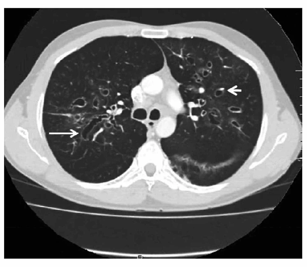
Long description
CT image labeled with arrows showing 'signet ring' and 'string of pearls' appearances indicative of central bronchiectasis, with mucoid impaction and dilated bronchi also seen.

High-resolution CT of the chest is the standard test. The following features indicate bronchiectasis:
- Broncho-arterial ratio (BAR) greater than 1: the lumen of a bronchus exceeds the diameter of the adjacent pulmonary artery. On axial CT a dilated bronchus sitting next to its smaller artery produces the signet ring sign.
- Lack of normal bronchial tapering, so that medium-sized bronchi remain visible close to the pleura.
- Tram-track sign: thickened, dilated bronchi running parallel to each other like railway lines.
- Visibility of airways within 1 cm of the costal pleural surface or abutting the mediastinum.
Chest radiography is often normal or nonspecific; when abnormal it may show tram-track opacities, ring shadows and increased bronchovascular markings. A chest radiograph can support a diagnosis in resource-limited settings, but cannot confirm or exclude bronchiectasis.
Severity of dilatation is classified as cylindrical (tubular), varicose (varicoid) or cystic (saccular). The number of lobes involved and the presence of varicose or cystic change correlate with severity.
- Sputum culture should be performed in every patient with confirmed bronchiectasis, including when the patient is clinically stable. Up to 70 percent of patients are chronically infected even when they feel well; a positive culture in a stable patient does not by itself require antibiotics.[1]
- Send sputum for acid-fast bacilli (NTM) culture at diagnosis and before starting long-term macrolides, because macrolide monotherapy can select resistant mycobacteria.
- Mycobacterium avium complex is the most common non-tuberculous mycobacterial pathogen; it presents classically in tall, thin women with chronic cough or weight loss and needs prolonged combination therapy.[1]
- Screen also for Pseudomonas aeruginosa. It is isolated in about 22 percent of European patients with bronchiectasis who have sputum sent (up to about 33 percent in southern Europe) and predicts a three-fold increase in mortality and a seven-fold increase in hospitalisations.[1] It forms biofilms and is hard to eradicate once established.
A standard panel at diagnosis, to find a treatable cause and establish the baseline, is: full blood count, immunoglobulins, serum protein electrophoresis, ABPA serology and sputum culture including NTM.[1] Many of these are simple and available even in primary care.
Other useful tests:
- Spirometry. There is no single bronchiectasis pattern: spirometry may be normal, obstructive, restrictive or mixed. An obstructive pattern is the most common, and FEV1 percent of predicted forms part of severity assessment.[1]
- Total IgE and Aspergillus-specific IgE and IgG with eosinophil count for ABPA (see Aetiology).
- Serum immunoglobulins (IgG, IgA, IgM) for antibody deficiency.
- Sweat chloride and CFTR genetics when cystic fibrosis is possible.
- Alpha-1 antitrypsin level, with phenotyping/genotyping.
- Laboratory tests for connective tissue disease and serum inflammatory markers when clinically indicated.
Approach to a patient with suspected bronchiectasis
- Compatible historyChronic productive cough, recurrent chest infections, haemoptysis, breathlessness, or clubbing
- Chest X-ray and spirometryOften normal or nonspecific; spirometry rarely diagnostic
- Arrange HRCT when availableSignet ring sign, tram-track sign, lack of tapering, visibility of peripheral airways
- Question: Bronchiectasis confirmed or strongly suspected?
Yes
- Standard aetiological panelFull blood count, immunoglobulins, serum protein electrophoresis, ABPA serology, sputum culture including NTM
- Cause-specific testsSweat chloride and CFTR if cystic fibrosis possible; alpha-1 antitrypsin; PCD testing; rheumatology screen
- Refer to respiratory specialistAirway clearance training, vaccination, consideration of macrolides, inhaled antibiotics, brensocatib
No
- Reconsider asthma and COPDObjective testing for airflow obstruction and reversibility; review whether ICS are appropriate
- Treat the alternative diagnosisReassess if symptoms persist or recur
Severity scores combine age, FEV1 percent predicted, hospitalisation or exacerbation count, colonisation with P. aeruginosa and other clinical markers into an index (such as the Bronchiectasis Severity Index). The score is useful for prognosis and for deciding how intensively to treat and follow the patient. FEV1 percent of predicted is measured routinely and forms part of these scores.[1]
The aims are to improve symptoms and quality of life, reduce exacerbations and preserve lung function, and to treat the underlying cause.
- Airway clearance techniques are the foundation of treatment and should be taught by a specialist respiratory physiotherapist.[1] No single technique suits everyone, so individual assessment (oscillating positive expiratory pressure devices, active cycle of breathing, postural drainage with percussion, autogenic drainage) is needed.
- Pulmonary rehabilitation should be offered to patients with breathlessness or impaired exercise capacity, and works at least as well as in COPD.[1]
- Hydration and humidity to keep secretions thin.
- Vaccination: influenza vaccine annually and pneumococcal vaccination according to local guidance.
- Smoking cessation where relevant.
- Sputum self-management: patients should be taught to recognise their own exacerbation (change in cough, sputum volume or consistency, purulence, breathlessness, fatigue, haemoptysis).
Adherence to airway clearance is poor in many patients; education and review help.
Mucoactive drugs are used when airway clearance alone does not control symptoms.[1]
- Carbocisteine and N-acetylcysteine may reduce sputum viscosity but evidence is mixed; a supervised trial with planned review, and a trial of stopping if no benefit, are reasonable.
- Hypertonic saline improves airway hydration and stimulates cough. Bronchospasm can occur, sometimes unpredictably even after prolonged use, so pre-treatment with a nebulised bronchodilator (for example, salbutamol 2.5 mg) is recommended and hypertonic saline stopped if symptoms persist.[1]
- Recombinant DNase (dornase alfa) is not recommended in bronchiectasis (evidence of benefit is lacking and harm is possible).[1]
Bronchodilators. Bronchiectasis itself does not always cause reversible airflow obstruction, but many patients are breathless and many have overlapping asthma or COPD. A trial of dual long-acting bronchodilation is reasonable in patients with significant breathlessness, particularly where spirometry shows obstruction.[1]
Inhaled corticosteroids (ICS). ICS are not indicated in bronchiectasis without coexisting asthma or COPD with an ICS indication.[1] They increase the risk of pneumonia and may promote airway bacterial infection, including non-tuberculous mycobacterial disease; high doses carry systemic risks. In patients with possible bronchiectasis, review any asthma or COPD label carefully: a patient whose clinical history is not suggestive of asthma and who has never had objective evidence of it, and who has not responded to appropriate inhaled steroid therapy, is unlikely to have asthma. Similarly, airflow obstruction in a never-smoker without exposure should not be labelled COPD even when bronchiectasis is present (the ROSE criteria set out the exposure and spirometry thresholds required).[1]
Eosinophilic bronchiectasis is a distinct subgroup in a proportion of patients (peripheral eosinophils at least 300 per microlitre or exhaled nitric oxide at least 25 parts per billion in up to about 20 percent). In this subgroup ICS may reduce exacerbations even without asthma, and monoclonal antibodies such as anti-IL-5 or anti-IL-5 receptor agents are being studied.[2]
Macrolides. For patients at high risk of exacerbations despite standard care, long-term macrolide therapy is strongly recommended.[1] Treatment is usually with azithromycin 250 mg three times weekly to 250 mg once daily, with clarithromycin or erythromycin substituted for clinical or availability reasons.[1] Azithromycin has both anti-inflammatory and antimicrobial activity and reduces exacerbation rates by about 50 percent.[1] Important cautions:
- Gastrointestinal side effects (diarrhoea, nausea) occur in up to 1 in 5 patients, more at higher doses; alternative-day or three-times-weekly dosing helps.
- Macrolides can prolong the QT interval; obtain an ECG before starting and monitor if QT-prolonging drugs are added.
- Tinnitus and hearing loss are rare but recognised; stop the drug and arrange assessment.
- Liver enzyme elevations can occur; many clinicians check baseline liver function and repeat at 2 to 4 weeks.
- Always obtain NTM culture before starting, and avoid macrolides alone when NTM is present.[1]
Macrolides are prescribed off-label in many countries; prescribing responsibility and funding vary, and stable patients may be discharged to primary care after a successful period of treatment.
Inhaled antibiotics (for example, nebulised colistin, tobramycin or gentamicin, or a dry-powder formulation) are strongly recommended for patients at high risk of exacerbations with chronic Pseudomonas aeruginosa infection, and conditionally recommended for high-risk patients with other chronic pathogens.[1] They are usually started and tested in secondary care, with airway clearance and bronchodilator before each dose to improve tolerability. Bronchospasm may occur immediately after inhalation.
Pseudomonas eradication. Where P. aeruginosa is newly isolated (even a scanty growth on a single culture), it should prompt repeat culture and urgent discussion with secondary care. Eradication is usually attempted promptly, with oral ciprofloxacin 500 to 750 mg twice daily for two weeks, often combined with or followed by inhaled or intravenous antibiotics under specialist guidance.[1] Treating as early as possible after first isolation gives the best chance of success. Other organisms such as Haemophilus influenzae are not usually eradicated.
Long-term oral non-macrolide antibiotics are not recommended first-line for high-risk exacerbators.[1]
A bronchiectasis exacerbation is a worsening of symptoms beyond normal day-to-day variation that requires a change in management. Core symptoms include change in cough, sputum volume or consistency, purulence, dyspnoea or reduced exercise tolerance, fatigue or malaise and haemoptysis; fever, wheeze, anorexia, weight loss and pleuritic pain may also occur.[1] Presentation is heterogeneous and patients often report subtle early changes; their judgement should be trusted.
Features of a severe exacerbation (requiring hospitalisation or intravenous antibiotics) include tachypnoea, acute or acute-on-chronic respiratory failure, a significant fall in oxygen saturation, hypercapnia, haemoptysis, cyanosis, new cor pulmonale, haemodynamic instability or impaired cognition.[1]
Management of exacerbations:
- Obtain a sputum sample for culture before starting antibiotics when possible; repeat culture if there is no response.
- Prescribe antibiotics guided by previous microbiology, local susceptibility and severity.
- A 14-day antibiotic course is considered standard, especially in severe exacerbations or P. aeruginosa infection; shorter courses may be appropriate in mild disease, sensitive organisms such as Streptococcus pneumoniae, or rapid return to baseline.[1]
- Airway clearance should be intensified and adapted.
- Oral corticosteroids are not routinely used unless the patient has asthma or COPD with bronchoconstriction.
- In patients with overlapping asthma or COPD, it can be useful to give antibiotics and steroids separately to identify which helps.
- New haemoptysis is a red flag requiring imaging and discussion with secondary care.
- Non-responders need reassessment, change of antibiotic or intravenous therapy and hospitalisation.
Supply selected patients with a self-management plan and a rescue pack of antibiotics (and steroids where indicated), with clear advice on when to start, what to take and when to contact the team.
Surgery. Localised, irreversible disease that remains disabling despite optimal medical management (for example, a single lobe with recurrent disabling haemoptysis or persistent purulent sputum after maximal medical therapy) may be considered for surgical resection. Surgery is now uncommon and the decision and timing are made by a thoracic surgery and bronchiectasis multidisciplinary team. Pre-operative optimisation of nutrition and airway clearance is essential. End-stage disease that is not surgically resectable may be referred for evaluation for lung transplantation.
Bronchial artery embolisation is the first-line intervention for significant or recurrent haemoptysis in bronchiectasis, when the bleeding is not controlled by antibiotics alone or is life-threatening. It is performed by interventional radiology. Re-bleeding can occur, and the procedure may need repeating; surgery is reserved for embolisation failures or unreachable bleeding.
Haemoptysis is usually treated with antibiotics in bronchiectasis, as it commonly reflects infection, but a new bleed (or any large-volume or recurrent haemoptysis) is a red flag requiring chest imaging and involvement of secondary care.[1]
Review at least annually in stable disease, more often in moderate or severe disease. Reassess aetiology and comorbidities, repeat spirometry, review microbiology, exacerbation frequency and symptom burden, and consider escalating to long-term antibiotics. Deteriorating patients (increasing exacerbation frequency or severity, worsening symptoms or rapid decline in lung function) should be referred for specialist review, after excluding pneumonia, pulmonary embolism, malignancy and cardiac disease, and considering adherence and mental health.[1]
- Recurrent and increasingly severe exacerbations, with hospitalisation.
- Respiratory failure, often with cor pulmonale in advanced cases.
- Haemoptysis and, rarely, massive life-threatening bleed.
- Empyema, lung abscess and pneumonia.
- Secondary amyloidosis in very longstanding suppuration (uncommon now).
- Malnutrition and growth failure in children and cystic fibrosis.
- Depression, anxiety and reduced quality of life from cough and sputum.
- Disturbed sleep from nocturnal cough and post-nasal drip.
Antibiotic resistance is an increasing problem in chronic infection, particularly P. aeruginosa, and should inform empiric choices.
Bronchiectasis is chronic and progressive in many patients but stable for years in others. The factors that worsen prognosis are frequent exacerbations, P. aeruginosa or NTM infection, lower FEV1, wider radiological disease, and comorbid disease. Early diagnosis, airway clearance and infection control improve outcomes. Once dilatation is fixed, lung function decline can be slowed but rarely reversed.
There is no population screening test for bronchiectasis. Prevention lies in:
- prompt and complete treatment of tuberculosis and childhood respiratory infections;
- immunisation (influenza, pneumococcal, pertussis, measles) according to national schedules;
- awareness that chronic productive cough is not "just a smoker's cough", especially in non-smokers, women under 60 and those with recurrent chest infections;
- early evaluation of chronically coughing children and young adults for cystic fibrosis or PCD.
Children. The commonest cause of bronchiectasis in children globally is post-infective (post-measles, post-pertussis, post-tuberculosis) plus congenital causes such as cystic fibrosis and PCD. Congenital causes should be sought early. Growth and development need special attention.
Pregnancy. Bronchiectasis is usually compatible with pregnancy. Exacerbations should be treated promptly with pregnancy-safe antibiotics; chest physiotherapy continues. Very severe disease warrants specialist assessment before conception.
Immunocompromised patients. Management includes treatment of the underlying immunodeficiency where possible (immunoglobulin replacement, control of HIV), together with standard bronchiectasis care.
Elderly patients. Comorbidities, aspiration risk and frailty change the balance of treatment; airway clearance must be realistic and tailored.
East Africa
In settings where high-resolution CT, bronchoscopy or specialised microbiology are scarce, a chest radiograph showing tram-track or ring shadows, recurrent purulent sputum and compatible symptoms can support the diagnosis. Sending sputum early, before antibiotics are started and by the quickest route to the laboratory, is the single highest-yield test; acid-fast culture should be requested whenever TB or NTM is a possibility. Airway clearance can be taught with written or online resources and by trained nurses where physiotherapy is unavailable, although professional assessment is better. Hypertonic saline, carbocisteine and inhaled bronchodilators are often the only drugs available and should be used judiciously.
Post-tuberculosis bronchiectasis deserves particular emphasis. Tuberculosis is the most common attributable cause of non-cystic bronchiectasis in many high-TB-burden settings; in one Johannesburg cohort 77.0 percent of cases were attributed to prior TB.[3] Bronchiectasis develops after an estimated 16 to 65 percent of tuberculosis episodes, and in 44 percent of people completing TB treatment in one Malawi cohort.[3] With millions of TB survivors, the burden is enormous.[3]
Features that should prompt aggressive evaluation in a person with a history of TB include persistent or recurrent cough, haemoptysis, progressive breathlessness and recurrent chest infections after TB treatment has finished. Upper-lobe traction bronchiectasis and bronchial stenosis from tuberculous lymphadenopathy add to the complexity and may not respond to usual bronchiectasis therapy. Inhaled corticosteroids should not be started for bronchiectasis unless there is coexisting asthma, because they may be associated with worse infection outcomes, mycobacterial infection and mortality.[3] Macrolides, bronchodilators and airway clearance need local evaluation of their benefit and cost in this phenotype.
Finally, in low-resource settings a wait-and-see approach when Pseudomonas aeruginosa is first isolated is never appropriate; any isolation should prompt repeat culture and urgent discussion with secondary care.[1]
- Bronchiectasis is permanent dilatation of the bronchi with a compatible clinical picture; HRCT shows signet ring, tram-track and non-tapering airways.
- Treatable causes must be sought: immunodeficiency, ABPA, cystic fibrosis, PCD, obstruction, rheumatoid arthritis, alpha-1 antitrypsin deficiency and non-tuberculous mycobacteria.
- The vicious vortex links mucus retention, infection and neutrophilic inflammation to progressive wall damage; once dilated, the airway does not return to normal.
- Chronic productive cough, recurrent infections, haemoptysis and clubbing are typical; diagnostic delay is common.
- Sputum culture, including for NTM, should be taken in every patient, even when stable.
- Airway clearance, vaccination and exacerbation treatment are the foundations; hypertonic saline and N-acetylcysteine are adjuncts and dornase alfa is not recommended.
- ICS are not indicated without asthma or COPD; they can worsen infection risk.
- Frequent exacerbators benefit from long-term macrolides (azithromycin 250 mg three times weekly to daily) and, with chronic Pseudomonas infection, inhaled antibiotics.
- Pseudomonas eradication is attempted promptly with oral ciprofloxacin 500 to 750 mg twice daily for two weeks, often plus inhaled or intravenous therapy.
- Brensocatib, a DPP-1 inhibitor taken once daily at 10 or 25 mg, reduces exacerbations in adults and adolescents aged 12 years and older with non-cystic fibrosis bronchiectasis (ASPEN: annualised exacerbation rate 1.29 with placebo versus 1.02 with 10 mg and 1.04 with 25 mg; median time to first exacerbation 36.7 versus 49.0 and 50.7 weeks). Common adverse effects include upper respiratory infection, headache, rash, dry skin, hyperkeratosis and hypertension, with dental and dermatological monitoring advised.[5]
- Significant or recurrent haemoptysis warrants bronchial artery embolisation; focal disabling disease may be resected.
- Post-TB bronchiectasis is common in high-TB-burden settings and needs its own appraisal of benefit and risk.
Answer each one in your head first, then open the answer.
What three things must be present for the diagnosis of bronchiectasis, and what imaging sign is most characteristic?
Show answerHide answer
A compatible clinical history (chronic productive cough, recurrent infections, haemoptysis or breathlessness), plus evidence of permanent bronchial dilatation. The structural change is confirmed on high-resolution CT, where the signet ring sign (a dilated bronchus next to its smaller pulmonary artery) is the most characteristic finding; tram-track opacities and lack of bronchial tapering are also typical.
A 19-year-old has bronchiectasis with recurrent sinusitis, otitis media and situs inversus. Which diagnosis should be considered first and what test is usually abnormal?
Show answerHide answer
Primary ciliary dyskinesia, in which defective ciliary function impairs mucociliary clearance from early life. Nasal nitric oxide is characteristically low, and the diagnosis is confirmed by high-speed videomicroscopy, electron microscopy or genetics.
Why does a patient with *Pseudomonas aeruginosa* infection and bronchiectasis have a three-fold mortality risk even when reasonably well?
Show answerHide answer
Pseudomonas forms biofilms, persists in damaged airways and drives neutrophilic inflammation and exacerbations. Its isolation marks structurally advanced, chronically infected disease with frequent severe exacerbations and hospitalisations, hence the higher mortality. Management therefore aims at eradication when first isolated and at suppression with inhaled antibiotics when chronic infection is established.
How should an exacerbation of bronchiectasis be treated, and for how long?
Show answerHide answer
With antibiotics guided by previous sputum culture, local susceptibility and severity, plus intensified airway clearance. A 14-day course is considered standard, especially in severe exacerbations or with P. aeruginosa; shorter courses may be used if the infection is mild or the organism is sensitive and recovery is rapid. New sputum should be sent before antibiotics, and oral corticosteroids are reserved for coexisting asthma or COPD with bronchoconstriction.
A patient with newly confirmed bronchiectasis and no asthma or COPD asks about starting an inhaled corticosteroid. What do you advise?
Show answerHide answer
Inhaled corticosteroids are not indicated in bronchiectasis without asthma or COPD. They increase the risk of pneumonia and may promote airway bacterial infection, including non-tuberculous mycobacterial disease. Advise against starting them and record a clear review date for the asthma or COPD diagnosis if either is suspected.
A patient with chronic daily sputum, intermittent haemoptysis and recurrent infections is referred. At bronchoscopy a focal bleeding point from a bronchiectatic area is found and antibiotics fail to stop the bleeding. What is the next step?
Show answerHide answer
Bronchial artery embolisation, performed by interventional radiology, is the first-line treatment for significant or recurrent haemoptysis when antibiotics alone are insufficient. Surgery is reserved for embolisation failures or inaccessible bleeding, and focal disabling disease may be considered for surgical resection after multidisciplinary review.
What is the place of brensocatib in bronchiectasis, and in whom is it used?
Show answerHide answer
Brensocatib is a once-daily oral DPP-1 inhibitor (10 or 25 mg) that reduces activation of neutrophil serine proteases. It is used in adults and adolescents aged 12 years and older with non-cystic fibrosis bronchiectasis who have frequent exacerbations. It reduces exacerbation rates but requires monitoring for skin changes, gum and dental problems, and avoidance of live attenuated vaccines while taking it.
Chronic obstructive pulmonary disease (COPD)
Chronic lung disease with persistent airflow obstruction: clinical features, diagnosis, staging, management and complications.Asthma
A chronic inflammatory airway disease causing wheeze and breathlessness, with reversible obstruction; modern stepwise management and acute care.Pulmonary function tests
How lung volumes, airflow, gas transfer and blood gases are measured and what their patterns mean.
Another topic links here.
- Mosgrove F, Kocks JWH, Kompatsiari E, et al. The European respiratory society guideline for adult bronchiectasis 2025: summary and implementation guide for primary care. NPJ Primary Care Respiratory Medicine 2026;36:62. doi:10.1038/s41533-026-00528-z. Accessed .
- Raboso B, Pou C, Abril R, et al. Bronchiectasis. Open Respiratory Archives 2024;6(3):100339. doi:10.1016/j.opresp.2024.100339. Accessed .
- Perumal R. The hidden epidemic of post-tuberculosis bronchiectasis. African Journal of Thoracic and Critical Care Medicine 2023;29(4):e1728. doi:10.7196/AJTCCM.2023.v29i4.1728. Accessed .
- Patel AR, Patel AR, Singh S, Singh S, Khawaja I. Diagnosing Allergic Bronchopulmonary Aspergillosis: A Review. Cureus 2019;11(4):e4550. doi:10.7759/cureus.4550. Accessed .
- Insmed Incorporated. BRINSUPRI (brensocatib) tablets, for oral use: Prescribing Information. DailyMed (US National Library of Medicine). Revised 03/2026.. Accessed .
- Farrell PM, Rosenstein BJ, White TB, et al. Guidelines for Diagnosis of Cystic Fibrosis in Newborns through Older Adults: Cystic Fibrosis Foundation Consensus Report. Journal of Pediatrics 2008;153(2):S4-S14. doi:10.1016/j.jpeds.2008.05.005. Accessed .
Figure credits
Educational content for medical students and doctors. It is not a substitute for clinical judgement or local guidelines.