Acute kidney injury
Rapid loss of kidney function: KDIGO definition and staging, causes, urinary indices, assessment, complications, and management.
- Acute kidney injury (AKI) is a rapid fall in kidney function over days to weeks, producing a rise in nitrogenous waste in the blood, with or without a fall in urine output. It is a syndrome, not a single disease: prerenal, intrinsic renal and postrenal mechanisms can all produce it, and more than one may coexist.
- KDIGO defines AKI as a rise in serum creatinine of at least 0.3 mg/dL (26.5 micromol/L) within 48 hours, a rise to at least 1.5 times baseline within 7 days, or urine output under 0.5 mL/kg per hour for 6 hours. [1][2]
- The first two questions in every patient are "Is there reduced perfusion?" and "Is there obstruction?" Both are frequently reversible if found early, and both must be excluded before labelling the problem intrinsic renal disease.
- A small panel of urine tests separates the major types: urine sodium, fractional excretion of sodium (FeNa), fractional excretion of urea (FeUrea), the BUN to creatinine ratio, urine osmolality, and the sediment, in which brown granular casts point to acute tubular injury.
- Management is cause-directed and supportive: stop nephrotoxins, restore circulating volume when it is low, relieve obstruction, protect the patient from hyperkalaemia, acidosis, fluid overload and uraemia, and start dialysis promptly when those complications are not controlled by medical treatment.
- Most patients recover at least partial function, but even a mild episode raises the long-term risk of chronic kidney disease, so kidney function is rechecked after recovery.
By the end of this page you should be able to:
- State the KDIGO definition and stage AKI from serum creatinine and urine output.
- Place a case into the prerenal, intrinsic or postrenal group from the history, volume status, imaging and urine findings, including hepatorenal syndrome and drug-induced disease.
- Use urine sodium, FeNa, FeUrea, the BUN to creatinine ratio, urine osmolality and sediment to separate prerenal states from acute tubular injury, allergic interstitial nephritis, glomerulonephritis and vasculitis.
- Assess a patient with AKI: history, volume status, bladder scan, renal ultrasound and urinalysis, with the imaging and referral timings recommended by NICE.
- Recognise and treat the dangerous complications: hyperkalaemia, metabolic acidosis, fluid overload, uraemia and hyponatraemia, and state the indications for urgent dialysis in plain words.
- Explain recovery and the risk of chronic kidney disease, and outline care when tests, fluids or dialysis are not available.
DefinitionAcute kidney injury (AKI)
A rapid decrease in kidney function over days to weeks, producing accumulation of nitrogenous products in the blood (azotaemia), with or without a reduction in urine output.[1]
AKI is a syndrome, not a disease. KDIGO, in its 2012 Clinical Practice Guideline, defines it as any of the following:[2]
- an increase in serum creatinine of at least 0.3 mg/dL (26.5 micromol/L) within 48 hours;
- an increase in serum creatinine to at least 1.5 times baseline, known or presumed to have occurred within the prior 7 days;
- urine volume under 0.5 mL/kg per hour for 6 hours.
This definition deliberately includes small, even reversible, rises in creatinine, because epidemiological evidence links even apparently trivial AKI with worse outcomes, including death.[2] Because its manifestations are similar regardless of where the insult began, the syndrome covers both direct injury to the kidney and acute impairment of function from outside stresses such as hypoperfusion or obstruction.[2]
The older names, acute renal failure and acute renal insufficiency, are largely replaced by AKI in modern practice, because kidney dysfunction often becomes apparent before filtration falls enough to be called "failure".
Once volume status is optimised and obstruction is excluded, AKI is staged by serum creatinine and urine output. Stage matters for risk stratification, response to treatment and follow-up, although management is driven by the patient's condition and the cause, not by stage alone.[1][2]
| Stage | Rise in serum creatinine | Decline in urine output |
|---|---|---|
| 1 | 1.5 to 1.9 times baseline or a rise of at least 0.3 mg/dL (26.5 micromol/L) | under 0.5 mL/kg per hour for 6 to 12 hours |
| 2 | 2.0 to 2.9 times baseline | under 0.5 mL/kg per hour for at least 12 hours |
| 3 | at least 3.0 times baseline or at least 4.0 mg/dL (353.6 micromol/L) or starting renal replacement therapy (or, in patients under 18 years, a fall in eGFR to under 35 mL/min per 1.73 m2) | under 0.3 mL/kg per hour for at least 24 hours or anuria for at least 12 hours [2] |
KDIGO staging is reproduced from the KDIGO 2012 guideline, cited in Reference 1 and Reference 2. Do not use creatinine clearance formulas (such as estimating equations) while creatinine is actively rising, because they become inaccurate during a rapid change.[1]
AKI is classified by where the problem lies:
- Prerenal: inadequate perfusion of an otherwise functioning kidney.
- Intrinsic (renal): damage to vessels, glomeruli, tubules or interstitium.
- Postrenal: obstruction of the urinary tract, from tubules to urethra.
AKI very often has more than one component. A patient with sepsis may be hypovolaemic, hypotensive, receiving nephrotoxic drugs and obstructed by a catheter, all at once. Searching for every contributing cause, rather than stopping at the first, is central to management.
AKI is common in hospitalised patients and in community settings, especially where fluid access is unreliable. It is increasingly recognised as a global problem, and mild or quickly reversed episodes still carry excess risk of death and of future kidney disease.[2] It is less common than hyponatraemia but is more dangerous when it occurs, largely because it marks serious underlying illness and because potassium, acid-base and volume abnormalities escalate quickly.[1]
Among adults admitted to hospital in the United States in 2023, death or discharge to hospice occurred in 5.4% of admissions without AKI, 13.8% of admissions with AKI not requiring dialysis, and 38.3% of admissions requiring dialysis.[1] These figures measure how sick the population is as much as how dangerous AKI is, but they make the point that outcome depends heavily on severity and on whether dialysis is needed.
Non-oliguric AKI (urine output above 500 mL per day) carries a better prognosis than oliguric or anuric disease, and rising urine output, with or without a diuretic, suggests recovering function or a milder insult.[1]
Prerenal AKI comes from inadequate renal perfusion. The mechanisms fall into categories:[1]
- Extracellular fluid volume depletion: inadequate intake, diarrhoea, vomiting, haemorrhage, burns, third spacing (ascites, peritonitis, pancreatitis, burns), excessive diuresis, renal salt and water wasting.
- Low cardiac output: myocardial infarction with shock or worsening heart failure, cardiomyopathy with reduced left ventricular function, cardiac tamponade, pulmonary embolism, pulmonary hypertension.
- Low systemic vascular resistance: septic shock, anaphylaxis, liver failure, vasodilating drugs.
- Afferent arteriolar vasoconstriction: NSAIDs, calcineurin inhibitors (cyclosporin, tacrolimus), hypercalcaemia.
- Efferent arteriolar vasodilation: ACE inhibitors and ARBs reduce glomerular transcapillary pressure, particularly with bilateral renal artery stenosis.
- Decompensated liver disease (hepatorenal syndrome): in advanced cirrhosis, splanchnic vasodilation lowers effective circulating volume; the kidney is structurally normal at first but underperfused. The picture is one of prerenal physiology with very low urine sodium and a rising creatinine, despite aggressive volume repletion.
Intrinsic causes involve structural disease of vessels, glomeruli, tubules or interstitium.[1]
- Acute tubular necrosis (ATN): the commonest intrinsic cause. It follows prolonged or severe ischaemia (the extreme of a prerenal state) or nephrotoxins: aminoglycosides, vancomycin, amphotericin, radiocontrast media, ethylene glycol, methotrexate, ifosfamide, foscarnet, heavy metals, and endogenous pigments, namely myoglobin (rhabdomyolysis) and haemoglobin (haemolysis). Several of these mechanisms are usually active together, so "ATN" is a convenient rather than precise label.
- Rhabdomyolysis deserves emphasis: crushed muscle, extreme exertion, prolonged immobilisation, statin or fibrate myopathy and channelopathies can release myoglobin, which precipitates in tubules and also vasoconstricts. The presentation is classically aching muscles, very dark or cola-coloured urine, enormously elevated creatine kinase and a very high serum creatinine relative to the creatinine rise, with hyperkalaemia, hyperphosphataemia, hypocalcaemia and metabolic acidosis.
- Acute interstitial nephritis (AIN): an immunological or allergic response, most often to a drug. The typical culprits are beta-lactams, proton pump inhibitors, immune checkpoint inhibitors, NSAIDs, sulphonamides, ciprofloxacin, thiazide diuretics, furosemide, cimetidine, phenytoin and allopurinol; infections (pyelonephritis) and infiltrative diseases can also do it. Fever, rash, eosinophilia and sterile pyuria point to it, but these clues are often absent.
- Acute glomerulonephritis and vasculitis: ANCA-associated disease (granulomatosis with polyangiitis, microscopic polyangiitis, anti-glomerular basement membrane disease/Goodpasture syndrome), immune-complex disease (lupus nephritis, post-infectious glomerulonephritis, cryoglobulinaemia), and thrombotic microangiopathies. Haematuria with red cell casts, proteinuria and an active urinary sediment are the renal hallmarks.
- Vascular causes: malignant hypertension, atheroembolism, renal artery occlusion or thrombosis, systemic sclerosis, thrombotic microangiopathy.
- Infiltrative diseases: leukaemia, lymphoma, sarcoidosis.
Infections can injure the kidney by sepsis-associated inflammation or shock, and kidney-directed injury has been reported after COVID-19.[1]
Obstruction can occur anywhere from tubules to urethra; to produce AKI both ureters must be obstructed unless only one kidney functions.[1] Major causes include:
- Tubular precipitation: uric acid (tumour lysis), calcium oxalate (ethylene glycol, very high vitamin C intake), methotrexate, acyclovir, indinavir, sulphonamides, triamterene, myoglobin and myeloma light chains.
- Ureteral obstruction: calculi, sloughed papilla, blood clots, strictures, tumours, retroperitoneal fibrosis, fungus balls, congenital defects, and iatrogenic injury during pelvic surgery.
- Bladder outlet obstruction: benign prostatic hyperplasia, prostate cancer, bladder cancer, phimosis or paraphimosis, urethral stricture or valves, bladder stones, blood clots, and neurogenic bladder from anticholinergic drugs or upper or lower motor neuron lesions.
- Obstructed indwelling urinary catheter: a surprisingly common and easily reversed cause in hospitalised patients.
Obstruction raises pressure in the urinary space of the glomerulus and drops GFR. Recovery is more likely when obstruction is relieved early; weeks to months of obstruction may leave permanent damage.[1]
Major causes are summarised in the table below, drawn from the Merck Manual and adapted.
| Category | Examples |
|---|---|
| Prerenal | ECF volume depletion (diarrhoea, vomiting, haemorrhage, burns, third spacing); low cardiac output (MI, heart failure, tamponade, PE, pulmonary hypertension); low systemic vascular resistance (sepsis, anaphylaxis, liver failure); afferent vasoconstriction (NSAIDs, calcineurin inhibitors, hypercalcaemia); efferent vasodilation (ACE inhibitors, ARBs); hepatorenal syndrome |
| Intrinsic (renal) | Ischaemic or toxic ATN; rhabdomyolysis and haemolysis; allergic interstitial nephritis; glomerulonephritis and vasculitis; vascular disease (malignant hypertension, atheroembolism, thrombotic microangiopathy); infiltrates (leukaemia, lymphoma, sarcoidosis) |
| Postrenal | Tubular crystal or protein precipitation (uric acid, oxalate, methotrexate, acyclovir, myoglobin, myeloma light chains); ureteral obstruction (stones, tumours, retroperitoneal fibrosis, clots); bladder outlet obstruction (prostate, cancer, stricture, clots, neurogenic bladder); obstructed urinary catheter |
AKI is the result of an exposure acting on a susceptible patient.[2] Common exposures are sepsis, critical illness, circulatory shock, burns, trauma, cardiac or major non-cardiac surgery, nephrotoxic drugs, radiocontrast and poisonous plants or animals. Susceptibility factors include dehydration or volume depletion, older age, chronic kidney disease, chronic heart, lung or liver disease, diabetes, cancer, anaemia, female sex and Black race.[2] Previous AKI, chronic kidney disease and recent contrast exposure are especially important incremental risks.[4]
| Exposures | Susceptibilities |
|---|---|
| Sepsis, critical illness, shock | Dehydration or volume depletion |
| Burns, trauma, surgery | Older age, female sex, Black race |
| Nephrotoxic drugs, radiocontrast | Chronic kidney disease |
| Poisonous plants and animals | Chronic heart, lung or liver disease; diabetes; cancer; anaemia |
The kidney receives about a fifth of the cardiac output, most of it directed to high-metabolism tubules. Underperfusion triggers a sequence: afferent vasoconstriction and efferent (angiotensin II-dependent) vasodilation normally maintain glomerular pressure; when they fail, GFR falls. At the same time, enhanced tubular reabsorption of sodium and water produces oliguria, a high urine osmolality and a low urine sodium, all features of a prerenal state.[1]
If hypoperfusion is severe or prolonged, ischaemia injures the tubular epithelium directly (ATN). Tubular cells slough, obstruct the lumen and back-leak filtrate into the interstitium. The result is an abrupt rise in creatinine and urea, impaired excretion of potassium and acid, and a fall in urine-concentrating ability, often producing a diuresis of dilute, sodium-rich urine. Recovery requires regeneration of the tubular basement membrane, which takes days to weeks, and during this "post-oliguric" phase tubular dysfunction can persist as polyuria, sodium wasting or hyperchloraemic metabolic acidosis.[1]
Nephrotoxic drugs, contrast and endogenous pigments cause injury through several convergent mechanisms: direct tubular toxicity, vasoconstriction, cast formation and oxidative injury. Obstruction adds a hydrostatic back-pressure on the glomerulus and, in acute tubular deposition (crystals, light chains, myoglobin), direct chemical injury as well.
The nitrogenous rise is only part of the problem. The failing kidney cannot excrete potassium, acid or water, so hyperkalaemia, metabolic acidosis and fluid overload appear; phosphate rises and calcium falls; and urea itself impairs platelet function and can produce encephalopathy and pericarditis when very high.[1]
Urine output alone does not separate the causes, but measuring it detects AKI before the creatinine rises and helps stratify risk and prognosis.[1] Three phases are described:
- Prodromal phase: normal urine output, variable duration, depending on the insult.
- Oliguric phase: typically 50 to 500 mL of urine per day. Duration is unpredictable and depends on the cause and on how quickly treatment begins; many patients are never oliguric, and non-oliguric disease has lower mortality, morbidity and dialysis need. [1]
- Post-oliguric (polyuric) phase: urine output returns towards normal, but creatinine and urea may not fall for several more days, and tubular dysfunction (sodium wasting, polyuria unresponsive to vasopressin, hyperchloraemic metabolic acidosis) can persist.
Urine may be dark brown, reddish-brown or tea-coloured in glomerulonephritis or in rhabdomyolysis/myoglobinuria.[1]
As nitrogenous products accumulate, the features of uraemia develop:[1]
- anorexia, nausea and vomiting;
- weakness, confusion, myoclonic jerks, seizures, coma;
- asterixis and hyperreflexia;
- pericarditis, which may present as chest pain, a friction rub or tamponade;
- dyspnoea and crackles from pulmonary oedema.
Other findings depend on the cause: a palpable bladder with outlet obstruction, costovertebral angle tenderness with acute hydronephrosis, a purpuric rash or haemoptysis with ANCA-associated vasculitis, oedema and marked proteinuria with glomerulonephritis, and the constitutional features of severe infection.
The clinical picture depends more on the underlying condition than on the kidney numbers. Volume depletion produces tachycardia, postural hypotension, flat veins, reduced skin turgor, dry mucous membranes and low urine output. Volume overload produces raised jugular venous pressure, oedema and pulmonary congestion. In euvolaemic AKI the examination can be surprisingly normal except for polyuria and a dilute urine. Weight trends, fluid balance charts and the trend in creatinine are often more informative than any single sign.
The first distinction is between reduced perfusion, obstruction and intrinsic disease. Several conditions can masquerade as or complicate AKI:
- Prerenal states without structural damage (hypovolaemia, heart failure, hepatorenal syndrome) are reversible with perfusion.
- Nephrotoxin-induced injury looks intrinsic but improves once the offending drug is stopped.
- Obstruction can be silent until anuria occurs; a post-void residual volume above 100 mL already suggests inadequate emptying.[1]
- Glomerulonephritis or vasculitis presents with haematuria and red cell casts rather than brown granular casts.
- Rhabdomyolysis may be mistaken for acute viral infection or exercise-induced illness until the dark urine and the creatine kinase are checked.
- Acute interstitial nephritis is often labelled "unexplained AKI" in a patient taking several drugs.
- Chronic kidney disease with an acute-on-chronic presentation can look like AKI; kidney size on imaging helps (small kidneys suggest chronic disease, normal or enlarged kidneys favour reversibility, although cystic, infiltrative and diabetic disease can enlarge kidneys).[1]
AKI is often diagnosed from the history: fluid losses, reduced intake, recent illness, drugs, contrast exposure, symptoms of obstruction, weight change and baseline weight. Record all prescription and over-the-counter drugs, herbal remedies and recreational drugs, because many patients do not volunteer NSAIDs, diuretics or proton pump inhibitors. A social and travel history covers tropical infections; for dependent, cognitively impaired or intubated patients, "did not drink" is a recurring cause.
Assess volume status (pulse, lying and standing blood pressure, jugular venous pressure, skin turgor, mucous membranes, urine output), signs of cardiac, hepatic and respiratory disease, rashes, arthritis, haemoptysis, fever and a palpable or percussed bladder. Check costovertebral angle tenderness.
Blood tests usually include a full blood count, creatinine, BUN/urea, electrolytes (including potassium, calcium and phosphate), glucose, and bicarbonate or full acid-base status.[1] Useful additional tests when the cause is unclear include creatine kinase or myoglobin (for rhabdomyolysis), liver function, complement and antistreptolysin-O, antinuclear and double-stranded DNA antibodies, ANCA, anti-glomerular basement membrane antibodies, serum and urine protein electrophoresis, and uric acid (tumour lysis or prerenal disease).[1]
Expected trends when creatinine is rising are a creatinine rise of up to 2 mg/dL per day (180 micromol/L per day) and a BUN rise of 10 to 20 mg/dL per day (3.6 to 7.1 mmol urea per litre per day); BUN is easily confounded by catabolism, steroid therapy, bleeding or gastrointestinal losses.[1]
Other typical laboratory findings in established AKI:[1]
- metabolic acidosis, usually moderate, with plasma bicarbonate 15 to 20 mmol/L (worse with sepsis or ischaemia); [1]
- hyperkalaemia, the rise depending on intake, metabolic state, drugs and cell breakdown;
- hyponatraemia, usually moderate, with serum sodium 125 to 135 mmol/L, related to excess water intake; [1]
- normochromic-normocytic anaemia with haematocrit 25 to 30%; [1]
- hyperphosphataemia and hypocalcaemia, which can be profound in rhabdomyolysis or tumour lysis; rhabdomyolysis produces hypercalcaemia later during recovery.
Figure 1RTE cast, muddy granular cast, WBC cast and RBC cast in urine
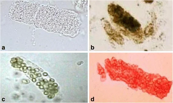
Long description
Composite urine microscopy image of casts in acute kidney injury, including a muddy brown granular cast.

Figure 2WBC cast in urine
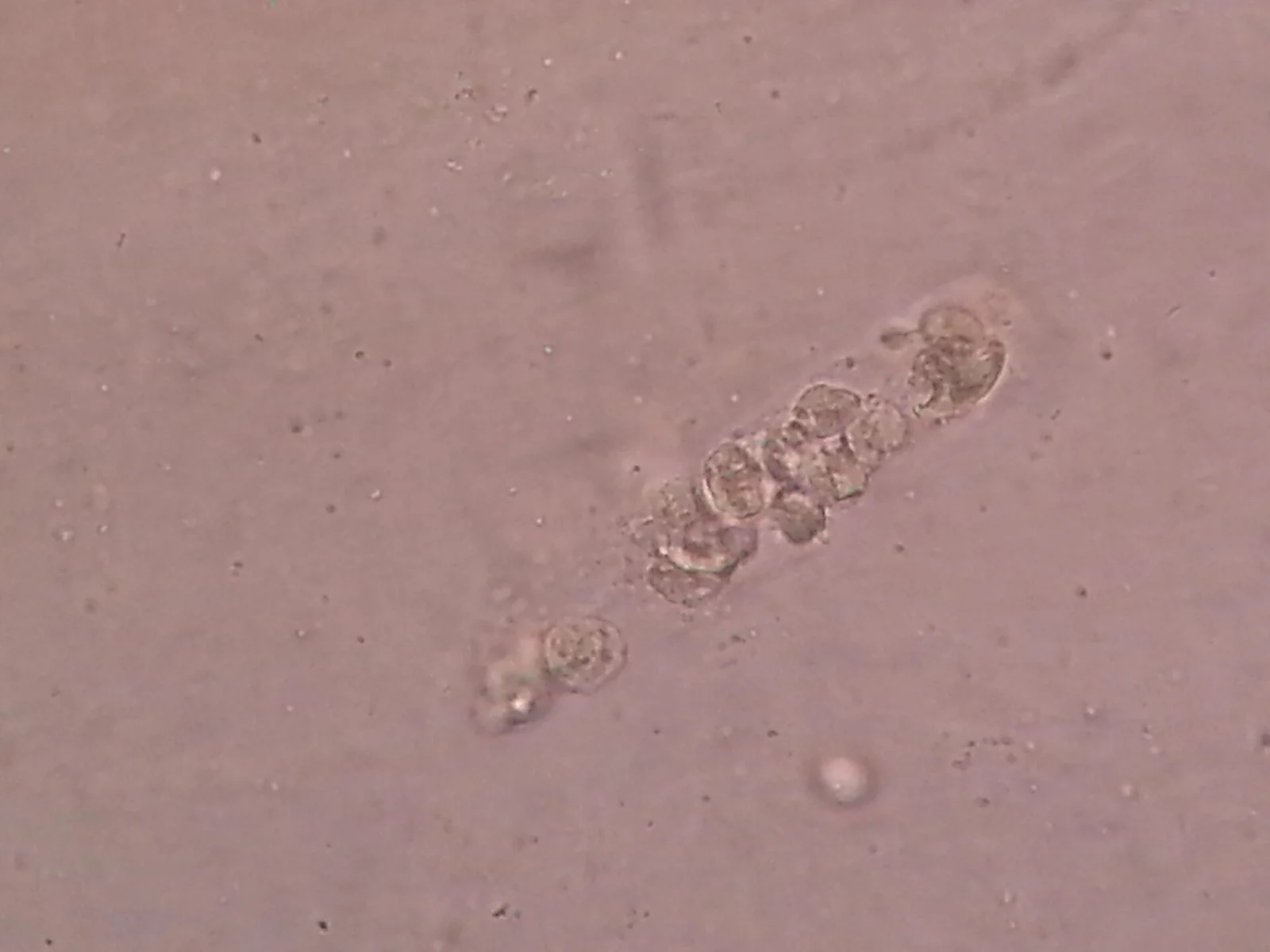
Long description
White blood cell (WBC) casts in urine are shown as abnormal microscopic findings, with the description stating they form when white blood cells clump together and become embedded in a protein matrix within the renal tubules.

Figure 3Hyaline Cast in Urine Microscopy
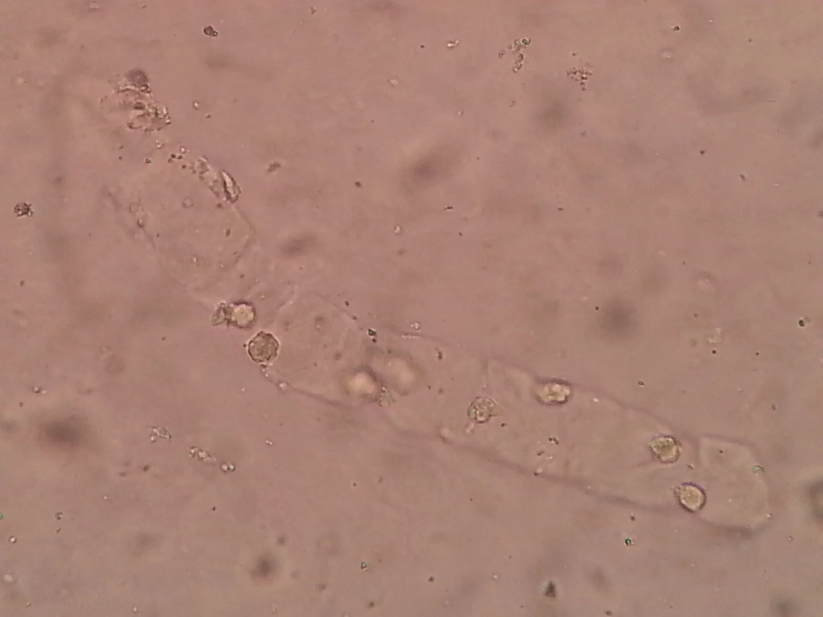
Long description
Hyaline casts described as a type of urinary cast that can be observed during urine microscopy, forming cylindrical structures in the renal tubules.

Perform a dipstick test for blood, protein, leucocytes, nitrites and glucose in everyone with suspected or detected AKI, and act on abnormalities.[4] Red cell casts or dysmorphic red cells point to glomerulonephritis or vasculitis; white cell casts to tubulointerstitial or parenchymal infection; granular or "muddy brown" casts and tubular epithelial cells to acute tubular injury; urinary eosinophils suggest allergic interstitial nephritis, although this finding is neither sensitive nor specific.[1] A normal sediment does not exclude disease and can occur in prerenal AKI and obstructive uropathy.
Urinary diagnostic indices help separate prerenal states from acute tubular injury in oliguric patients, although they are not reliable when the patient is not oliguric or has received diuretics.[1] Interpret them alongside, not instead of, the clinical picture.
| Index | Prerenal | Acute tubular injury |
|---|---|---|
| Urine to plasma osmolality | above 1.5 | 1 to 1.5 |
| Urine sodium (mmol/L) | under 10 | above 40 |
| Fractional excretion of sodium (FeNa) | under 1% | above 1% |
| BUN to creatinine ratio | above 20 | under 10 |
FeNa is the ratio of sodium excreted to sodium filtered. In specialist settings it has pooled sensitivity around 90% and specificity around 82% for separating prerenal from intrinsic AKI at a cut-off of under 1% versus above 1%, but these fall to about 83% and 66% in patients with chronic kidney disease or recent diuretic use, and rise to about 95% and 91% in oliguric patients with normal baseline kidneys and no recent diuretics.[3]
Fractional excretion of urea (FeUrea) is the better test after recent diuretic use, because urea reabsorption occurs mainly in the proximal tubule and is less affected by loop and thiazide diuretics. A FeUrea under 35% favours a prerenal state (positive predictive value about 98% in one prospective comparison), while intrinsic injury tends to produce higher values.[3] Neither test performs well in non-oliguric AKI or in chronic kidney disease, and neither replaces clinical judgement; for example, a low FeNa can be seen in pigment nephropathy, and a high FeNa can be seen with diuretic use.[3]
Ultrasound is the principal imaging test. Bedside bladder ultrasound (or catheterisation) after the patient voids measures the post-void residual; a residual volume above 100 mL suggests inadequate emptying, for example from bladder outlet obstruction or detrusor weakness. Renal ultrasound traces more proximal obstruction, although it can be falsely normal in very acute obstruction, when the ureter is encased by tumour or retroperitoneal fibrosis, or when the patient is volume depleted. If obstruction is strongly suspected, non-contrast CT localises the site and guides therapy.[1]
NICE recommends that ultrasound be performed within 6 hours when pyonephrosis is suspected and within 24 hours when there is no identified cause or a risk of obstruction. It should not be requested routinely when the cause is already clear.[4] Nephrostomy or stenting for upper tract obstruction is carried out as soon as possible and within 12 hours of diagnosis.[4]
Contrast studies should be avoided where possible. Where gadolinium-enhanced MRI is used, the eGFR should be considered, because gadolinium is implicated in nephrogenic systemic fibrosis when renal function is below 30 mL/min per 1.73 m2. Iodinated contrast should be minimised in at-risk groups (older patients, chronic kidney disease with GFR under 30 mL/min per 1.73 m2, volume depletion, diabetes, heart failure), using the lowest necessary volume, low-osmolal or iso-osmolal agents and normal saline before, during and after the study; intravenous sodium bicarbonate is an alternative in patients with concurrent metabolic acidosis.[1]
Kidney size on imaging supports reversibility: normal or enlarged kidneys favour reversible disease; small kidneys suggest chronic renal insufficiency. Atypical enlargement occurs in polycystic disease, HIV-associated nephropathy, diabetic nephropathy, sarcoidosis and infiltrative lymphoma.[1]
Biopsy is reserved for cases where the diagnosis remains uncertain after clinical assessment, imaging and blood tests, or where histology would change treatment, as in suspected glomerulonephritis, vasculitis or acute interstitial nephritis. It is not performed before surgery when a cardiac-type lesion is obvious; the analogy here is that biopsy and the initiation of immunosuppression are decisions for the nephrology team. NICE recommends discussing any potential indication for biopsy-level disease with the nephrology team as soon as possible and within 24 hours of detection in stage 3 AKI, when the cause is unclear, when there is inadequate response to treatment, or when there are complications.[4]
Approach to acute kidney injury
- AKI confirmed by KDIGO criteriaCreatinine rise within 48 hours or 7 days, or oliguria
- Treat life-threatening complications firstHyperkalaemia: calcium, dextrose and insulin; pulmonary oedema: oxygen, vasodilators, diuretics, dialysis if needed
- Restore circulating volume and perfusionIsotonic crystalloids, not colloids; vasopressors in vasomotor shock; treat heart failure, sepsis or tamponade
- Exclude and relieve obstructionBladder scan (residual above 100 mL is significant), ultrasound within 6 or 24 hours, catheter or nephrostomy / stent within 12 hours
- Question: Does kidney function improve after these measures?
Yes
- Prerenal cause likelyContinue fluids, stop nephrotoxins, reassess daily
No
- Intrinsic or continuing postrenal AKIUrinalysis and sediment, urinary indices, autoantibodies, imaging, consider biopsy
- Manage complications and dialyse if indicated
- Recheck kidney function at about 3 months and rationalise medicines
The KDIGO definition and the staging table appear in the Definition section above. In words, AKI is present when creatinine rises by at least 0.3 mg/dL (26.5 micromol/L) within 48 hours, rises to 1.5 times baseline within 7 days, or urine output falls below 0.5 mL/kg per hour for 6 hours.[2] NICE endorses this definition and notes the paediatric urine-output threshold of 8 hours rather than 6.[4]
The table below maps abnormal tests to their diagnostic implications.[1]
| Test | Finding | Suggested cause |
|---|---|---|
| Anti-GBM antibodies | Positive | Anti-GBM disease (Goodpasture) |
| ANCA | Positive | Small-vessel vasculitis (GPA or microscopic polyangiitis) |
| Antinuclear / anti-dsDNA antibodies | Positive | Systemic lupus erythematosus |
| Antistreptolysin-O / streptokinase antibodies | Positive | Post-streptococcal glomerulonephritis |
| Creatine kinase or myoglobin | Markedly elevated | Rhabdomyolysis |
| Complement titers | Low | Cholesterol embolism, post-infectious GN, SLE, endocarditis |
| Serum protein electrophoresis | Monoclonal spike | Multiple myeloma |
| Uric acid | Elevated | Tumour lysis, prerenal AKI |
Threats to life are treated before the diagnostic hunt is finished. In practice this means two things on arrival.[1]
- Hyperkalaemia: give 10 mL of 10% calcium gluconate intravenously, 50 g of dextrose and 5 to 10 units of insulin, as a membrane-stabilising and redistributive combination; this does not remove potassium, so binders, diuretics or dialysis must follow. Calcium can be repeated if ECG changes persist. [1]
- Pulmonary oedema: oxygen, intravenous vasodilators such as nitrates, cautious diuretics, which are often ineffective when filtration is very low, and dialysis if it does not improve.
Metabolic acidosis with pH below 7.20 is often treated with intravenous sodium bicarbonate: isotonic bicarbonate (150 mEq sodium bicarbonate in 1 litre of 5% dextrose) at 50 to 200 mL per hour when the patient is not volume-overloaded, or a concentrated hypertonic solution (50 mEq in 50 mL) over 1 to 2 minutes when volume-overloaded or in arrest. The dose is titrated against serial blood gases rather than calculated, because buffer and acid-production vary too much.[1]
- Stop nephrotoxins (NSAIDs, aminoglycosides, radiocontrast, and reduce calcineurin inhibitors where they are the cause) and review all drugs excreted by the kidneys; nephrology or pharmacy advice on dosing is valuable.[1][4]
- Aminoglycosides deserve special caution: the risk of drug-induced AKI is up to 25% in some series, so they should no longer be standard empirical therapy unless no suitable alternative exists.[2]
- Replace water carefully: daily intake is restricted to the previous day's urine output plus measured extrarenal losses plus 500 to 1000 mL per day for insensible loss. Intake can be relaxed for hypernatraemia or tightened for hyponatraemia; weight gain signals fluid excess, but normal serum sodium means sodium, not water, is the problem.[1]
- Restrict sodium and potassium except where there is a documented deficiency or gastrointestinal loss, and provide adequate protein at about 0.8 g per kg per day.[1][2]
- Control phosphate with calcium, iron-based or synthetic phosphate binders before meals, aiming for serum phosphate under 5.5 mg/dL (1.8 mmol/L), and use cation-exchange resin where delayed-onset potassium control is acceptable to keep serum potassium under 6 mmol/L. Sodium polystyrene sulfonate, oral or rectal, is avoided in patients at high risk of bowel dysmotility (obstruction, impaction, post-operative ileus); patiromer and sodium zirconium cyclosilicate are oral only.[1]
- Avoid unnecessary bladder catheterisation because of infection risk; when a catheter is genuinely needed for accurate output monitoring, it should be removed promptly.[1]
- Avoid diuretics to treat AKI except for volume overload or oedema, and do not use low-dose dopamine, fenoldopam, or atrial natriuretic peptide; loop diuretics do not prevent AKI, may precipitate it, and have not been shown to improve outcome.[2][4]
- The KDIGO approach (formerly called the care bundle in many services): KDIGO and NICE both recommend a structured set of actions tailored to stage and cause, beginning at the identification of risk, not at diagnosis: optimise volume status and haemodynamics (isotonic crystalloids initially, not colloids; vasopressors with fluids in vasomotor shock; protocolised goal-directed resuscitation in high-risk perioperative or septic patients); control glucose without treating below 110 mg/dL, targeting not exceeding 149 mg/dL on average in critically ill patients; meet energy needs (20 to 30 kcal per kg per day) with adequate enteral protein; stop nephrotoxic drugs; avoid radiocontrast where possible; and reassess stage daily with creatinine and urine output.[2]
- Relieve obstruction promptly: refer upper tract obstruction to a urologist immediately for pyonephrosis, an obstructed solitary kidney, bilateral obstruction or complications of AKI from obstruction, and perform nephrostomy or stenting within 12 hours.[4]
- After relief, expect a post-obstructive diuresis: replace urine output with 0.45% saline at about 75% of output, monitoring potassium, sodium and magnesium closely, because over-replacement prolongs the diuresis.[1]
- Nutritional support: 0.8 to 1.0 g/kg per day protein in non-catabolic AKI without dialysis, 1.0 to 1.5 g/kg per day on dialysis, up to 1.7 g/kg per day on continuous renal replacement therapy or in hypercatabolic patients; prefer the enteral route. Do not restrict protein with the aim of delaying dialysis.[2]
Dialysis is not started from a single creatinine or urea number. It is started when a complication of AKI does not respond to medical treatment. In plain words, the indications are:[1][4]
- potassium that will not stay under control (severe, rising or ECG changes);
- fluid in the lungs (pulmonary oedema) that does not improve;
- acidosis that does not respond to treatment (especially with pH below 7.20);
- symptoms or complications of uraemia such as persistent vomiting thought to be due to uraemia, confusion or encephalopathy, pericarditis, seizures or asterixis;
- fluid overload not controllable with diuretics.
NICE adds that the decision must be based on the patient as a whole, and that nephrology or critical care should be involved immediately when any of these appear.[4] NICE recommends immediate referral for AKI from reduced perfusion not responding to initial volume resuscitation, urological obstruction, intrinsic renal disease needing specialist treatment (vasculitis, glomerulonephritis, interstitial nephritis, myeloma), AKI with no clear cause, stage 3 disease, a renal transplant or significant CKD.[4]
Choice of modality (intermittent haemodialysis, continuous haemofiltration/haemodiafiltration, peritoneal dialysis) depends on haemodynamics, availability and local practice; patients in shock are often better served by continuous techniques. Vascular access is a practical constraint in low-resource settings, where temporary catheter dialysis is more common.
- Hyperkalaemia: the most immediately dangerous. Risk rises with cell breakdown, acidosis and oliguria; levels can move quickly.
- Metabolic acidosis: moderate, with bicarbonate typically 15 to 20 mmol/L, worse with sepsis or ischaemia.
- Fluid overload and pulmonary oedema: common when a patient receives liberal fluids with a falling urine output.
- Uraemia: impaired platelet function, pericarditis, encephalopathy, seizures.
- Hyponatraemia: usually mild to moderate, serum sodium 125 to 135 mmol/L, driven by excess water.
- Hyperphosphataemia and hypocalcaemia: often profound in rhabdomyolysis and tumour lysis, with hypercalcaemia possible in recovery.
- Anaemia: normochromic-normocytic, haematocrit 25 to 30%.
- Infection: an indwelling catheter and uraemia both raise risk.
Recovery correlates with premorbid kidney function; patients with chronic kidney disease are more likely to need dialysis and to progress to end-stage kidney disease.[1] Non-oliguric AKI has better outcomes than oliguric or anuric disease. Even when kidney function returns to normal before discharge, patients remain at increased risk of future chronic kidney disease and end-stage disease, so testing at three months is recommended, with nephrology review where eGFR is 30 mL/min per 1.73 m2 or less.[2][4]
A practical plan after AKI is: recheck creatinine before discharge, arrange outpatient renal function testing and review all medicines, advise the patient about the risks of dehydration and about over-the-counter NSAIDs, and put a chronic kidney disease label on the patient's list when GFR is low. Children who recover may still need nephrology follow-up if there is hypertension, impaired renal function or proteinuria on an early-morning sample.[4]
For in-patient survival, severity matters: in a large 2023 US series, admissions with AKI not requiring dialysis and those requiring dialysis had markedly higher death or hospice-discharge rates than admissions without AKI (about 14% and 38% respectively, against about 5%).[1]
Prevention is often straightforward and high-yield.[1][4]
- Maintain fluid balance, blood volume and blood pressure in trauma, burns, severe haemorrhage and major surgery.
- Minimise radiocontrast in at-risk groups, using volume expansion with isotonic saline or sodium bicarbonate, lowest volume of a low-osmolal or iso-osmolal agent, and temporary interruption of ACE inhibitors and ARBs before a contrast study in chronic kidney disease with eGFR under 30 mL/min per 1.73 m2, discussed case by case.
- Temporarily withhold ACE inhibitors, ARBs and diuretics during diarrhoea, vomiting or sepsis until the patient is stable.
- Avoid NSAIDs, and be vigilant with aminoglycosides, vancomycin, amphotericin and calcineurin inhibitors, with drug levels where available.
- Keep water reachable and daily intake documented for dependent patients, older adults, infants and post-operative patients; add a few hundred millilitres to the feed or convert a dry diet to wet when a patient cannot drink.
- Monitor high-risk in-patients: creatinine at least daily and more often after an exposure, and urine output in the critically ill; KDIGO noted that many emergency admissions miss opportunities to prevent or recognise AKI because high-risk patients are not tracked.[2]
- Children and adolescents: a urine output threshold of under 0.5 mL/kg per hour for more than 8 hours, or a 25% or greater fall in eGFR within 7 days, also defines AKI in this age group under NICE. Fluid balance is best tracked by twice-daily weights and measured output. Renal replacement therapy is discussed with a paediatric nephrologist.[4]
- Older adults: the most common group with AKI, through impaired thirst, dependence on carers, and diminished renal reserve; hypernatraemia and AKI often coexist.
- Critical illness: protocolised resuscitation (blood pressure, oxygen delivery, fluids, vasopressors, inotropes) reduces postoperative AKI in high-risk groups.[2]
- Neonates after perinatal asphyxia: a single dose of theophylline may be considered in high-risk neonates, because adenosine-mediated vasoconstriction contributes to early renal dysfunction; evidence of late benefit is limited.[2]
- Pregnancy: pregnancy-related AKI includes pre-eclampsia, HELLP syndrome, acute fatty liver, severe infection and obstruction. These cases are managed by obstetric, nephrology and intensive care specialists together, and drugs are chosen with pregnancy safety in mind.
When laboratory, imaging or dialysis capacity is limited, the priorities areclinical, not technological:
- Assess volume first: pulse, postural blood pressure, jugular venous pressure, skin turgor, mucous membranes and urine output. Repeated weight and a simple fluid balance chart are the cheapest monitors there are.
- Measure creatinine if at all possible; where it is unavailable, urine output trends and signs of obstruction carry the diagnosis.
- Urine dipstick and a plain ultrasound (including a bedside bladder scan or even a catheter) detect obstruction. A post-void residual above 100 mL is a prompt to relieve it; urethral catheterisation, when obstruction is responsible, can be both diagnostic and therapeutic.
- A urinalysis by eye and sediment by microscopy, where available, separate brown granular casts of tubular injury from red cell casts of glomerulonephritis.
- Fluids: oral and enteral hydration are adequate for many prerenal cases; intravenous normal saline restores perfusion in shock, then hypotonic fluid replaces the free water deficit. Where only normal saline exists, restore volume first and add water by mouth.
- Drug access (diuretics, binders, vasopressors, bicarbonate) is often limited; use what is on the essential list, and avoid aminoglycosides if at all possible.
- Obstruction can usually be relieved by catheterisation or temporary nephrostomy, bought time with fluids, with definitive surgery arranged.
- Dialysis requires a machine, clean water, dialysate and trained staff. Where none is available, manage the dangerous complications medically: restrict potassium and protein, remove potassium with binders if they exist, control acidosis with bicarbonate where available, and refer early; peritoneal dialysis with locally prepared solution is sometimes the more available option.
- Referral thresholds can be applied clinically: anuria, no response to fluids, suspected intrinsic or obstetric AKI, stage 3 disease, hyperkalaemia or acidosis that will not correct, and the need for dialysis.
- AKI is a rapid fall in kidney function over days to weeks, defined by KDIGO as a creatinine rise of at least 0.3 mg/dL in 48 hours, a rise to 1.5 times baseline within 7 days, or urine output under 0.5 mL/kg per hour for 6 hours.
- It is a syndrome with three mechanism groups: prerenal, intrinsic renal and postrenal; more than one often coexist.
- Prerenal causes include hypovolaemia, low cardiac output, hepatorenal syndrome and drugs that reduce perfusion (NSAIDs, ACE inhibitors, ARBs, calcineurin inhibitors); intrinsic causes include ischaemic or toxic ATN (contrast, aminoglycosides, rhabdomyolysis), interstitial nephritis (drug allergy), glomerulonephritis and vasculitis, vascular and infiltrative disease.
- Postrenal AKI comes from tubular crystals or light chains, ureteric obstruction, bladder outlet obstruction (prostate, stones, tumours, catheter) or neurogenic bladder.
- Prerenal urine shows high osmolality and low sodium (under 10 mmol/L), FeNa under 1% and a BUN to creatinine ratio above 20; tubular injury shows urine sodium above 40 mmol/L, FeNa above 1%, ratio under 10, and muddy brown granular casts.
- FeUrea under 35% is the more reliable index when the patient has recently had diuretics.
- Assessment centres on history, volume status, bladder scan, ultrasound (within 6 hours if infection is suspected, 24 hours if the cause is unclear) and urinalysis.
- Complications to take seriously: hyperkalaemia, metabolic acidosis, fluid overload, uraemia and hyponatraemia.
- Management is cause-directed: stop nephrotoxins, restore perfusion, relieve obstruction, avoid unnecessary diuretics, follow the KDIGO structured approach and provide adequate nutrition.
- Dialyse when potassium, acidosis, fluid overload, pulmonary oedema or uraemic symptoms do not respond to medical treatment; start from the patient's overall condition, not from a number.
- Recovery is common, but even mild AKI raises the long-term risk of chronic kidney disease; recheck kidney function around 3 months.
- Most cases are preventable by protecting perfusion, avoiding nephrotoxins and keeping water available to patients who cannot drink.
Answer each one in your head first, then open the answer.
A 72-year-old man develops anuria within 24 hours of starting ibuprofen for back pain. What is the most likely category of acute kidney injury, what urine index would support it, and what is the first treatment?
Show answerHide answer
Most likely prerenal AKI from NSAID-induced afferent vasoconstriction combined with probable hypovolaemia. Expect urine sodium under 10 mmol/L, FeNa under 1% and a BUN to creatinine ratio above 20. First steps are to stop the ibuprofen, assess and restore volume status, and re-examine for other nephrotoxins; if oliguria persists after volume replacement, reassess for intrinsic injury or obstruction.
What three urine findings point away from prerenal azotaemia and towards established acute tubular injury, and what does an FeUrea under 35% imply?
Show answerHide answer
Acute tubular injury is suggested by urine sodium above 40 mmol/L, FeNa above 1%, a BUN to creatinine ratio under 10, and a sediment containing granular or muddy brown casts. An FeUrea under 35% favours a prerenal state despite any of the other indices, and is the more reliable index when the patient has recently received diuretics, with a reported positive predictive value for prerenal disease of about 98%.
State the KDIGO definition of AKI as serum creatinine and as urine output, and give the KDIGO stage 3 creatinine and urine-output thresholds.
Show answerHide answer
AKI is present if serum creatinine rises by at least 0.3 mg/dL (26.5 micromol/L) within 48 hours, rises to at least 1.5 times baseline within 7 days, or urine output falls below 0.5 mL/kg per hour for 6 hours. Stage 3 is reached at a creatinine rise of at least 3 times baseline or to 4.0 mg/dL (353.6 micromol/L), or if dialysis is started; urine output under 0.3 mL/kg per hour for at least 24 hours or anuria for at least 12 hours also qualifies. [1]
A patient has AKI with severe hyperkalaemia and peaked T waves on ECG but is still passing urine and is euvolaemic. What are your first actions, and would you give diuretics or dopamine?
Show answerHide answer
First stabilise the cardiac membrane with 10 mL of 10% calcium gluconate intravenously, then move potassium into cells with 50 g of dextrose and 5 to 10 units of insulin, and arrange potassium removal (binder, and dialysis if necessary). Do not give low-dose dopamine (no proven renal benefit) or diuretics unless there is volume overload; diuretics do not treat AKI itself.
A patient with AKI, severe acidosis below pH 7.20, and no uraemic symptoms is oliguric and not responding to bicarbonate. Why might dialysis still be appropriate, and how should this decision be made?
Show answerHide answer
Severe acidosis (pH below 7.20) not responding to bicarbonate, uncontrolled hyperkalaemia, pulmonary oedema or refractory fluid overload are all indications for dialysis even before symptoms of uraemia appear. The decision should be made with nephrology or critical care, based on the patient as a whole rather than on the creatinine alone.
An older patient has AKI after a hip fracture repair, with normal urine output, and microscopy shows muddy brown casts. Is the cause prerenal or intrinsic, and what complication would you watch for first?
Show answerHide answer
Muddy brown granular casts indicate acute tubular injury, usually from per-operative ischaemia and possibly NSAID exposure, rather than simple prerenal azotaemia. Watch first for hyperkalaemia and accumulating fluid overload, because tubular concentrating and excretory failure progress quickly and the diuresis of recovery may not begin for days.
Why should kidney function be rechecked about 3 months after an episode of AKI, and what does small or normal-sized kidneys on imaging suggest?
Show answerHide answer
[2]Even mild or recovered AKI increases the long-term risk of chronic kidney disease and end-stage kidney disease, so creatinine and eGFR are rechecked, with nephrology review if eGFR is 30 mL/min per 1.73 m2 or less. Small kidneys on imaging suggest underlying chronic renal insufficiency; normal or enlarged kidneys favour a reversible or acute process.
- Chronic kidney disease and end-stage kidney disease
- Hyperkalaemia
- Hyponatraemia
- Metabolic acidosis
- Rhabdomyolysis
- Benign prostatic hyperplasia and prostate cancer
- Urinary calculi
- Acute tubular necrosis and tubulointerstitial nephritis
- Sepsis and septic shock
- Cardiorenal syndrome and heart failure
- Hepatorenal syndrome and cirrhosis
- Renal replacement therapy (dialysis)
- Malkina A (peer review: Jaipaul N). Acute Kidney Injury (AKI). Merck Manual Professional Version, Full Review Feb 2026, last updated Oct 2026. Accessed .
- Kellum JA, Lameire N, for the KDIGO AKI Guideline Work Group. Diagnosis, evaluation, and management of acute kidney injury: a KDIGO summary (Part 1). Crit Care. 2013;17(1):204. doi:10.1186/cc11454. Accessed .
- Hamadah A, Gharaibeh K. Fractional Excretion of Sodium and Urea are Useful Tools in the Evaluation of AKI: PRO. Kidney360. 2023;4(6):e725-e727. doi:10.34067/KID.0002492022. Accessed .
- National Institute for Health and Care Excellence. Acute kidney injury: prevention, detection and management (NG148). NICE guideline NG148, published 18 Dec 2019, last updated 16 Oct 2024. Accessed .
Figure credits
Educational content for medical students and doctors. It is not a substitute for clinical judgement or local guidelines.