Bladder cancer
Bladder cancer: painless visible haematuria, cystoscopy and TURBT, with stage-based care from intravesical BCG to cystectomy and immunotherapy.
- Bladder cancer is most often a cancer of the urothelium, the cell layer lining the bladder (and the renal pelvis, ureters and urethra), and it is the disease to suspect in any adult with unexplained blood in the urine.
- The classic clue is painless visible haematuria: about three-quarters of patients present this way, and even an episode that clears up must be investigated until a cause is proven.
- It is roughly 4 times more common in men than in women, about 9 out of 10 cases occur after the age of 55, and a history of smoking is the biggest single cause.
- Most patients (about 70 to 75%) have non-muscle-invasive disease at diagnosis: tumour confined to the inner lining or to the layer just beneath it, treated by resection through the urethra (TURBT) plus risk-based adjuvant therapy.
- Muscle-invasive disease needs radical treatment (neoadjuvant cisplatin-based chemotherapy plus radical cystectomy, or bladder-sparing chemoradiotherapy) and carries a much worse outlook; advanced disease is now also treated with immunotherapy and antibody-drug conjugates.
By the end of this page you should be able to:
- Explain why tobacco and certain industrial chemicals lead to urothelial cancer, and distinguish the main histological types.
- Recognise the presentation of bladder cancer and say when microscopic haematuria is a concern. [2]
- Choose and interpret the first investigations: cystoscopy, TURBT (with detrusor muscle in the specimen), urine cytology, and imaging (including CT urography for the upper tracts).
- Stage bladder cancer using the TNM system and separate non-muscle-invasive (Ta, Tis, T1) from muscle-invasive (T2 to T4) disease.
- Plan treatment by risk group: surveillance or single-dose intravesical chemotherapy for low risk; BCG induction and maintenance for intermediate and high risk; radical cystectomy or bladder-sparing chemoradiotherapy for muscle-invasive disease; systemic therapy for locally advanced or metastatic disease.
- Outline prognosis, recurrence and surveillance, and the approach when tests or cisplatin/imaging are not available.
DefinitionBladder cancer
A malignant tumour of the bladder. Most (about 90 to 95%) are urothelial (transitional cell) carcinomas arising from the urothelium lining the bladder; the rest are mainly squamous cell carcinoma, adenocarcinoma and small cell or neuroendocrine carcinoma. Classification into non-muscle-invasive and muscle-invasive disease, based on whether the tumour has reached the bladder's detrusor muscle, is what drives treatment. [1]
Urothelial carcinoma often originates not only in the bladder but anywhere lined by urothelium, so the renal pelvis, ureters and urethra can also be involved; one tumour can be followed by another along the urinary tract. The depth of invasion (TNM stage) correlates with behaviour, but grade (low versus high grade) also matters, particularly in non-muscle-invasive disease. Carcinoma in situ is flat, high-grade urothelial cancer confined to the lining and is clinically aggressive despite being non-invasive.
The World Health Organization (WHO), in its 5th edition, recognises many urothelial subtypes (for example nested, micropapillary, plasmacytoid, sarcomatoid and others); some, such as micropapillary and plasmacytoid disease, are biologically aggressive even at a shallow stage. [2]
GLOBOCAN 2020 data describe bladder cancer as one of the ten most common cancers worldwide: about 573,000 new cases and 213,000 deaths, roughly 3% of all new cancer diagnoses, and a five-year prevalence of about 1.72 million cases. Incidence is highest in higher-income regions such as Southern and Western Europe and North America; within Europe, Greece has the highest age-standardised rate in men. About three-quarters of cases occur in men; the male-to-female ratio is about 4:1, so bladder cancer is the sixth most common cancer in men (and the fourth in men in the USA) but the seventeenth in women. [1][5] [2][3]
- Age. The average age at diagnosis is about 73 years, and more than 90% of cases occur in people over 55 years. [5] [1]
- Mortality. Bladder cancer is the thirteenth most common cause of cancer death. The cumulative probability of dying of it between birth and age 74 is about 0.3% in males and 0.08% in females. [1]
- Early diagnosis. Five-year survival in the United States is reported as about 96% for in-situ disease, about 69% for localised disease, and much lower for regional (about 36%) and metastatic (about 4%) disease. [1]
- Stage at presentation. About 70 to 75% of patients have non-muscle-invasive disease, about 20 to 25% have muscle-invasive disease, and about 5% have metastatic disease at first presentation. [2]
Incidence mirrors the history of tobacco use and the prevalence of industrial chemical exposure; rates are falling in some countries that have succeeded in tobacco control. [1]
Smoking is the dominant risk factor. It accounts for a large share of cases (estimates range from about 30 to 40% up to about half or more of all bladder cancer, depending on the source). Smokers have a two- to three-fold higher chance of developing bladder cancer, and they develop it about six years earlier than non-smokers; the risk falls after quitting (to a relative risk of about 1.1 after more than 15 years smoke-free, against 2.6 at 1 to 3 years). Even pipes and cigars are implicated, as is second-hand smoke. [1][5]
Occupational exposure to carcinogenic aromatic amines (2-naphthylamine, 4-aminobiphenyl, benzidine) used in dye, paint, rubber, metal, petroleum and leather manufacture accounts for about 5 to 10% of bladder cancer. Hairdressers, painters, chimney sweeps, firefighters and rubber or textile workers are among the higher-risk groups; diesel fumes and hair dyes are also implicated. [1][5] Arsenic in drinking water and chronic exposure to pesticides are environmental contributors. [2]
In northern Africa and the Middle East the most important risk factor for squamous cell carcinoma of the bladder is chronic infection with Schistosoma haematobium. The parasite deposits eggs in the bladder wall, producing granulomatous inflammation and fibrosis; calcified eggs and chronic inflammation drive squamous metaplasia and, in time, squamous cell carcinoma. In endemic regions squamous cell carcinoma is the second most common cancer after hepatocellular carcinoma. Control programmes (sanitation, safe water, and mass treatment with praziquantel) have lowered incidence in countries such as Egypt. [1][5]
Persistent or recurrent urinary tract infection, stones, diverticula, chronic inflammation (including that from long-term indwelling catheters) and recurrent gonorrhoea are associated with squamous carcinoma of the bladder, although confounding is strong. [1]
- Cyclophosphamide induces bladder cancer. The risk is dose-dependent and is greatest in those who have received more than 20 g total, with a relative risk of roughly 4.5. [1]
- Pelvic radiotherapy (for example, for cervical cancer) carries a two- to four-fold increased risk of later bladder cancer, with a latency of 15 to 30 years. [1]
- Pioglitazone has been associated with an increased risk and should be avoided in people with previous or active bladder cancer. [3]
NAT2 slow acetylation and GSTM1 deletion reduce detoxification of aromatic amines. Lynch syndrome (mismatch repair gene mutations) increases the risk of urothelial cancer, Cowden syndrome (PTEN) is associated with genitourinary neoplasia, and first-degree relatives of patients have roughly a two-fold increase in risk. Bladder cancer is rare in children (incidence 0.1 to 0.4%). Body mass index above 30 has also been linked to progression. [1][5]
Tobacco carcinogens (such as beta-naphthylamine) and industrial aromatic amines are absorbed, partly detoxified in the liver, and excreted in the urine, where they contact the urothelium directly; DNA adducts and mutations (activation of oncogenes, loss of tumour suppressors) follow. Chronic bladder epithelial turnover from inflammation (schistosomiasis, stones, catheters) adds a promoting stimulus. [1]
Two broad pathways are recognised:
- Papillary non-muscle-invasive pathway. Genetically relatively stable tumours arise from normal-appearing (or hyperplastic) urothelium. They are characterised by chromosome 9 deletion (in about half of cases) and by activating FGFR3 and RAS/PIK3CA mutations, with RAS and FGFR3 mutations being mutually exclusive. Low-grade Ta tumours often recur but progress rarely.
- Flat/carcinoma in situ pathway. Inactivation of TP53, RB1 and PTEN precedes flat urothelial dysplasia and carcinoma in situ, which share molecular features with high-grade and invasive cancer and progress to muscle-invasive disease.
Most non-muscle-invasive tumours show activation of the RAS-MAPK pathway in about 80% of cases. [2]
Clinical pearlField effect
Bladder cancer often behaves as a field disease: carcinogen-damaged urothelium can give rise to multiple tumours over years. This is why removing one tumour does not end the risk, and why surveillance (watching for new tumours) is as important as treating the first one.
Figure 1Papillary urothelial carcinoma (low-grade), low mag
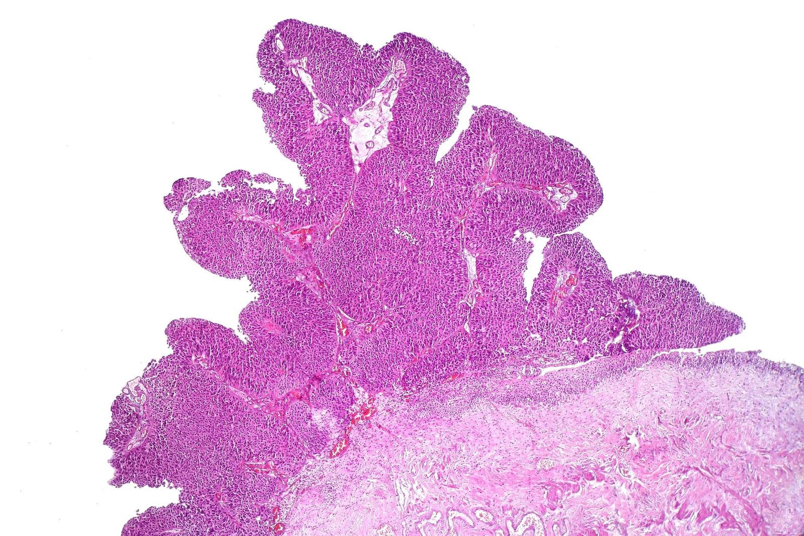
Long description
Low-power histology of a papillary urothelial carcinoma of the bladder, with fronds of tumour above the bladder wall.

Histology. About 90 to 95% are urothelial carcinomas. Mixed/variant histology and non-urothelial types (squamous, adenocarcinoma, small cell or neuroendocrine) make up the rest. Squamous cell carcinoma is the dominant type in endemic schistosomiasis regions. [5]
Grading. Low grade versus high grade. All muscle-invasive urothelial cancers are high grade. [2]
Staging (TNM, 8th edition). Non-muscle-invasive disease comprises Ta, Tis and T1; muscle-invasive disease is T2 to T4. [3] See the stage table below.
Tumour sampling. TURBT removes visible lesions and is both diagnostic and therapeutic. En-bloc resection is possible for small tumours; larger ones come out in fragments. A detrusor muscle sample must be present in the specimen so that invasion depth can be judged. Repeat TURBT is recommended within 2 to 6 weeks when resection was incomplete, when the tumour invades the lamina propria (T1), and should be considered for high-grade non-invasive disease (other than carcinoma in situ alone), because up to 40% of T1 tumours upstage to T2 if muscle was not present in the first specimen. [2][3]
- Painless visible (gross) haematuria is the presenting symptom in roughly 75 to 85% of patients. In a large haematuria clinic series, roughly 10 to 22% of patients with visible haematuria were found to have bladder cancer, rising from 4.7% under age 35 to 35% over age 75. [5]
- Non-visible (microscopic) haematuria, often found on health checks, leads to a bladder cancer diagnosis in about 3.3 to 5.2% of people; such cancers are more often low stage. [5]
- Irritative voiding symptoms (urgency, frequency, dysuria) are more common with carcinoma in situ than with papillary Ta/T1 tumours; they should prompt urological assessment, although urinary tract infection must be excluded (it mimics and co-exists with cancer). [2]
- Advanced disease can present with pelvic pain, symptoms of obstruction, hydronephrosis or systemic symptoms.
Eighty-five percent of patients with a new diagnosis have painless gross haematuria; haematuria is often intermittent and can follow effort (Valsalva). [1]
Rectal and vaginal bimanual examination under anaesthesia (before or after TURBT) assesses whether a tumour is palpable or fixed to the pelvic wall, but clinical staging by examination is error-prone in both directions. [3]
NICE groups NMIBC into low, intermediate and high risk, using tumour stage, grade, size, number and the presence of carcinoma in situ; high-risk non-muscle-invasive cancer includes pTaG3, pT1G2, pT1G3, pTis (CIS) and aggressive variants such as micropapillary or nested disease. [4] [2]
| Condition | Features that point to it | How bladder cancer is separated |
|---|---|---|
| Urinary tract infection or stones | Dysuria, fever, colic, haematuria; infection can co-exist | Culture negative for persistent haematuria; cystoscopy and cytology |
| Benign prostatic hyperplasia | Older man, voiding symptoms, benign-feeling gland | Cystoscopy shows no tumour; ultrasound features |
| Renal cell carcinoma | Haematuria with flank pain/mass | Upper-tract imaging (CT urography) |
| Squamous carcinoma from schistosomiasis | Endemic exposure, sandy patches, SCC histology | Cystoscopy with biopsy |
| Chronic interstitial cystitis | Pain, urgency, no mass on vision | Cystoscopy and biopsy, negative cytology |
| Inflammatory lesion mimicking CIS | Erythematous, sometimes multifocal | Biopsy shows no malignancy |
Non-visible haematuria in a patient over 50 with risk factors is a red flag that should not be attributed to age alone; the look-alike urgent condition is an upper tract urothelial cancer or renal cancer, which is why upper-tract imaging is part of the work-up. [3][5]
Figure 2Blue light and white light cystoscopy images
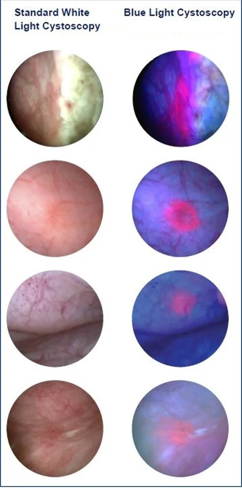
Long description
Bladder lesions seen at cystoscopy under standard white light (left column) and under blue light (right column), where tumour areas stand out in pink against the blue background.

White-light cystoscopy remains the gold standard for initial diagnosis and examination of the whole urethra and bladder, with documentation of site, size, appearance (papillary or solid) and mucosal abnormality. Blue-light (photodynamic, using hexaminolevulinate) or narrow-band imaging, which improve detection of carcinoma in situ, should be used when available. [2][3]
Urine cytology is the most cost-effective urine-based method for high-grade disease; its sensitivity is modest but its specificity is high. The Paris System (2016) reports high-grade urothelial carcinoma as the key category, and a diagnosis in that category carries a risk of cancer of more than 90%. NICE and EAU guidance agree that no urinary biomarker (including NMP22, BTA and ImmunoCyt) should substitute for cystoscopy, either for investigation or for follow-up. [4][5]
CT or MRI of the abdomen and pelvis stage known muscle-invasive or high-risk non-muscle-invasive disease, and MRI is better than CT for local staging, although neither can reliably detect microscopic invasion of perivesical fat. CT urography has the highest diagnostic accuracy for upper tract urothelial cancer (sensitivity 0.67 to 1.0, specificity 0.93 to 0.99) and is used to detect upper-tract involvement in high-risk or muscle-invasive disease. Chest imaging (at least CT in smokers) is added because of the strong smoking association. Bone or brain imaging is added only when metastasis is suspected. [2][3][4]
TURBT, performed by an experienced urologist, is the most reliable way to obtain histology and stage. A single intravesical dose of a chemotherapeutic agent (gemcitabine or mitomycin C) should be given during TURBT or within 24 hours of it, unless there is concern about bladder perforation. [3][4]
Staging uses the TNM 2017 (8th edition) classification applied to the depth of invasion into the bladder wall. [3][5]
| Stage (TNM) | Meaning | Typical management |
|---|---|---|
| Ta | Non-invasive papillary carcinoma (NMIBC) | TURBT; single intravesical dose in low risk; BCG for intermediate/higher |
| Tis (CIS) | Flat carcinoma in situ, high grade (NMIBC) | BCG induction + maintenance; radical cystectomy or alternative if BCG fails |
| T1 | Invasion of the lamina propria (NMIBC) | Re-TURBT within 2-6 weeks; BCG; radical cystectomy for high-risk features |
| T2a / T2b | Invasion of superficial / deep detrusor muscle (MIBC) | Neoadjuvant cisplatin chemotherapy + radical cystectomy, or trimodal therapy |
| T3a / T3b | Microscopic / macroscopic perivesical invasion (MIBC) | As above; chemoradiotherapy if unfit for cystectomy |
| T4a | Invasion of prostate, seminal vesicle, uterus or vagina (MIBC) | Radical cystectomy or chemoradiotherapy depending on fitness and extent |
| T4b | Invasion of pelvic or abdominal wall (locally advanced) | No curative option; palliative chemotherapy, radiotherapy or chemoradiotherapy |
| N1-N3 | Pelvic or common iliac lymph-node metastasis | Cisplatin-based systemic therapy; consider cystectomy if responding |
| M1a / M1b | Non-regional node / other distant metastasis | Cisplatin-based combination or immune/ADC therapy |
Lymph-node groups: N1 single true-pelvic node; N2 multiple true-pelvic nodes; N3 common iliac node. Distant spread: M1a non-regional nodes, M1b bone, lung, liver or other viscera. [3]
Cystoscopic and bimanual examination under-estimate or over-estimate T stage; completeness of TURBT (detrusor muscle in the specimen) and cross-sectional imaging improve accuracy. [3]
- TURBT removes all visible tumour and gives the histological stage and grade.
- Low risk (solitary small Ta G1/G2 or papillary urothelial neoplasm of low malignant potential): TURBT alone is enough if all disease is visibly resected; cystoscopy at 3 months and 12 months, then discharge to primary care if no recurrence; no urinary cytology is needed. A single intravesical dose of mitomycin C is given with the first TURBT. [4]
- Intermediate risk: a course of at least six instillations of intravesical mitomycin C after TURBT. [4]
- High risk (pTa G3, T1, CIS, or aggressive variant): TURBT is repeated when indicated; the patient is offered either intravesical BCG or radical cystectomy. BCG is given with induction and maintenance: up to one year for intermediate risk and up to three years for high risk, if tolerated. [2][4]
- Recurrent small, low-grade papillary tumours in patients with no high-risk history and a disease-free interval of 6 months or more can be fulgurated in the clinic without biopsy. [4]
- BCG-unresponsive disease (persistent or progressive disease during or after adequate BCG) is best treated by radical cystectomy in those fit for it; alternatives (intravesical chemotherapy such as gemcitabine/docetaxel combinations, intravesical gene therapy, or clinical trials) are used when cystectomy is declined or unsafe. [2]
- Intravesical chemotherapy given within 24 hours of TURBT reduces one-year recurrence by about 40% and five-year recurrence by about 15%. [2]
- Neoadjuvant cisplatin-based chemotherapy plus radical cystectomy is the standard curative approach for T2 to T4a disease with no distant metastasis, when the patient is fit for cisplatin. Neoadjuvant therapy adds roughly a 5 to 8% absolute survival advantage at five years over surgery alone. Regimens are gemcitabine plus cisplatin or dose-dense MVAC (methotrexate, vinblastine, doxorubicin, cisplatin); six cycles of dose-dense MVAC have shown better progression-free survival at three years than four cycles of gemcitabine-cisplatin in the GETUG/AFU V05 VESPER trial, at the cost of more toxicity. [3][4] [2]
- Cisplatin fitness. Consensus criteria (Galsky) require a glomerular filtration rate of 60 ml/min/1.73 m2 or more; some experts accept creatinine clearance as low as 40 for curative treatment. If the clearance is 40 to 60, split-dose cisplatin (35 mg/m2 on each of two days rather than 70 mg/m2 on one day) improves tolerability. Gemcitabine plus carboplatin is the fallback when cisplatin is unsuitable. Up to half of patients with bladder cancer are cisplatin-ineligible because of renal dysfunction. [2][4]
- Radical cystectomy is performed within 3 months of the initial resection, includes bilateral pelvic lymph-node dissection, and requires a urinary diversion (stoma or continent diversion). Minimally invasive (robotic or laparoscopic) approaches are increasingly used; open cystectomy remains the reference standard. ERAS protocols (early feeding, mobilisation, opioid-sparing analgesia) shorten recovery. [3][4]
- Bladder-sparing trimodality therapy (maximum TURBT, then radiotherapy with a radiosensitiser such as mitomycin plus 5-fluorouracil or carbogen plus nicotinamide) is offered to selected patients instead of cystectomy. Doses such as 64 Gy in 32 fractions over 6.5 weeks or 55 Gy in 20 fractions over 4 weeks are used. Five-year cancer-specific survival is 50 to 82%, but 25 to 30% of patients later need salvage cystectomy, and trimodality therapy reduces the chance of a future orthotopic neobladder. [2][3][4]
- Partial cystectomy is an option for a small, solitary tumour without CIS, away from the trigone, usually in a diverticulum. Radical TURBT alone can suit highly selected patients who cannot have cystectomy or chemoradiotherapy.
- Adjuvant therapy. Adjuvant cisplatin combination chemotherapy is considered after cystectomy for pT3 or greater or node-positive disease when neoadjuvant therapy was not given. Adjuvant nivolumab improves disease-free survival after radical resection in high-risk muscle-invasive disease (FDA approval for high-risk post-resection disease; European approval only where tumours are PD-L1 positive at 1% or more). Adjuvant radiotherapy is considered for adverse pathological features. [2][4]
- Cisplatin-based chemotherapy (cisplatin plus gemcitabine, or accelerated high-dose MVAC with G-CSF) is offered to patients who are physically fit (ECOG 0 to 1) with renal function of 60 ml/min/1.73 m2 or more; carboplatin plus gemcitabine is used when cisplatin is unsuitable (including patients with ECOG 0 to 2 and glomerular filtration under 60). [4]
- First-line platinum chemotherapy is increasingly being supplanted by enfortumab vedotin (an antibody-drug conjugate against nectin-4) plus pembrolizumab, which NICE recommends for untreated unresectable or metastatic urothelial cancer when platinum therapy is suitable, with a median survival advantage over platinum chemotherapy. [2][4]
- Other options include immune checkpoint inhibitors (atezolizumab as first-line for PD-L1 positive disease if cisplatin is unsuitable; avelumab maintenance until 5 years or progression; atezolizumab or pembrolizumab after platinum failure; nivolumab in selected adjuvant settings), erdafitinib for tumours with susceptible FGFR3 alterations, and durvalumab with gemcitabine/cisplatin in the perioperative setting. [2][4]
- Relapse within 12 months of prior cisplatin therapy counts as platinum refractory; gemcitabine plus cisplatin, accelerated MVAC, or carboplatin/paclitaxel combinations are used, with response monitoring and active management of toxicity. [2][4]
- Symptom control includes palliative hypofractionated radiotherapy for haematuria, dysuria, frequency or nocturia; percutaneous nephrostomy or stenting for ureteric obstruction; and hypofractionated radiotherapy or embolisation for intractable bleeding. [4]
- After TURBT for NMIBC: cystoscopy at 3 months and 12 months (low risk); at 3, 9 and 18 months and annually thereafter (intermediate risk); and at 3, 6, 9 then 6-monthly intervals as above, for 2 years, then 6-monthly for 2 years, then annually (high risk). [2][4]
- After radical cystectomy: imaging and renal function (including glomerular filtration) at least annually, with CT of chest, abdomen and pelvis at 6, 12 and 24 months for local or distant recurrence, plus annual metabolic acidosis screening and vitamin B12 and folate levels. If a urethra is left in situ, urethral washing for cytology or urethroscopy annually for 5 years. [3][4]
- After radical radiotherapy: cystoscopy at 3 months and then 3-monthly for 2 years, 6-monthly for a further 2 years and annually thereafter; upper-tract imaging annually for 5 years; CT at 6, 12 and 24 months. [3][4]
- Progression. Untreated carcinoma in situ progresses to muscle-invasive disease at a rate of 40 to 80% within five years; untreated or under-treated high-grade T1 disease also progresses in a substantial minority (progression of non-muscle-invasive disease overall is 1 to 45% at five years). [2]
- Recurrence after cystectomy. About 30% of patients recur, on average 12 months after surgery, and survival after cystectomy correlates closely with stage (for pT0 without nodal disease the 10-year recurrence-free survival is 86%; with lymph-node involvement it falls to 34%). [2]
- Metastasis spreads mainly to lymph nodes, bone, lung and liver; muscle-invasive disease is associated with a median survival of about 15 months when metastatic. [5]
- Treatment toxicity. Radical cystectomy carries anaesthetic, infection and wound risk; BCG causes local and systemic inflammatory reactions (seek specialist advice when bladder toxicity is uncontrolled); chemoradiotherapy can produce cystitis and bladder toxicity requiring specialist review. [2][4]
Survival falls steeply with stage and grade: in-situ disease has a 5-year survival of about 96%; localised (muscle-invasive) disease about 69%; regional about 36%; and metastatic about 4%. [1] Five-year recurrence of non-muscle-invasive disease is reported at 31 to 78%, and progression at 1 to 45%; carcinoma in situ progresses at a five-year rate of about 50%; for high-grade T1 cancer five-year recurrence is 42%, progression 21% and cancer-specific survival 87%. [2] [3]
Long-term outcomes after cystectomy, by final pathology, include ten-year recurrence-free survival of 86% for pT0, 76% for T1 to pT3a, 61% for T3b and 45% for T4 (pT0 to T4 without lymph-node involvement); about 34% once lymph nodes are involved regardless of T stage. Neoadjuvant chemotherapy adds about a 5% absolute advantage in five-year overall survival and a 22% relative improvement in disease-free survival. [2]
No major organisation recommends routine population screening for bladder cancer. A high proportion of cases (about 82%) are attributable to modifiable factors: smoking cessation is the most effective; occupational hygiene with protective equipment reduces exposure to aromatic amines; schistosomiasis prevention (safe water, sanitation and mass praziquantel) is effective in endemic regions; and weight control and physical activity may have modest benefit. High-risk groups (prior bladder cancer, certain congenital bladder conditions, marked occupational exposure) may be offered periodic urine testing and cystoscopy. [1][2]
- Older adults. Over 90% of patients are over 55; early cystectomy is supported even beyond 80 years with attention to frailty and centre expertise. [2][3]
- Children. Bladder cancer is rare; when it occurs (mostly low-grade Ta) it is usually low stage and post-treatment follows the same principles. [5]
- High-risk genetic groups (Lynch, Cowden syndromes) merit surveillance tailored to their risk. [5]
Where imaging, endoscopy or oncology services are limited:
- Recognise the clinical pattern: painless visible haematuria in an adult (especially a smoker or an older man) is bladder cancer until proven otherwise; microscopic haematuria with risk factors also warrants assessment.
- Cystoscopy, even with simple white-light equipment, is the single most useful diagnostic test. Where it is unavailable, persistent haematuria without an obvious infection should lead to referral.
- TURBT is both diagnostic and therapeutic. A single intravesical dose of gemcitabine or mitomycin (if obtainable) within 24 hours is cheap and reduces recurrence substantially. Keep basic histology reports stating grade and whether detrusor muscle was present.
- CT urography requires contrast and a scanner; where unavailable, ultrasound plus careful endoscopy and clinical judgement, or referral before spread occurs, are the pragmatic options.
- For non-muscle-invasive high-risk disease without BCG, intravesical chemotherapy (or re-TURBT plus careful surveillance) is an acceptable alternative, with referral for BCG or cystectomy where possible.
- For muscle-invasive disease without cisplatin, carboplatin schemes, radiotherapy, or radical cystectomy as locally available; palliative diversion or nephrostomy relieve obstruction. Where radical treatment is impossible, palliative hypofractionated radiotherapy and symptomatic care are still worthwhile.
- For schistosomiasis-endemic communities, prevention (safe water, sanitation, snail control, mass praziquantel) is the most cost-effective intervention.
- Bladder cancer is predominantly a cancer of the urothelium (about 90 to 95% of cases), about 4 times more common in men than women, and diagnosed at a mean age of about 73. [1]
- Painless visible (gross) haematuria is the classic presenting symptom (roughly 75 to 85% of patients); even an episode that resolves demands investigation, since 10 to 20% of people presenting with gross haematuria have bladder cancer. [2]
- Tobacco is the dominant cause (a large share of cases, estimated from about 30% to half or more); occupational aromatic amines account for about 5 to 10%; schistosomiasis causes squamous cell carcinoma in endemic regions. [1]
- Stage determines everything: non-muscle-invasive disease (Ta, Tis, T1) is managed with TURBT, intravesical chemotherapy or BCG, and surveillance; muscle-invasive disease (T2 to T4) requires neoadjuvant cisplatin-based chemotherapy plus radical cystectomy, or bladder-sparing trimodality chemoradiotherapy.
- CIS is flat high-grade disease and a precursor lesion: it progresses at a 5-year rate of about 50% if untreated, so it merits aggressive treatment (BCG, with radical cystectomy if BCG fails).
- Re-TURBT with detrusor muscle in the specimen is essential: up to 40% of T1 tumours upstage when the initial specimen lacked muscle.
- For locally advanced or metastatic disease, cisplatin-based combinations remain standard where the patient is fit (GFR 60 or more, ECOG 0 to 1), with carboplatin combinations and, increasingly, enfortumab vedotin plus pembrolizumab. [4]
- Surveillance is long: cystoscopy and imaging schedules differ by risk group, and after cystectomy there is a 30% risk of recurrence (median 12 months).
- Outcomes are stage-dependent: about 96% five-year survival for in-situ disease, 69% localised, 36% regional and 4% metastatic.
Answer each one in your head first, then open the answer.
A 62-year-old man who smokes 20 cigarettes a day notices that his urine is pink for two days, without pain. He then feels well again. What is the most likely diagnosis and the correct next step?
Show answerHide answer
Bladder cancer until proven otherwise. Painless visible (gross) haematuria, even if it clears, should be investigated with flexible cystoscopy and upper-tract imaging. Urinary infection and stones are common mimics but must be excluded, not assumed.
A TURBT specimen from a 70-year-old shows high-grade papillary tumour invading the lamina propria, but no detrusor muscle is present in the specimen. What should happen next, and why?
Show answerHide answer
A repeat TURBT within 2 to 6 weeks. Without detrusor muscle in the first specimen, up to 40% of such T1 tumours are actually T2; the second resection determines the true stage and drives the decision between intravesical BCG and radical treatment.
Which patients with non-muscle-invasive bladder cancer are offered intravesical BCG rather than just TURBT, and for how long should maintenance be continued?
Show answerHide answer
Intermediate- and high-risk patients (including pT1, pTa grade 3 and CIS). Maintenance BCG should be continued for up to one year in intermediate-risk disease and up to three years in high-risk disease if tolerated, because maintenance adds protection against recurrence and progression. TURBT plus a single intravesical dose of chemotherapy is enough for genuine low-risk disease.
A patient with muscle-invasive bladder cancer (cT2N0M0) has a creatinine clearance of 45 ml/min. How should neoadjuvant chemotherapy be handled?
Show answerHide answer
Neoadjuvant cisplatin-based chemotherapy still adds survival benefit (roughly 5 to 8% at five years). With creatinine clearance 40 to 60, use split-dose cisplatin (35 mg per square metre on each of two days rather than 70 on one day) if a cisplatin regimen is used. If cisplatin is truly unsuitable (for example, clearance under 40 or poor performance status), proceed to radical cystectomy, or use gemcitabine plus carboplatin when appropriate. Check local protocols.
Which of the following best summarises the rationale for bladder-sparing trimodality therapy, and its main trade-off?
Show answerHide answer
Trimodality therapy (maximal TURBT plus chemoradiotherapy) offers 5-year cancer-specific survival of 50 to 82% for selected patients with muscle-invasive disease, with 25 to 30% later requiring salvage cystectomy. The trade-off is that prior pelvic radiotherapy greatly complicates any future cystectomy or orthotopic neobladder reconstruction.
A 75-year-old woman in a rural clinic has visible haematuria and a positive cystoscopy for papillary tumour, but CT urography and BCG are unavailable. What pragmatic plan is defensible?
Show answerHide answer
Perform the best possible TURBT with documentation of grade and detrusor invasion; give a single intravesical dose of the cheapest available chemotherapeutic agent (gemcitabine or mitomycin) within 24 hours if obtainable; arrange histopathology; institute interim surveillance (urine cytology and whatever endoscopy is possible), treat any suspected schistosomiasis in an endemic area, and refer for BCG, radiotherapy or cystectomy as the stage indicates. Palliative radiotherapy and symptomatic care apply for incurable disease. Safe water and sanitation remain the long-term prevention.
- Haematuria (visible and non-visible): investigation and differential diagnosis
- Urinary tract infection in adults
- Renal cell carcinoma
- Upper tract (urothelial) carcinoma of the renal pelvis and ureter
- Prostate cancer
- Erectile dysfunction after pelvic surgery
- Acute kidney injury
- Hydronephrosis
References are held in the front matter (entries 1 to 5) and are cited throughout as Ref 1 to Ref 5.
- Halaseh SA, Halaseh S, Alali Y, Ashour ME, Alharayzah MJ. A Review of the Etiology and Epidemiology of Bladder Cancer: All You Need To Know. Cureus 2022;14(7):e27330. Accessed .
- Lopez-Beltran A, Cookson MS, Guercio BJ, Cheng L. Advances in diagnosis and treatment of bladder cancer. BMJ 2023;384:bmj-2023-076743. Accessed .
- Omorphos NP, Piedad JCP, Vasdev N. Guideline of guidelines: Muscle-invasive bladder cancer. Turkish Journal of Urology 2020;47(Suppl 1):S71-S78. Accessed .
- National Institute for Health and Care Excellence (NICE). Bladder cancer: diagnosis and management. NICE guideline NG2, 2015 (accessed 2026-10-28). Accessed .
- Dyrskjøt L, Hansel DE, Efstathiou JA, et al. Bladder cancer. Nature Reviews Disease Primers 2023;9:58. Accessed .
Figure credits
Educational content for medical students and doctors. It is not a substitute for clinical judgement or local guidelines.