Renal cell carcinoma and the approach to haematuria
Clear cell and other renal cell cancers, from risk and pathology to staging, surgery and systemic therapy, plus a practical approach to haematuria.
- Renal cell carcinoma (RCC) is a cancer of the renal tubular epithelium. It is the commonest solid kidney cancer in adults and represents about 2% of all cancers worldwide.[1]
- Clear cell RCC is the dominant type (about 70% of cases) and arises from proximal tubule cells after loss of the Von Hippel-Lindau (VHL) tumour suppressor; papillary, chromophobe and collecting duct variants each carry a different biology.[1]
- Most RCCs are now found incidentally on imaging done for something else. The classic triad of flank pain, visible haematuria and a palpable abdominal mass is rare (under 1%).[1][3]
- Contrast-enhanced CT of the abdomen (and chest for staging) is the central investigation: it shows the tumour, its venous extension, lymph nodes and distant disease. A change of about 15 Hounsfield units after contrast confirms enhancement.[3]
- Small organ-confined tumours are cured by partial nephrectomy where possible; advanced disease is treated with immune checkpoint combinations and tyrosine kinase inhibitors. Conventional chemotherapy is generally ineffective in RCC.[4]
By the end of this page you should be able to:
- Describe the common risk factors for RCC and name the principal inherited syndromes associated with it.
- Explain how proximal tubule cell VHL loss produces clear cell RCC, and list the main RCC subtypes.
- Recognise the common and paraneoplastic presentations, including the classic triad and haematuria.
- Plan the diagnostic work-up of a renal mass, including how to look for tumour thrombus in the renal vein and vena cava.
- Stage RCC with the TNM system and choose management for localised versus advanced disease.
- Outline the common causes of haematuria by age and work through a flowchart for visible and non-visible blood in the urine.
- State the low-resource and safety considerations for both parts of the topic.
DefinitionRenal cell carcinoma
A malignant epithelial tumour arising from the renal tubules. The term now covers a family of related cancers defined by the 2022 WHO classification of urogenital tumours, of which clear cell, papillary and chromophobe RCC are the three main types.[1]
RCC is not a single disease. The 2022 WHO classification separates tumours into clear cell, papillary and oncocytic/chromophobe groups, plus collecting duct and other rare tumours.[1] Tumour subtype, grade, stage and the patient's general condition all drive management, so the pathologist's report matters as much as the scan.
The principal malignant types differ in origin and frequency:
- Clear cell RCC (ccRCC) - about 70% of RCC, derived from proximal tubule epithelium, with loss of chromosome 3p and mutation of the VHL gene.[1]
- Papillary RCC (pRCC) - 13 to 20% of renal epithelial tumours, with papillary or tubulopapillary architecture and trisomy of chromosomes 7 and 17. The 2022 classification dropped the old type I and type II split.[1]
- Chromophobe RCC (chRCC) - grouped with oncocytic tumours; relatively indolent, with five-year recurrence-free survival of 94.3%. Rare hereditary forms include Birt-Hogg-Dube and Cowden syndromes.[1]
- Collecting duct carcinoma - rare and aggressive, often presenting with nodal or metastatic disease; five-year overall survival 44% in reported series.[1]
Other renal cortical tumours (oncocytoma, angiomyolipoma, mixed epithelial and stromal tumours, cystic neoplasms) are sometimes mistaken for RCC on imaging and make up roughly 15% of excised renal masses.[1] Many do not need cancer treatment at all.
RCC ranks 14th among cancers and represents roughly 2% of all cancers. In 2022 there were an estimated 434 840 new cases worldwide and 155 953 deaths.[1] Incidence is highest in Western countries and is projected to keep rising with population ageing. Men are affected more than women (age-standardised incidence rates of about 13.7 versus 6.4 per 100 000 person-years), and most patients are in the sixth to eighth decades.[1]
A high human development index is associated with higher incidence and age-standardised mortality. Mortality is nevertheless falling in several European countries, probably because more tumours are now found early.[1] The useful mental model is this: RCC is increasingly a disease discovered by accident before symptoms appear, and earlier discovery is what improves outcomes.
There is no single cause. RCC arises from an interplay of environmental and genetic factors, most of them only partly modifiable.
Lifestyle and medical factors in current guidance and cohort studies:[1]
- Smoking: hazard ratio 1.23 to 1.58. About half of RCC patients are current or former smokers, ranging from 38% in chromophobe RCC to 61.9% in collecting duct and medullary carcinoma. [1]
- Obesity: body mass index over 35 versus under 25 carried a hazard ratio of 1.71. [1]
- Hypertension: hazard ratio 1.70. [1]
- Metabolic syndrome: relative risk 1.62. [1]
- Diabetes: associated with increased risk.
- First-degree relative with kidney cancer: roughly doubles risk, with a somewhat greater increase in women. [1]
- Occupational exposure to specific carcinogens has been suggested but the evidence is low quality.
- Protective associations: moderate alcohol intake (relative risk 0.85), physical activity, and diets rich in vegetables, coffee and tea.
End-stage renal disease deserves special emphasis: the lifetime risk of RCC in patients with end-stage kidney disease is at least ten times higher than in the general population, and acquired cystic kidney disease in long-term dialysis patients is itself a risk state.[1]
Hereditary and syndrome-specific disease accounts for an estimated 5 to 8% of RCC. Median age at onset is 37 years, and 70% of hereditary RCCs occur in people aged 46 years or younger. The main syndromes, with lifetime risk of RCC, are:[5]
- Von Hippel-Lindau disease (VHL; 3p25.3): 30 to 40%. Clear cell RCC, with retinal or central nervous system haemangioblastomas, pancreatic cysts and neuroendocrine tumours, pheochromocytoma, epididymal cystadenomas and endolymphatic sac tumours.[1][5]
- Hereditary papillary RCC (MET; 7q31): essentially 100%. Type (classic) papillary RCC, with no consistent extrarenal feature.
- Birt-Hogg-Dube syndrome (FLCN; 17p11.2): about 30%. Hybrid oncocytic and chromophobe tumours, with fibrofolliculomas, lung cysts, pneumothorax and parotid oncocytomas.
- Fumarate hydratase-deficient RCC (1p42.1): 15 to 32%. Aggressive tumours, with uterine and cutaneous leiomyomas and leiomyosarcomas. [5]
- Tuberous sclerosis complex (TSC1/TSC2): under 5%. Angiomyolipoma of the kidney, plus cortical dysplasia, subependymal giant cell astrocytoma, lymphangioleiomyomatosis and seizures.
- SDH-deficient (1p36 and others): under 10%. ccRCC or unclassified RCC, with pheochromocytoma or paraganglioma and gastrointestinal stromal tumour. [5]
- Hereditary leiomyomatosis and RCC-associated with fumarate hydratase deficiency is the most aggressive of these.
- Clear cell papillary renal cell tumour is seen in acquired cystic kidney disease and carries a roughly 100-fold increased risk compared with the general population.[1]
Hereditary RCC usually presents with bilateral, multifocal or repeatedly new tumours and needs nephron-sparing strategies or surveillance rather than one operation.[4][5]
Clear cell RCC is the place to understand the whole topic. The VHL protein is part of an E3 ubiquitin ligase complex that tags hypoxia-inducible factor (HIF) for destruction when oxygen is normal. Loss of VHL (by mutation, deletion or promoter methylation) lets HIF accumulate even in normal oxygen. HIF is a transcription factor that switches on genes for new blood vessels (VEGF), for glycolysis and for survival, so the cell becomes hypervascular, glycolytic and resistant to apoptosis. That is why clear cell tumours are so vascular on imaging and why anti-VEGF tyrosine kinase inhibitors and HIF-2alpha inhibitors such as belzutifan have antitumour activity.[1][5]
The loss of 3p (where VHL sits) also removes other tumour suppressors (PBRM1, SETD2, BAP1), which helps explain why clear cell tumours progress and metastasise.[1]
Papillary RCC follows a different route: gains of chromosomes 7 and 17 and activating mutations of MET in the classic form drive proliferation of papillae. Chromophobe tumours lose multiple whole chromosomes and are cytogenetically distinct. Collecting duct and renal medullary carcinoma, by contrast, are aggressive rare tumours driven by SMARCB1 loss and are linked to sickle haemoglobinopathies in young adults of African ancestry (renal medullary carcinoma: under 0.5% of RCCs, median age 28 years).[1]
RCC spreads first into the renal vein and inferior vena cava (tumour thrombus), and then to lung, bone, liver, brain and adrenal gland. Tumour thrombus occurs in 4 to 10% of cases: renal vein in about 78%, subdiaphragmatic vena cava in about 16% and supradiaphragmatic vena cava in about 5%. Five-year survival for patients without other metastases despite thrombus is 36 to 57%.[4]
Gross appearance. Clear cell tumours are usually well circumscribed with a pseudocapsule; the cut surface is golden-yellow, often with haemorrhage and necrosis. Papillary tumours are often friable with haemorrhagic areas and have a characteristic "minced meat" consistency on section. Chromophobe tumours are pale tan, homogeneous and tough.[1]
Microscopy. Clear cell: nests and acini of cells with clear cytoplasm (abundant lipid and glycogen) and a delicate branching vascular network. Papillary: cuboidal cells lining papillae with fibrovascular cores. Chromophobe: large cells with prominent cell membranes, raisinoid nuclei and abundant pale cytoplasm. Grade is now assigned with the four-tier WHO/ISUP system, which has replaced the older Fuhrman grading system.[1]
Histology also records sarcomatoid features, vascular invasion, necrosis and invasion of perirenal fat or the collecting system, all of which affect stage and prognosis.[1]
Presentation. RCC is now most often (about 60% of patients overall, 87% in stage T1a) found incidentally on imaging requested for unrelated reasons.[1][3] When symptomatic it tends to be aggressive.
The classic triad of flank pain, visible haematuria and a palpable abdominal mass is now rare (about 0.6%).[1] When it does occur, the disease is usually advanced. The presence of local symptoms, or of systemic symptoms, is associated with aggressive histology, advanced stage and poorer outcome.[1]
- Haematuria (visible blood in the urine) is the commonest red-flag symptom. It may be intermittent and is more likely with tumours that invade the collecting system.
- Flank or abdominal pain - a dull ache or heaviness; acute pain suggests haemorrhage into the tumour.
- A palpable mass in the loin or upper abdomen (late).
- Varicocele that is left-sided, non-reducing and of recent onset: a left renal mass can obstruct the left renal vein (where the gonadal vein drains) and produce a varicocele that does not empty when the patient lies down.[3]
- Paraneoplastic syndromes occur in about one third of symptomatic RCCs and can dominate the picture: polycythaemia from ectopic erythropoietin, hypercalcaemia, hypertension (from renin secretion), non-metastatic hepatic dysfunction (Stauffer syndrome, with raised alkaline phosphatase and a prolonged prothrombin time that resolves after nephrectomy), fever and weight loss, amyloidosis, and neuromyopathies. These resolve in about half of patients after nephrectomy.[1]
- Metastatic symptoms: bone pain, pathological fracture, persistent cough from lung metastases, or focal neurological signs.
Examination. Physical examination has a limited role in diagnosis. A palpable abdominal mass, cervical lymphadenopathy, a non-reducing varicocele or bilateral lower-limb oedema (suggesting venous involvement) should prompt imaging.[3]
Laboratory findings. Baseline tests are serum creatinine and estimated glomerular filtration rate, full blood count, CRP or erythrocyte sedimentation rate, liver function (including alkaline phosphatase), lactate dehydrogenase, coagulation studies and urinalysis.[3] For centrally placed tumours abutting the collecting system, urinary cytology and possibly ureteroscopy are needed to exclude urothelial cancer.[3] Split renal function testing (renography) is recommended when renal function is impaired, or clinically important (solitary kidney, multiple or bilateral tumours).[3]
The question is rarely "RCC or nothing"; it is "what is this renal mass?" The main alternatives and distinguishing features are:[1]
- Oncocytoma and other benign tumours (about 15% of resected masses): cannot be reliably told apart from RCC on CT or MRI without biopsy.
- Angiomyolipoma: fat visible on CT or MRI identifies the classic type; fat-poor angiomyolipoma remains a diagnostic problem.
- Urothelial carcinoma of the renal pelvis or upper ureter: often presents with haematuria; urine cytology and ureteroscopy separate it.
- Metastasis or lymphoma in the kidney, in a patient with a known primary elsewhere.
- Complicated or haemorrhagic renal cyst (Bosniak III or IV) mimicking a solid tumour.
- Renal abscess or xanthogranulomatous pyelonephritis.
- Pseudo-tumour in a hypertrophied column: a normal variant that enhancement shows to be perfused normally.
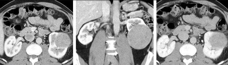
Figure 1Corticomedullary phase CT in axial and coronal plane, and parenchymal phase, of renal cell carcinoma
Long description
Contrast CT of a renal cell carcinoma in the left kidney: axial and coronal corticomedullary phase and axial parenchymal phase (left to right).

Imaging. Most tumours are found on ultrasound or CT performed for other reasons. For a known renal mass, multiphasic contrast-enhanced CT of the abdomen (and chest for staging) is the standard. Imaging is done unenhanced, in an early arterial phase and in a parenchymal phase; a change of 15 Hounsfield units or more after contrast demonstrates enhancement and therefore viable tumour.[3] CT also defines the contralateral kidney, primary tumour extension, venous involvement, locoregional lymph nodes and the adrenal glands and other solid organs.[3]
MRI is used when CT is inconclusive, when there is allergy to iodinated contrast, in pregnancy, or when the extent of an inferior vena cava tumour thrombus is unclear; T2-weighted imaging delineates the uppermost extent of thrombus well.[3] For cystic masses, MRI is preferred: CT has poor sensitivity (36%) and specificity (76%) for Bosniak IIF to III lesions, whereas MRI reaches 71% sensitivity and 91% specificity, and contrast-enhanced ultrasound 100% sensitivity and 97% specificity.[3] FDG-PET CT is not recommended for primary staging.[3]
Staging metastases. Chest CT is accurate for chest staging; a nomogram based on tumour size, stage, systemic symptoms, anaemia and thrombocythaemia suggests chest CT can be omitted in incidentally found cT1a, cN0 disease without systemic symptoms because lung metastases occur in less than 1%.[3] Routine bone imaging is not generally indicated. Brain CT or MRI is offered in metastatic disease, particularly when systemic therapy or cytoreductive surgery is planned (occult brain metastases occur in about 4.3% of metastatic patients at baseline).[3]
Renal mass biopsy is selectively useful: before ablative or systemic therapy without prior histology, before active surveillance, and for indeterminate masses. Core biopsy (preferred over fine-needle aspiration, with a coaxial technique) reaches 99.1% sensitivity and 99.7% specificity for malignancy in experienced centres, although 8% of cores are non-diagnostic and repeat biopsy is diagnostic in 83 to 100% of cases.[3] Cystic masses are not biopsied unless a solid component is present. Biopsy is not needed when surgery is already planned, nor in frail patients on watchful waiting.[3] The 2022 WHO classification does not allow a definitive diagnosis of oncocytoma on needle core, so a "benign" biopsy result must be interpreted with caution.[3]
RCC is diagnosed and staged pathologically after resection, but pre-operative staging uses the 2017 TNM classification.[2]
T - primary tumour[2]
- T1: tumour up to 7 cm, limited to the kidney (T1a up to 4 cm, T1b 4 to 7 cm).
- T2: tumour over 7 cm, limited to the kidney (T2a 7 to 10 cm, T2b over 10 cm).
- T3: tumour extends into major veins or perinephric tissues, but not into the ipsilateral adrenal gland or beyond Gerota fascia. T3a: renal vein or segmental branches, pelvicalyceal system, or perirenal and renal sinus fat. T3b: vena cava below the diaphragm. T3c: vena cava above the diaphragm or wall invasion.
- T4: beyond Gerota fascia, including contiguous extension into the ipsilateral adrenal gland.
N and M. N0/N1 for regional nodal status; M0/M1 for metastasis. Stage groupings are I (T1 N0 M0), II (T2 N0 M0), III (T3 N0 M0, or T1-T3 with N1), IV (T4, or any T any N M1). Note that a T1 versus T1b split at 4 cm is debated for nephron-sparing surgery, and that renal sinus fat invasion may carry a worse prognosis than perirenal fat despite sharing the T3a category.[2]
Clinical anatomy matters alongside stage: scores such as RENAL and PADUA standardise description of position and predict surgical complexity.[2]
Surgery is the only curative treatment.[4]
- Partial nephrectomy (nephron-sparing surgery) is the treatment of choice for T1 tumours, and is offered for T2 tumours when there is a solitary kidney or chronic kidney disease, if technically feasible.[4]
- Radical nephrectomy (laparoscopic or robotic) is offered for T2 tumours and localised masses not amenable to partial nephrectomy.[4]
- Ipsi-lateral adrenalectomy is avoided unless there is clinical evidence of adrenal invasion.[4]
- Lymph node dissection is not performed routinely in organ-confined disease.[4]
- For T3 disease, partial nephrectomy may still be considered if technically feasible but must be balanced against oncological risk.[4]
For patients unfit for surgery, or with non-resectable disease, embolisation controls symptoms such as massive haematuria and flank pain.[4] Watchful waiting (no imaging follow-up, symptom-focused) and active surveillance (serial imaging with delayed intervention) are appropriate for small renal masses in older or comorbid patients: metastasis occurs in only about 1.8 to 2.1% during follow-up of small masses.[4]
Tumour ablation (cryoablation, radiofrequency ablation) is an alternative to surgery for small masses, provided a biopsy is taken first. Pre-ablation biopsy has significantly increased and has reduced treatment of benign or unknown histology.[4]
Systemic therapy in the adjuvant setting. Adjuvant pembrolizumab improves disease-free and overall survival in high-risk resected clear cell RCC (intermediate-high: pT2 grade 4 or sarcomatoid N0 M0, or pT3 any grade N0 M0; high: pT4, or any pT N+; and M1 with no evidence of disease after resection of oligometastatic sites within one year). Adjuvant VEGF-targeted therapy (sunitinib sorafenib, pazopanib, axitinib and everolimus) does not improve overall survival.[4]
Clear cell metastatic RCC is treated with immune checkpoint combinations, not with single-agent agents where combination is possible. First-line options are nivolumab plus ipilimumab (intermediate and poor risk), and pembrolizumab plus axitinib, lenvatinib plus pembrolizumab, or nivolumab plus cabozantinib (all IMDC risk groups); for favourable-risk disease these combinations improve progression-free survival and response rates without an overall survival benefit, so TKI monotherapy (sunitinib, pazopanib) remains an additional choice. Sunitinib or pazopanib are also offered to any patient who cannot receive or tolerate immune checkpoint inhibition.[4]
For second- and third-line therapy after failure of a checkpoint-containing regimen, cabozantinib or another VEGF-targeted TKI is used; cabozantinib is the agent with survival advantage data in VEGF-refractory disease. Nivolumab monotherapy improves overall survival (median 25.0 versus 19.6 months, hazard ratio 0.73) compared with everolimus after one or two lines of VEGF-targeted therapy.[4]
Important accompanying numbers: nivolumab plus ipilimumab was associated with 46% grade 3 to 4 toxicity and 1.5% treatment-related deaths; TKI plus immune checkpoint combinations with 61 to 72% grade 3 to 5 toxicity and about 1% treatment-related deaths.[4]
For VHL disease specifically, belzutifan (a HIF-2alpha inhibitor) has an objective response rate of 67% (complete response 11%) in tumours not needing immediate surgery, with median time to response of 11.1 months.[4][5]
Hereditary syndromes need nephron-sparing or surveillance approaches rather than radical surgery, with an exception for aggressive fumarate hydratase-deficient and high-grade SDH-deficient tumours, which need immediate surgery. In non-aggressive VHL and other syndromes, surveillance until the largest tumour reaches 3 cm limits the number of operations.[4][5]
Cytoreductive nephrectomy of the primary in metastatic disease is individualised (select patients with good performance status and without brain or bone metastases) alongside systemic therapy.[4]
Chemotherapy is generally ineffective in RCC and should not be offered in metastatic disease; it is reserved for collecting duct and renal medullary carcinoma.[4]
Symptom control. Embolisation or radiotherapy relieve bleeding or pain in unfit patients; palliative radiotherapy may control bone pain.[4]
- Direct complications: spontaneous haemorrhage into the tumour, pain, obstruction of the renal pelvis and collecting system with hydronephrosis, tumour thrombus with vena cava syndrome, and metastases.
- Treatment complications: bleeding, urinary leak after partial nephrectomy, and the substantial systemic therapy toxicities listed above. Positive surgical margins occur in 2 to 8% of partial nephrectomies for T1 disease and are not usually an indication for re-operation.[4]
- Long-term: chronic kidney disease after radical nephrectomy, which is why partial nephrectomy is preferred when feasible.
Five-year overall survival for non-metastatic disease by subtype is 91% for chromophobe, 82% for papillary, 81% for clear cell and 44% for collecting duct RCC.[1] Localised clear cell disease treated curatively does well; advanced clear cell disease is incurable but modern combinations produce long-term remissions in a minority (nivolumab plus ipilimumab about 31% overall survival probability at 111 months (9 years) of follow-up in intermediate and poor risk).[4]
The main primary prevention is smoking avoidance and weight reduction. Consumption of a vegetable-rich diet, coffee and tea, and physical activity are associated with lower risk.[1] There is no evidence to support population screening for primary RCC (urinary dipstick is inadequate and CT carries radiation and incidental findings). Screening is considered only in selected high-risk groups, such as end-stage renal disease (ten-fold risk), or families with two first-degree relatives affected.[1]
- Pregnancy: surgery in the second trimester and individualised multidisciplinary care. Many renal masses in pregnancy are benign.
- Children: RCC is rare in childhood; Xp11 translocation RCC occurs. Renal medullary carcinoma occurs in adolescents and young adults with sickle cell trait or disease.
- Older and frail patients: small masses can often be observed. The decision to operate balances tumour biology, competing mortality and comorbidity.
- Hereditary disease: as above; offer genetic counselling and testing for patients aged 46 years or younger, with bilateral or multifocal tumours, or a first- or second-degree relative with RCC.[3][5]
- Renal ultrasound and intravenous urography are affordable; a solid enhancing renal mass cannot be fully characterised without CT. Consider referral of imaging to a regional centre rather than operating on uncertain cysts.
- Surgery for large, symptomatic or locally advanced tumours is often delayed. Early referral of confirmed or suspected masses avoids crisis presentation with haemorrhage.
- Embolisation may not be available; bulky tumours with bleeding can instead be palliated with radiotherapy or analgesia.
- Abdominal mass in an adult is more likely to be advanced RCC; accept that late diagnosis is common and focus on symptom relief and accessible surgery.
- Hereditary cancer is under-recognised without genetic services; a family history is free information and should be taken in every patient with RCC.
- For haematuria, urine microscopy and ultrasound are usually available; cystoscopy and CT urography may require referral and planning.
Blood in the urine is alarming but not always malignant. The first distinction is between visible (macroscopic) haematuria (the patient sees red or brown urine) and non-visible (microscopic) haematuria (detected on a dipstick or microscopy). The second is whether it is explained (a recognised cause such as a current infection, kidney stone or recent exercise) or unexplained (no clear cause).
A practical definition of significant microscopic haematuria is blood seen on standard microscopy in a properly collected mid-stream urine. Dipstick positives must be confirmed on microscopy, because dilute urine, myoglobin, food pigments and contamination can all cause a false positive. Menstrual contamination, exercise and sexual intercourse are common benign causes; a repeat test after the provoking factor has stopped is appropriate. A useful clinical rule: one episode of visible unexplained haematuria is never ignored in an adult.
Causes of haematuria, broadly grouped by age of the patient. Children and young adults more often have glomerular disease or infection; older adults more often have urothelial cancer.
| Age group | Common causes | Helpful clues |
|---|---|---|
| Children and adolescents | Urinary tract infection; glomerular disease (post-streptococcal, IgA nephropathy, thin basement membrane disease, Alport syndrome); sickle cell trait or disease; exercise; congenital urinary tract anomalies | Cola-coloured urine after an upper respiratory tract infection; RBC casts; family history of kidney disease |
| Young adults | Infection; kidney stones; glomerular disease; exercise; trauma; sickle cell; tuberculosis | Dysuria and frequency; unilateral colic; haematuria after exertion that resolves with rest |
| Middle-aged adults | Stones; urinary tract infection; benign prostatic hyperplasia; medications (anticoagulants, NSAIDs, cyclophosphamide); polycystic kidney disease; hypertension-related glomerular disease | Stone pain; obstructive urinary symptoms; medication review |
| Older adults (45 and over) | Urothelial (bladder, renal pelvis, ureter) or renal cell cancer; stones; BPH; urinary tract infection; anticoagulation | Painless visible haematuria; smoking history; possible mass on imaging |
| Any age | Bleeding disorders or anticoagulation; trauma (blunt or penetrating); runner's haematuria; menses (contamination); schistosomiasis in endemic regions | History of bruising, liver disease or anticoagulant use; recent injury |
Exam trapAnticoagulants do not 'cause' haematuria
Drugs that impair coagulation (warfarin, direct oral anticoagulants, antiplatelets) unmask an underlying lesion. A patient bleeding while anticoagulated still needs a full urological work-up once the drug has been stabilised. Do not dismiss the blood as a drug effect alone.[6]
Cancer risk by age. In the United Kingdom, NICE advises a suspected cancer referral for visible unexplained haematuria in people aged 45 and over, and for non-visible unexplained haematuria with dysuria or a raised white-cell count in those aged 60 and over, because the urological cancer yield is highest in these groups.[6]
Core investigations that apply at every level of care:
- Urinalysis and microscopy of a mid-stream specimen: confirm blood; look for protein and casts. RBC casts point to glomerular disease.
- Urine culture to exclude infection; if positive, treat and re-test.
- Renal function and full blood count (haemoglobin falls with brisk or chronic bleeding; the platelet count matters).
- Imaging of the upper tract: renal ultrasound is the first, widely available test; CT urography (or MRI urography) is the more sensitive study for stones, masses and urothelial tumours.
- Flexible cystoscopy in adults with visible haematuria, and in non-visible haematuria with risk factors, to inspect the bladder.
- Renal function tests and, where indicated, cystoscopy with biopsy, CT of the chest and staging.
Imaging should precede cystoscopy when there is suspicion of an upper-tract lesion, so that the whole urinary tract is assessed in one pathway.
Evaluating visible and non-visible haematuria
- Blood detectedVisible red or brown urine, or dipstick positivity confirmed on microscopy
- Confirm and characteriseMid-stream urinalysis, microscopy (RBC casts), urine culture; renal function and full blood count
- Question: Is there an obvious provoking cause?
Yes (infection, stone, exercise, trauma, contamination)
- Treat the causeAntibiotics for infection; fluids and analgesia for stones; repeat urinalysis after recovery
- Question: Repeat urinalysis 6 weeks after treatment
Yes
- Discharge with safety-nettingRe-test if symptoms recur; document a baseline
No, or recurs
- Proceed as unexplained haematuria
No obvious cause
- Risk-stratify by age and risk factorsAge 45+ with visible unexplained haematuria: suspected cancer referral; age 60+ with non-visible unexplained haematuria plus dysuria or raised WCC: suspected cancer referral
- Question: Visible haematuria?
Yes
- Imaging of the upper tractUltrasound first; CT urography if higher risk or stones suspected
- Flexible cystoscopyTo exclude bladder and urethral lesions
- Manage findingsStones, strictures, masses, tumours: treat per diagnosis
No (microscopic only)
- Assess cancer riskAge, smoking, occupational exposure, persistent unexplained blood
- Question: High risk or persistent?
Yes
- CT urography and cystoscopy
No
- Conservative follow-upAnnual urinalysis and blood pressure; repeat investigation if visible haematuria develops
Low-resource notes for haematuria. Microscopy is needed to confirm a dipstick positive; without it, avoid exposing patients to unnecessary investigations. Renal ultrasound is adequate initial imaging when CT is unavailable, but cannot exclude small urothelial lesions. Urine cytology, although imperfect, can be added where cystoscopy is not promptly available. In schistosomiasis-endemic regions, terminal haematuria in young people requires uroparasitology and treatment as well as cancer exclusion.
- RCC is the commonest solid kidney cancer in adults (about 2% of all cancers) and is increasingly found incidentally; the classic triad is rare (under 1%).[1][3]
- Clear cell RCC (about 70%) follows VHL loss, with HIF accumulation, VEGF-driven angiogenesis, and therefore a clear rationale for anti-VEGF and HIF-2alpha therapy.[1][5]
- The common subtypes differ in prognosis: non-metastatic five-year survival about 91% (chromophobe), 82% (papillary), 81% (clear cell) and 44% (collecting duct).[1]
- Contrast-enhanced CT is the central diagnostic and staging investigation; a tumour-thrombus change in the renal vein or vena cava upstages to T3 and occurs in 4 to 10% of cases.[2][3][4]
- Partial nephrectomy is preferred for T1 tumours; radical nephrectomy for larger masses not amenable to nephron-sparing surgery.[4]
- Advanced clear cell disease is treated with immune checkpoint combinations and tyrosine kinase inhibitors; conventional chemotherapy is generally ineffective.[4]
- End-stage renal disease and dialysis-acquired cystic disease raise RCC risk at least ten-fold.[1]
- Hereditary RCC (5 to 8%) presents young (median 37 years) and needs nephron-sparing or surveillance strategies.[5]
- Haematuria splits into visible and non-visible, explained and unexplained; the cancer yield rises steeply with age, with a suspected-cancer referral at 45 for visible and at 60 for non-visible haematuria with dysuria or raised white cells.[6]
- Anticoagulation exposes but does not cause haematuria; the underlying lesion still needs investigating.[6]
- Low-resource practice rests on ultrasound, mid-stream urine and clinical judgement, with referral for cystoscopy and cross-sectional imaging.
Answer each one in your head first, then open the answer.
A 58-year-old smoker is found to have an 8 cm solid left renal mass with a thrombus extending into the renal vein, but no distant metastases. What is the T stage and what is the planned definitive treatment?
Show answerHide answer
Stage T3a (tumour extends into the renal vein). Definitive treatment is surgical removal of the kidney and tumour thrombus, with lymph node dissection only if nodes are clinically involved, followed by adjuvant therapy according to risk. The patient should be counselled that a positive margin is uncommon and that partial nephrectomy is not feasible for a tumour of this size with venous involvement.[2][4]
Why does loss of the VHL gene make clear cell RCC both hypervascular on imaging and responsive to anti-VEGF or HIF-2alpha therapy?
Show answerHide answer
VHL normally tags hypoxia-inducible factor for destruction in normal oxygen. Loss of VHL lets HIF accumulate, switching on VEGF and glycolytic survival genes; hence the abundant tumour vessels seen on CT, and the efficacy of tyrosine kinase inhibitors (which block VEGF signalling) and of belzutifan (which blocks HIF-2alpha).[1][5]
Which patients with RCC should be offered genetic assessment, and which renal cancer syndrome carries the highest lifetime risk of renal tumour?
Show answerHide answer
Genetic assessment is offered to patients aged 46 years or younger, those with bilateral or multifocal tumours, and those with a first- or second-degree relative with RCC or a known pathogenic variant, or with suggestive histology. Hereditary papillary RCC (MET mutation) carries an essentially 100% lifetime risk; VHL carries 30 to 40%.[3][5]
A 45-year-old woman reports one episode of painless visible haematuria that has resolved. She takes warfarin and has a normal renal ultrasound. What is the next step?
Show answerHide answer
Anticoagulation exposes but does not cause haematuria, so a visible unexplained episode at 45 years or over needs a suspected-cancer referral and a complete upper-tract assessment including CT urography, plus flexible cystoscopy to exclude a urothelial lesion. Repeating the urinalysis after the warfarin is stabilised is not sufficient.[6]
Name the three subtypes of RCC expected in a sporadic adult, and which one has the best five-year survival in non-metastatic disease.
Show answerHide answer
Clear cell (about 70%), papillary (13 to 20%) and chromophobe (the remainder); chromophobe RCC has the best five-year survival at about 91%.[1]
In an adult with microscopic haematuria only, what is a reasonable conservative plan when imaging is normal and risk is low?
Show answerHide answer
Repeat urinalysis periodically (for example annually) with blood pressure, reinforce safety-netting, and re-investigate promptly if visible haematuria develops or risk factors accumulate. Cancer is unlikely but the natural history of isolated microscopic haematuria is benign in most.[6]
A patient on long-term dialysis presents with cystic kidney changes and a solid 3 cm renal mass. How is their cancer risk different from the general population, and what is the tumour most likely to be?
Show answerHide answer
The lifetime risk of RCC in end-stage renal disease is at least ten times higher than in the general population, with acquired cystic disease-associated RCC a recognised subtype. Papillary tumours occur relatively more often in end-stage kidneys. Management is by nephron-sparing or radical surgery according to function and comorbidity.[1]
Renal cell carcinoma is discussed here in relation to acute kidney injury, chronic kidney disease, nephrolithiasis, polycystic kidney disease, hypercalcaemia and hyponatraemia topics elsewhere in this library, in plain text rather than as links until those pages are verified.
EOF
- EAU Guidelines Panel on Renal Cell Carcinoma. Renal cell carcinoma - EAU Guidelines: Epidemiology, aetiology and pathology (2026). European Association of Urology 2026. Accessed .
- EAU Guidelines Panel on Renal Cell Carcinoma. Renal cell carcinoma - EAU Guidelines: Staging and classification systems (2026). European Association of Urology 2026. Accessed .
- EAU Guidelines Panel on Renal Cell Carcinoma. Renal cell carcinoma - EAU Guidelines: Diagnostic evaluation (2026). European Association of Urology 2026. Accessed .
- EAU Guidelines Panel on Renal Cell Carcinoma. Renal cell carcinoma - EAU Guidelines: Disease management (2026). European Association of Urology 2026. Accessed .
- EAU Guidelines Panel on Renal Cell Carcinoma. Renal cell carcinoma - EAU Guidelines: Hereditary and syndrome-specific RCC (2026). European Association of Urology 2026. Accessed .
- National Institute for Health and Care Excellence. Suspected cancer: recognition and referral (NG12) - recommended actions organised by symptom and findings (urological symptoms: haematuria). NICE guideline NG12 (last updated 2026). Accessed .
Figure credits
Educational content for medical students and doctors. It is not a substitute for clinical judgement or local guidelines.