Hypokalaemia
Low serum potassium: causes, assessment with urine potassium and acid-base status, ECG changes, and safe replacement.
- Hypokalaemia is a serum potassium concentration below 3.5 mmol/L. Severity is graded mild (3.0 to 3.4), moderate (2.5 to 3.0) and severe (under 2.5 mmol/L).
- It is usually caused by renal or gastrointestinal potassium loss; intracellular shifts (insulin, beta-2 agonists, refeeding) can also drop the serum level rapidly.
- The diagnostic approach uses a 24-hour or spot urine potassium to separate renal from extrarenal loss, then acid-base status and blood pressure to narrow the cause.
- Muscle weakness, ileus, polyuria and dangerous arrhythmias are the main clinical problems; on the ECG look for ST sagging, flat T waves and prominent U waves.
- Oral replacement is preferred. For severe or symptomatic hypokalaemia, give IV potassium up to 40 mmol per litre peripherally, never faster than 10 mmol per hour routinely, after checking and correcting magnesium.
By the end of this page you should be able to:
- Define hypokalaemia and state its severity bands.
- Explain renal versus extrarenal potassium loss, and name the common causes of each.
- Use urine potassium, acid-base status and blood pressure to build a differential diagnosis.
- Recognise the ECG changes of hypokalaemia and the arrhythmia risk with digoxin.
- Plan safe oral and IV potassium replacement, including infusion rates, concentration limits and magnesium correction.
- Apply a practical approach to hypokalaemia in a low-resource setting.
DefinitionHypokalaemia
Hypokalaemia is a serum potassium concentration below 3.5 mmol/L (under 3.5 mEq/L), caused by a deficit in total body potassium stores or by abnormal movement of potassium into cells.[2]
Laboratory reference ranges are usually 3.5 to 5.0 mmol/L. Hypokalaemia may be present with normal or even high total body potassium stores, because most potassium sits inside cells: about 98% of the body's potassium is intracellular, and only about 2% in the extracellular fluid.[1] A small fall in the serum level can therefore reflect a large total body deficit, or simply a shift of potassium into cells.
Severity, based on the adult definitions used in current references, is:
| Band | Serum potassium | Typical features |
|---|---|---|
| Mild | 3.0 to 3.4 mmol/L | Usually asymptomatic |
| Moderate | 2.5 to 3.0 mmol/L | Muscle weakness; may progress to paralysis and respiratory failure |
| Severe | Under 2.5 mmol/L | ECG changes or severe symptoms; carries a risk of life-threatening arrhythmia [2] |
Severity bands and the under-2.5 mmol/L cut-off follow reference 2; the 3.5 to 5.0 mmol/L reference range follows reference 1. [1][2]
Severity should always be judged with the clinical picture and the speed of the fall, not the number alone. A rapid drop into the mild band can cause more trouble than a slow, long-standing value in the moderate band, particularly in patients taking digoxin or with significant heart disease, in whom even mild hypokalaemia can provoke conduction abnormalities.[2]
Hypokalaemia is common. Mild hypokalaemia is found in almost 14% of people undergoing outpatient laboratory testing, and up to 20% of hospitalised patients have some degree of hypokalaemia, although only 4 to 5% of those cases are judged clinically significant. Severe hypokalaemia is relatively uncommon. About 80% of patients taking diuretics become hypokalaemic. There is no consistent sex difference in prevalence.[1]
Potassium balance is kept by intake matching renal excretion (about 90% of the daily 100 mmol) and faecal loss (about 10%).[1] Hypokalaemia arises from one or more of four mechanisms: decreased intake, extrarenal (usually gastrointestinal) loss, renal loss, or movement of potassium into cells. The last two dominate clinical practice.
Any process that loses potassium-rich gut fluid (or exposes the mucosa to it) lowers serum potassium:
- Chronic diarrhoea, including chronic laxative abuse and bowel diversion.
- Protracted vomiting or nasogastric suction, which remove volume and hydrochloric acid: the resulting metabolic alkalosis and aldosterone drive renal potassium loss.[2]
- Rarely, a villous adenoma of the colon, which secretes large volumes of potassium-rich fluid.
- Clay (bentonite) ingestion, which binds potassium and greatly reduces absorption.[2]
Renal loss is the most common mechanism in practice. It arises from excess mineralocorticoid effect, inherited tubular defects, or drugs acting on the nephron.
Excess mineralocorticoid effect. Aldosterone binds mineralocorticoid receptors in the distal nephron, increasing sodium reabsorption and potassium secretion. This occurs in:
- Primary hyperaldosteronism (Conn syndrome), renovascular disease, or rare renin-secreting tumours.
- Cushing syndrome and glucocorticoid-remediable hyperaldosteronism.
- Congenital adrenal hyperplasia (11-beta and 17-alpha hydroxylase deficiency).
- Ingestion of substances such as glycyrrhizin in natural liquorice or chewing tobacco, which inhibits 11-beta-hydroxysteroid dehydrogenase and lets cortisol stimulate the mineralocorticoid receptor.
- Apparent mineralocorticoid excess, Geller syndrome (constitutive activation of the mineralocorticoid receptor) and familial glucocorticoid resistance.[1]
Inherited tubular defects. Bartter syndrome (defect in the thick ascending limb, loop of Henle) and Gitelman syndrome (defect in the distal convoluted tubule) both cause renal sodium and potassium wasting, with high renin and aldosterone, but normal blood pressure. Liddle syndrome (constitutive activation of the epithelial sodium channel, ENaC) gives severe hypertension with potassium wasting and suppressed aldosterone ("pseudoaldosteronism").[1][2]
Renal tubular acidosis and Fanconi syndrome can waste potassium, often with a metabolic acidosis. Hypomagnesaemia is a common correlate of hypokalaemia: part of the association is shared causes (diuretics, diarrhoea), but low magnesium itself increases renal potassium loss and makes hypokalaemia refractory to treatment.[1][2]
Potassium moves into cells in exchange for other cations, which can lower the serum level without changing total body stores:
- Insulin administration and glycogenesis during parenteral or enteral nutrition.
- Stimulation of the sympathetic nervous system, especially beta-2 agonists such as albuterol and terbutaline.
- Familial hypokalaemic periodic paralysis, with sudden shifts of potassium into cells causing transient weakness or paralysis.
- Thyrotoxicosis, via excessive beta-sympathetic stimulation (hypokalaemic thyrotoxic periodic paralysis).
- Refeeding after malnutrition or fasting, driven by insulin release.
- Gastrointestinal bicarbonate gain or alkalosis more generally.[1][2]
Diuretics are by far the commonest drug cause, especially loop and thiazide diuretics, followed by osmotic diuretics. Other culprits include amphotericin B, high-dose penicillin and antipseudomonal penicillins (e.g. piperacillin), laxative abuse and theophylline intoxication. Surreptitious use of diuretics or laxatives is a frequent cause of persistent hypokalaemia, particularly in patients preoccupied with weight loss and in healthcare staff with access to prescription medicines.[2]
"Decreased intake" alone is rarely the whole story in adults, but chronically inadequate intake becomes important where poverty, famine or severe alcohol misuse combine with other losses. Alcohol abuse can also precipitate life-threatening arrhythmias in the setting of hypokalaemia.[1]
The kidney is primarily responsible for total body potassium balance, but changes in extracellular potassium are initially buffered by movement into skeletal muscle, regulated by insulin and catecholamines. Aldosterone, stimulated by hyperkalaemia or volume depletion, drives renal potassium secretion in the principal cells of the collecting duct, dependent on sodium delivery, luminal potassium concentration and the electrical gradient. Aldosterone can signal salt retention without potassium loss in volume depletion, and potassium secretion without salt retention in hyperkalaemia ("the aldosterone paradox").[1]
In hypokalaemia, almost all filtered potassium has already been reabsorbed in the proximal tubule and loop of Henle, so the final urinary potassium is determined by secretion in the distal nephron. Hypomagnesaemia contributes through disinhibition of the ROMK channel, causing ongoing renal potassium wasting even after serum magnesium is partially corrected. Magnesium deficiency also renders hypokalaemia refractory to potassium replacement.[1][2]
A fall in serum potassium hyperpolarises cell membranes, producing non-responsiveness in muscle, and disrupts cardiac electrical conduction. In the heart, low extracellular potassium delays repolarisation, which is the basis of the electrocardiographic changes and arrhythmia risk. Hypokalaemia also participates in acid-base balance through intracellular exchange for hydrogen ions.
Symptoms generally do not appear until serum potassium is below 3.0 mmol/L, unless the fall is rapid or a potentiating factor such as digoxin use is present. Their severity tends to be proportionate to the degree and duration of the deficit.[1][2]
- Weakness, often beginning in the legs and proximal muscles.
- Cramps and fasciculations.
- With more severe depletion: paresis or ascending paralysis and respiratory failure from hypoventilation.[2]
- Constipation, reduced gut motility and, in severe cases, paralytic ileus.[2]
- Impaired concentrating ability causing polyuria and secondary polydipsia.
- Chronic hypokalaemia can cause tubulointerstitial disease, cyst formation and nephrogenic diabetes insipidus. Rhabdomyolysis may occur, particularly in severe hypokalaemia.[1][2]
Fundamentally this is where hypokalaemia can kill. ECG changes are usually minimal until serum potassium is below 3.0 mmol/L.[2] Hypokalaemia produces:
- Sagging (depression) of the ST segment.
- Flattening of the T wave.
- Elevation and prominence of the U wave.
As the deficit worsens, the T wave becomes progressively smaller and the U wave larger; sometimes a flattened or positive T wave merges with a positive U wave, which can be confused with QT prolongation. Arrhythmias include premature atrial and ventricular beats, ventricular and supraventricular tachyarrhythmias, and second- or third-degree atrioventricular block, becoming more severe as hypokalaemia deepens; ventricular fibrillation may eventually occur. Cardiac effects become increasingly severe with increasingly severe hypokalaemia. Patients with significant pre-existing heart disease, and patients taking digoxin, are at risk of conduction abnormalities even with mild hypokalaemia.[2]
Metabolic acidosis, rhabdomyolysis in severe hypokalaemia, and rarely chronic tubulointerstitial disease have been described. Constitutional symptoms (fatigue, polydipsia) may dominate chronic cases.[1]
Acute hypokalaemia is rarely a diagnostic puzzle in the emergency department: the cause is usually apparent from the history (vomiting, diarrhoea, diuretic therapy, refeeding, insulin). When it is not, the framework is:
- Renal versus extrarenal loss (urine potassium).
- Acid-base status (metabolic acidosis versus alkalosis).
- Blood pressure (normal versus hypertensive renal losses).
Persistently unexplained hypokalaemia should raise consideration of surreptitious vomiting or laxative or diuretic abuse, especially with metabolic alkalosis and normal or variable blood pressure.
Figure 1LowKECG
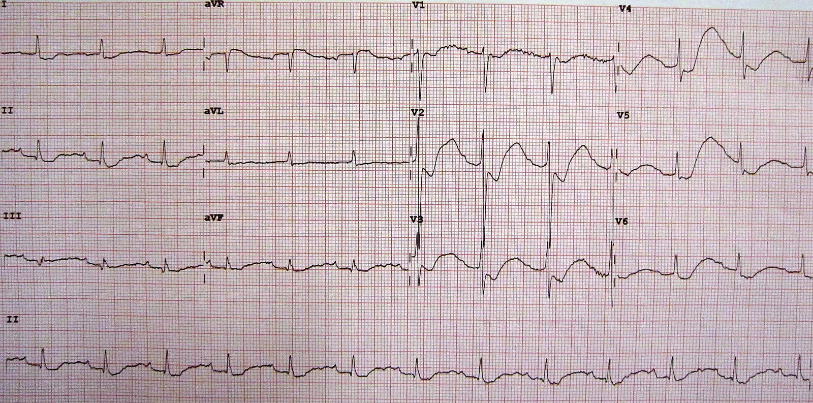
Long description
12-lead ECG from a patient with hypokalaemia (Wikimedia Commons file LowKECG): flattened T waves, ST depression and prominent U waves.

- Serum electrolytes (sodium, potassium, chloride, bicarbonate), glucose, urea/creatinine and pH if indicated.
- ECG for moderate to severe hypokalaemia (and earlier if the patient has heart disease or takes digoxin).
- Serum magnesium and calcium (magnesium is essential; phosphate if refeeding is suspected).
- Arterial or venous blood gas when the cause is not apparent, to detect metabolic acidosis or alkalosis.
- Serum digoxin level where relevant; drug screen for diuretics, amphetamines or sympathomimetics if abuse is suspected.
- TSH when there is tachycardia or clinical suspicion of thyrotoxic periodic paralysis.[1][2]
Urine potassium is the single best discriminator of mechanism.
- 24-hour urinary potassium excretion is the most accurate; in hypokalaemia, renal secretion is normally under 15 mmol per day. Excretion above 15 mmol per day indicates inappropriate renal potassium loss.[1][2]
- If a 24-hour collection is not feasible, a spot urine sample can be used: renal wasting is suggested when the spot urine potassium is above 15 mmol per litre, or the potassium-to-creatinine ratio is greater than 13 mmol per gram of creatinine (1.5 mmol per millimole).[1][2]
- Extrarenal loss or reduced intake is suspected when renal potassium secretion is under 15 mmol per day (or under 15 mmol per litre on a spot sample).[2]
Once renal versus extrarenal loss is established, acid-base and blood pressure further narrow the differential:[1][2]
- Metabolic acidosis with low urine potassium suggests lower gastrointestinal losses (laxatives, diarrhoea, villous adenoma).
- Metabolic acidosis with renal potassium wasting suggests renal tubular acidosis (types 1 and 2) or diabetic ketoacidosis.
- Metabolic alkalosis with low urine potassium can occur with surreptitious vomiting or diuretic use.
- Metabolic alkalosis with renal potassium wasting and normal blood pressure suggests diuretic use, vomiting, Bartter syndrome or Gitelman syndrome. Urine chloride helps here: it is generally normal (similar to intake) in Bartter or Gitelman syndrome, high or low with diuretics depending on duration of action, and low when bicarbonate is being excreted from vomiting.
- Metabolic alkalosis with renal potassium wasting and high blood pressure suggests primary aldosteronism, Liddle syndrome, renovascular disease, or other mineralocorticoid excess, or Cushing syndrome. In these patients a plasma aldosterone-to-renin ratio is the appropriate screen; adrenal CT and venous sampling follow if surgery is contemplated.[1]
Adrenal and pituitary imaging when mineralocorticoid or glucocorticoid excess is suspected. Urinary calcium distinguishes Bartter (hypercalciuria) from Gitelman (hypocalciuria) syndromes. Genetic testing is used for familial causes (primary aldosteronism before age 20, GRA, Liddle, Bartter, Gitelman, apparent mineralocorticoid excess) in appropriate cases.[1]
| Urine potassium | Typical causes | Useful clues |
|---|---|---|
| Low (under 15 mmol/day) | Inadequate intake; vomiting; diarrhoea and laxative abuse; intracellular shifts (insulin, beta-2 agonists, refeeding); periodic paralysis; thyrotoxic periodic paralysis | Low or high urine values depending on mechanism; metabolic acidosis with diarrhoea, alkalosis with vomiting |
| High, normal blood pressure, metabolic alkalosis | Thiazide/loop diuretics; Bartter syndrome; Gitelman syndrome; vomiting or diuretic abuse | Normal urine chloride in Bartter/Gitelman; high/low chloride with diuretics; hypocalciuria (Gitelman) versus hypercalciuria (Bartter) |
| High, high blood pressure | Primary hyperaldosteronism; renovascular disease; Cushing syndrome; Liddle syndrome; congenital adrenal hyperplasia; apparent mineralocorticoid excess | Elevated aldosterone/renin ratio; mineralocorticoid-driven |
| High, metabolic acidosis | Distal (type 1) and proximal (type 2) renal tubular acidosis; Fanconi syndrome; diabetic ketoacidosis | Inappropriate urinary acidification; bicarbonate wasting |
| High due to drugs | Loop, thiazide and osmotic diuretics; amphotericin B; high-dose penicillin; theophylline | Medication history usually reveals it |
Urine potassium thresholds (under 15 mmol/day or above 15 mmol/L) and the potassium-to-creatinine ratio cut-off (greater than 13 mmol/g creatinine, 1.5 mmol/mmol) follow references 1 and 2. [1][2]
Approach to hypokalaemia
- Serum potassium under 3.5 mmol/LConfirm on the analyser; note the sample (avoid haemolysis)
- Assess severityMild 3.0-3.4, moderate 2.5-3.0, severe under 2.5 mmol/L; rate of fall matters
- ECG and cardiac statusECG for moderate/severe or for heart disease/digoxin; look for ST sagging, flat T, U waves
- Question: ECG changes or severe symptoms?
Yes
- Give IV potassium chlorideMax 40 mmol/L peripherally; up to 40 mmol/h central with ECG monitoring; use 0.9% sodium chloride, not glucose
- Check and correct magnesiumHypomagnesaemia perpetuates renal wasting; treat first or alongside
- Continuous monitoringHourly or serial potassium; seek senior help
No
- Oral replacement preferred20 to 80 mmol per day in divided doses, with or after meals
- Identify and treat the causeStop offending drug, replace losses, control vomiting/diarrhoea
- Urine potassium and acid-base status24-hour (or spot) urine potassium; metabolic acidosis versus alkalosis
- Question: Renal or extrarenal loss?
Low urine potassium
- Look for GI loss or shiftVomiting, diarrhoea, laxatives, refeeding, insulin, beta-2 agonists, periodic paralysis
High urine potassium
- Use blood pressure and acid-baseNormal BP + alkalosis: diuretics, Bartter/Gitelman; high BP: aldosterone/Liddle/Cushing
- Correct magnesium and treat the causeMagnesium deficiency, primary aldosteronism, tubular disorder, endocrine cause
- Recheck potassiumMonitor daily while replacing; aim for gradual correction
Infusion limits, oral dosing and diuretic cautions in the flowchart are taken from references 1 and 2. [1][2]
Aims: reduce continuing losses, replenish stores, check for toxicity (especially digoxin and arrhythmia) and identify the cause to prevent recurrence. Where possible, treat the underlying driver rather than just the number: stop the offending diuretic or switch to a potassium-sparing agent, treat vomiting or diarrhoea, use H2-receptor antagonists with nasogastric suction, control hyperglycaemia if glycosuria is present, and address endocrine disease.[1][2]
- Mild to moderate hypokalaemia (3.0 to 3.4, and many at 2.5 to 3.0) in a patient who can eat and drink: give oral potassium. IV potassium is reserved for severe hypokalaemia (under 2.5), for ECG changes or severe symptoms, for inability to tolerate oral intake (severe nausea, vomiting, abdominal distress), for ongoing losses, or for hospitalised patients taking digoxin or with significant heart disease.[1][2]
- Severe or symptomatic hypokalaemia, or arrhythmia: replace intravenously with continuous ECG monitoring and serial potassium measurements.[1][2]
Oral potassium chloride is the preferred route whenever feasible. Potassium is usually given in divided doses, because high single doses irritate the gut and can cause bleeding. Liquid potassium chloride raises the serum level within 1 to 2 hours but tastes bitter and is poorly tolerated above 25 to 50 mmol per dose. Wax-impregnated and microencapsulated preparations are better tolerated. Potassium should be taken with plenty of water (100 to 250 mL depending on the preparation) and with or after meals.[1][2]
A useful rule of thumb for total body deficit: each 1 mmol/L fall in serum potassium corresponds to roughly 200 to 400 mmol of total body potassium deficit, but this can either overestimate or underestimate the true deficit depending on whether there has been a shift or ongoing loss.[1][2] Typical daily replacement is 20 to 80 mmol, split into divided doses; in one detailed regimen this corresponds to about 72 mmol per day for mild and 96 mmol per day for moderate hypokalaemia, with monitoring and dose adjustment.[1] Even with severe deficits it is rarely necessary to give more than 100 to 120 mmol in 24 hours unless losses are ongoing.[2]
- Dilute in 0.9% sodium chloride, which is the preferred fluid; 5% glucose (or glucose-containing solutions) can worsen hypokalaemia by stimulating insulin-driven cellular uptake. Use premixed infusions where possible.[1][2]
- Peripheral concentration should not exceed 40 mmol per litre (40 mmol/L), because concentrated potassium solutions irritate veins.[2]
- Routine infusion rates should not exceed 10 mmol per hour.[2] In life-threatening arrhythmia, up to 40 mmol per hour may be given, but only through a central vein or multiple peripheral veins simultaneously, with continuous cardiac monitoring and hourly serum potassium determinations.[2]
- Do not give glucose-containing diluents in these situations.
Potassium replacement often fails if magnesium deficiency coexists, because renal potassium wasting continues. Where hypomagnesaemia is present (or even suspected with diuretic use), correct both deficiencies. A practical schedule from one regimen is: give magnesium sulphate first (about 8 mmol magnesium, as 4 mL of 50% magnesium sulphate diluted in 10 mL 0.9% sodium chloride, over 20 minutes), then run the first 40 mmol potassium chloride infusion, followed by further magnesium replacement.[1] The core principle, supported by both references, is that potassium and magnesium must be replaced together in these patients.[1][2]
If diuretic therapy cannot be stopped (for example in heart failure), switching to a potassium-neutral or potassium-sparing diuretic (triamterene, spironolactone) avoids further renal potassium loss; spironolactone is usually given as 25 mg four times daily and triamterene 100 mg once daily in this context.[2] Aldosterone antagonists are also the treatment of choice for pumping-mediated potassium wasting (primary hyperaldosteronism, Liddle excepted) and are part of treating Bartter/Gitelman-related loss alongside supplementation and sodium chloride.[1]
- Diabetic ketoacidosis. When hypokalaemia occurs with a high serum potassium concentration (as in DKA), defer intravenous potassium until the serum potassium begins to fall, then replace aggressively as it drops.[2]
- Refeeding. In malnourished patients, potassium (and phosphate and magnesium) must be monitored and replaced alongside thiamine when feeding is restarted.
- Children. Inherited tubular disorders (Bartter, Gitelman) present in childhood; growth retardation, failure to thrive and hypocalciuria versus hypercalciuria distinguish the two.[1]
- Older adults and cardiac disease. These groups are least able to tolerate hypokalaemia, and even mild deficits predispose to arrhythmias.[1][2]
- Plan serum potassium checks daily (or more often) while replacing, with dose adjustment.[1]
- Digoxin: patients taking digoxin and those with significant heart disease are at risk of conduction abnormalities from even mild hypokalaemia, because digoxin and hypokalaemia both act on repolarisation. Monitor potassium closely whenever such patients are diuresed or unwell.[2]
- QT and U waves: the apparent QT prolongation seen on some ECGs in hypokalaemia is often a merged flat T wave and prominent U wave rather than true QT prolongation. Do not over-correct on the strength of a single tracing; use a 12-lead and the clinical picture, and monitor continuously during IV replacement.[2]
- Avoid glucose-containing diluents and overly rapid correction; both can cause rebound shifts.[2]
- Cardiac arrhythmia up to ventricular fibrillation, particularly with digoxin or pre-existing heart disease.
- Muscle paralysis, rhabdomyolysis and respiratory failure in severe depletion.
- Paralytic ileus and bowel obstruction-like picture.
- Chronic hypokalaemic nephropathy: impaired concentrating ability, polyuria, tubulointerstitial disease and cyst formation.
- Precipitation of arrhythmia in Bartter/Gitelman syndrome, especially with alcohol or other drugs, where chronic hypokalaemia is aggravated.[1][2]
Symptoms generally resolve with correction of the hypokalaemia. In hospital, the outcome depends chiefly on the underlying cause and on how quickly severe deficits and arrhythmias are recognised. Long-standing deficits may take days to replace because of the lag in potassium movement from extracellular fluid into cells.[2]
Routine potassium replacement is not necessary in most patients taking diuretics, but serum potassium should be monitored, particularly where the risk of complications is high: decreased left ventricular function, digoxin use, diabetes (fluctuating insulin) and asthma patients taking beta-2 agonists. When hypokalaemia develops, potassium supplementation, usually with oral potassium chloride, is indicated.[2]
- Where urine electrolytes and arterial blood gas are unavailable, diagnose from the history and examination: diarrhoea, vomiting, diuretic use, refeeding or insulin point to the mechanism, and basic renal function and volume status give the rest. Edema, hypertension and a simple urine dipstick can substitute for full investigation in many cases.
- Serum magnesium is often the first test to disappear from the panel: empirically replace magnesium when deficiency is likely and replacement therapy is available, because untreated hypomagnesaemia will frustrate potassium replacement.
- Prefer oral replacement (and potassium-containing foods) in stable patients; reserves IV potassium for severe or symptomatic cases.
- If IV potassium is needed, obey the same safety limits: maximum 40 mmol per litre peripherally and 10 mmol per hour by routine infusion, with as much cardiac monitoring as is available. Central venous access and continuous monitoring are not always available, so that correction in arrhythmia becomes a high-risk decision made with senior input.
- Send every complex or atypical case (hypertension with hypokalaemia, early-onset disease, polyuria with rhabdomyolysis) to a centre with endocrine and nephrology support when referral is possible.
- Treat the underlying cause: oral rehydration for diarrhoea, antiemetics for vomiting, and stopping the offending drug.
- Hypokalaemia is serum potassium below 3.5 mmol/L; severity bands are mild 3.0 to 3.4, moderate 2.5 to 3.0 and severe under 2.5 mmol/L.
- Renal or gastrointestinal loss accounts for most cases; intracellular shifts can drop serum potassium without changing total body stores.
- Urine potassium separates renal from extrarenal loss: over 15 mmol per day (or a spot value over 15 mmol per litre) suggests renal wasting.
- Acid-base status and blood pressure then narrow the diagnosis: alkalosis with hypertension suggests aldosterone-mediated loss, while acidosis suggests renal tubular acidosis or gastrointestinal/bicarbonate loss.
- ECG changes are ST sagging, flat T waves and prominent U waves, sometimes mimicking QT prolongation; cardiac effects are slight before 3.0 mmol/L.
- Even mild hypokalaemia is dangerous with digoxin or significant heart disease.
- Each 1 mmol/L fall in serum potassium corresponds to roughly 200 to 400 mmol of total body deficit; replace at 20 to 80 mmol per day and rarely more than 100 to 120 mmol in 24 hours.
- Oral potassium chloride is preferred; liquid forms act within 1 to 2 hours but are bitter and poorly tolerated above 25 to 50 mmol per dose.
- IV replacement: maximum 40 mmol per litre peripherally, routine rate 10 mmol per hour, up to 40 mmol per hour only in arrhythmia with central access and continuous monitoring, using 0.9% sodium chloride rather than glucose.
- Correct magnesium alongside potassium: hypomagnesaemia drives renal wasting and makes hypokalaemia refractory.
- Potassium-sparing diuretics help when diuretic therapy must continue.
- In diabetic ketoacidosis, defer IV potassium until serum potassium starts to fall.
Answer each one in your head first, then open the answer.
A 62-year-old man has diarrhoea for five days and a serum potassium of 2.8 mmol/L. How should you classify his hypokalaemia, and what is the likely mechanism?
Show answerHide answer
Moderate hypokalaemia (2.5 to 3.0 mmol/L). Prolonged diarrhoea causes gastrointestinal potassium loss; 24-hour urine potassium would usually be under 15 mmol per day, with a metabolic acidosis.
The same patient now has muscle weakness. His ECG shows flat T waves and prominent U waves. Why is hypokalaemia dangerous in a patient taking digoxin?
Show answerHide answer
Even mild hypokalaemia can produce cardiac conduction abnormalities in patients on digoxin or with significant heart disease, because both digoxin and low extracellular potassium alter ventricular repolarisation and predispose to arrhythmia.
A young woman with vomiting has hypokalaemia. Urine potassium is raised and blood pressure is normal. Which syndrome and which useful urine test are you thinking of?
Show answerHide answer
Bartter or Gitelman syndrome; both present with hypokalaemic, hypochloraemic metabolic alkalosis, high renin and aldosterone, and normal blood pressure. Urine chloride is usually normal (similar to intake) in these syndromes, and urinary calcium helps separate them (hypocalciuria in Gitelman, hypercalciuria in Bartter). Surreptitious vomiting or diuretic abuse must also be excluded.
A patient has lidocaine-resistant ventricular tachycardia and a serum potassium of 2.1 mmol/L. How rapidly may IV potassium be given, and what monitoring is required?
Show answerHide answer
Up to 40 mmol per hour, by central vein or several peripheral veins together, with continuous ECG monitoring and hourly serum potassium. The peripheral concentration must not exceed 40 mmol per litre; use 0.9% sodium chloride, not glucose.
Why might a patient fail to recover from hypokalaemia despite large potassium doses?
Show answerHide answer
Concurrent hypomagnesaemia: low magnesium increases renal potassium wasting via the ROMK channel and makes hypokalaemia refractory to potassium replacement. Both deficiencies must be corrected together.
A 45-year-old man with hypertension and hypokalaemia has a raised aldosterone-to-renin ratio. What is the most likely diagnosis, and what imaging follows?
Show answerHide answer
Primary hyperaldosteronism. Adrenal CT is recommended (to exclude large masses and adrenocortical carcinoma), and adrenal venous sampling by an experienced radiologist if surgery is feasible and the patient is willing.
On an ECG in hypokalaemia, a flattened T wave seems to merge with a prominent U wave and imitates a prolonged QT interval. Is the QT truly prolonged?
Show answerHide answer
Often no: the apparent prolongation is a merged T wave and U wave. Confirm with a 12-lead ECG and the clinical picture; monitor continuously during IV replacement rather than chase a single tracing.
Using the rule of thumb, estimate the potassium deficit for a serum potassium of 2.5 mmol/L in someone whose baseline is about 4.0 mmol/L.
Show answerHide answer
A fall of 1.5 mmol/L corresponds to roughly 300 to 600 mmol of total body deficit, but this is only a rough estimate and may over- or under-estimate the true deficit, especially with ongoing losses or shifts.
Related topics (plain text, linked once the discipline is complete): renal tubular acidosis and Fanconi syndrome; primary aldosteronism (Conn syndrome); Cushing syndrome; Bartter and Gitelman syndromes; Liddle syndrome; refeeding syndrome; thyrotoxicosis and periodic paralysis; acid-base disorders; digoxin and other drug causes; QT and arrhythmia disorders.
- Kardalas E, Paschou SA, Anagnostis P, Muscogiuri G, Siasos G, Vryonidou A. Hypokalemia: a clinical update. Endocrine Connections 2018;7(4):R135-R146. doi:10.1530/EC-18-0109. Accessed .
- Lewis JL III. Hypokalemia. Merck Manual Professional Version, reviewed June 2025, last updated December 2025. Accessed .
Figure credits
Educational content for medical students and doctors. It is not a substitute for clinical judgement or local guidelines.