Hyperkalaemia
Elevated serum potassium: why it matters, how it changes the ECG, and how to stabilise and remove it.
- Hyperkalaemia means too much potassium in the blood: a serum potassium of at least 5.5 mmol/L on the definition used by the UK Kidney Association, and levels above 5.0 mmol/L on many laboratory reference ranges. [1]
- It is usually silent until it disturbs the heartbeat, yet it can turn a stable patient into cardiac arrest within minutes, so the ECG matters as much as the number. [1][3]
- The commonest causes are reduced kidney excretion (acute or chronic kidney disease), drugs that block the renin-angiotensin-aldosterone system, aldosterone deficiency and type 4 renal tubular acidosis, and potassium shifting out of cells in acidosis or tissue breakdown. [3]
- The ECG changes track severity in a recognisable order: tall peaked T waves, then flattening of the P wave and widening of the QRS, then a sine wave. [1][3]
- Emergency care runs in sequence: protect the heart with intravenous calcium, shift potassium into cells with insulin and glucose, add nebulised salbutamol, treat acidosis, then remove potassium with binders, diuretics or dialysis, while monitoring both potassium and glucose for rebound and hypoglycaemia. [1][2]
- Longer term prevention is a drug review, diet, sick-day rules and, increasingly, a potassium binder so that heart and kidney protective drugs can be kept. [1]
By the end of this page you should be able to:
- Define hyperkalaemia with the threshold and severity bands used in current practice. [1]
- List the major causes under the three headings of reduced renal excretion, aldosterone or tubular problems, and redistribution of potassium. [3]
- Recognise the ECG progression of hyperkalaemia and say why a normal ECG does not exclude it. [1]
- Give the emergency sequence with doses, routes and monitoring for moderate and severe disease. [1][2]
- Explain the long-term plan to prevent recurrence without stopping renin-angiotensin blockade unnecessarily. [1]
- Describe safe management when tests or potassium binders are not available. [1][2]
DefinitionHyperkalaemia
An extracellular (usually serum) potassium concentration above the reference range. There is no single universally accepted cut-off: the UK Kidney Association adopted the European Resuscitation Council threshold of serum potassium at least 5.5 mmol/L, while many laboratories define hyperkalaemia as a value above 5.0 mmol/L. [1]
Potassium lives mainly inside cells: the intracellular concentration is about 100 to 150 mmol/L, while the plasma concentration is normally 3.5 to 5.5 mmol/L. [3] Because the resting membrane potential of cardiac and muscle cells depends on this gradient, even a modest rise in serum potassium can slow conduction and predispose to arrhythmia. [1][3]
Hyperkalaemia is often classified by severity using the UK Kidney Association bands: [1]
| Severity | Serum potassium (mmol/L) | Typical implications |
|---|---|---|
| Mild | 5.5 to 5.9 | Look for a cause; treat chronic causes in the community; no emergency treatment usually needed |
| Moderate | 6.0 to 6.4 | Hospital assessment and active treatment if the patient is unwell or ECG changes are present |
| Severe | 6.5 or more | Emergency treatment; continuous cardiac monitoring; urgent senior or renal review |
A normal ECG can be recorded when potassium is 6.5 mmol/L or higher, and ECG changes may appear in clusters rather than in the textbook sequence, so the ECG risk-stratifies but never rules hyperkalaemia out. [1]
Hyperkalaemia is common in hospital: the UK Kidney Association estimates an incidence of 1 to 10% in hospitalised patients, and it is independently associated with higher mortality, with risk rising as the potassium level worsens. [1] The higher prevalence in heart failure, chronic kidney disease, acute kidney injury and diabetes is largely explained by the widespread use of renin-angiotensin-aldosterone system (RAAS) blockers in these conditions. [1] In community series, hyperkalaemia is strongly concentrated in the elderly and in people with kidney disease. [3]
It helps to sort causes into three groups: too little potassium leaves the body, too much enters from outside, or potassium moves out of the cells. [3]
The kidneys excrete roughly 90% of the daily potassium load and maintain potassium balance until the glomerular filtration rate (GFR) falls below 15 mL/min/1.73 m2, so failure of excretion is the common final pathway. [3]
- Chronic kidney disease or acute kidney injury, especially with oliguria and low distal sodium delivery. [3]
- Hypoaldosteronism, primary (for example Addison disease) or secondary as the hyporeninaemic form seen in diabetic kidney disease and chronic renal disease. [3]
- Type 4 renal tubular acidosis: tubular resistance or aldosterone deficiency producing hyperkalaemia with a mild hyperchloraemic acidosis, often in the setting of diabetes or interstitial kidney disease. [3]
- Obstructive uropathy, which can produce a hyperkalaemic acidosis. [3]
- Constipation, which reduces the gut's compensatory potassium excretion, especially in advanced kidney disease. [3][1]
- Congestive heart failure, where renal impairment combines with potassium-retaining drugs. [3]
Many drugs cause hyperkalaemia by lowering aldosterone activity or blocking its effect (Table 9 of the UKKA guideline lists the full set): [1][3]
| Factor | Representative examples | Mechanism in brief |
|---|---|---|
| RAAS blockers | ACE inhibitors, angiotensin receptor blockers, renin inhibitors | Lower aldosterone and reduce glomerular filtration, so potassium is not excreted |
| Potassium-sparing and aldosterone antagonists | Spironolactone, eplerenone, amiloride, triamterene | Block aldosterone or the epithelial sodium channel in the collecting duct |
| Antibiotics | Trimethoprim, pentamidine, co-trimoxazole | Block sodium channels in the distal nephron and mimic amiloride. Trimethoprim can raise potassium by 0.36 to 1.21 mmol/L within 3 to 10 days [1] |
| NSAIDs | Ibuprofen, naproxen, indometacin | Induce hyporeninaemic hypoaldosteronism by inhibiting renal prostaglandins and reduce GFR |
| Calcineurin inhibitors | Cyclosporin, tacrolimus | Inhibit adrenal aldosterone synthesis and increase tubular potassium efflux |
| Heparin | Unfractionated or low molecular weight heparin | Inhibits adrenal aldosterone biosynthesis and reduces angiotensin receptor number |
| Beta-blockers | Especially non-selective agents | Inhibit renin secretion and reduce cellular potassium uptake |
| Other cardiac drugs | Digoxin toxicity, succinylcholine | Inhibit Na/K-ATPase or depolarise membranes so potassium leaves cells |
| Exogenous potassium | Supplements, salt substitutes, massive transfusion | Add potassium to an excretion system that cannot keep up |
The combination of a RAAS blocker with a potassium-sparing diuretic is a particularly risky pairing that needs close monitoring, especially in kidney disease or heart failure. [3]
Figure 1Potassium shifts between cells and the extracellular fluid
Long description
Two boxes are stacked. The upper box is the extracellular fluid, where serum potassium is measured. The lower box is the intracellular fluid, which holds most of the body's potassium. An upward arrow from the lower box to the upper box is labelled Out of cells, with six causes listed beside it: acidosis, insulin deficiency, hyperosmolality, exercise, cell breakdown and beta-blockers. A downward arrow from the upper box to the lower box is labelled Into cells, with three causes: insulin, beta-agonists and alkalosis.
Potassium moves from inside cells to the serum despite a normal total body content. [3]
- Acidosis: mineral acidosis (for example HCl excess or inorganic acid accumulation) shifts potassium out of cells more reliably than organic acidosis. [3]
- Insulin deficiency in diabetes mellitus. [3]
- Acute hyperosmolality from hyperglycaemia or mannitol infusion. [3]
- Tissue breakdown: rhabdomyolysis, tumour lysis, haemolysis, severe burns, crush injury. [3]
- Digoxin toxicity and beta-blockade. [3]
- Hyperkalaemic periodic paralysis. [3]
- Massive cell turnover or repeated exercise in anuric patients.
A person with normal kidneys and normal hormones can excrete large potassium loads, but even a modest supplement can cause severe hyperkalaemia when GFR is below 15 mL/min/1.73 m2. [3] Salt substitutes made with potassium chloride are a common hidden source, as are intravenous potassium infusions given too quickly. [3]
Pseudohyperkalaemia is a raised laboratory value that does not reflect the true plasma concentration: potassium leaks from cells during or after sampling. [3] Suspect it in a well, asymptomatic patient with moderate hyperkalaemia and no obvious cause.
- Haemolysis during a difficult draw, small needles, prolonged tourniquet application or vigorous fist clenching. [3]
- Excess alcohol left on the skin during capillary sampling. [3]
- Marked thrombocytosis or leucocytosis, where clotting releases potassium in the serum tube. [3]
- Very high white cell counts in leukaemia.
Management is simple: repeat the test on a free-flowing lithium heparin sample from a large vein, with gentle traction and prompt analysis. [1] Capillary blood gas measurement gives a rapid true plasma potassium while the formal sample is processed. [1]
Hyperkalaemia acts on the heart in two competing ways: it depolarises the resting membrane and inactivates sodium channels, slowing conduction, while calcium entry opposes the depolarisation to some extent. This is the physiological basis for giving calcium. [1] The atrial myocardium and the specialised conduction tissue are affected at different rates, which is why P wave amplitude falls early while the QRS may widen later. [1] Peaked T waves are the commonest and earliest sign, occurring in roughly a third of patients with moderate to severe hyperkalaemia. [1]
The kidneys compensate by increasing GFR, distal sodium delivery and aldosterone-stimulated secretion, but once GFR is low or aldosterone is blocked, the reserve disappears. [3] The gut excretes about 10% of the potassium load and can hypertrophy its secretory capacity in chronic kidney disease, which is why constipation matters. [3]
Hyperkalaemia is often asymptomatic, and a normal resting ECG is common even with markedly elevated serum potassium. [1] When symptoms occur they are non-specific and related to muscle or cardiac excitability: [3]
- Generalised weakness, fatigue or difficulty climbing stairs.
- Palpitations or an irregular pulse.
- Nausea or abdominal discomfort.
- Paraesthesia or muscle pain.
- In severe cases, flaccid paralysis.
- Syncope or sudden collapse.
Examination is usually normal except for warning signs of associated disease: signs of dehydration or shock, a postural drop in blood pressure, an enlarged kidney or bladder, or cardiac arrhythmia. [1][3] Bradycardia may be resistant to atropine, and in pacemaker-dependent patients hyperkalaemia widens the paced QRS and raises pacing thresholds, sometimes causing failure to capture. [1]
Other conditions mimic the arrhythmias of hyperkalaemia, but the principal diagnostic trap is pseudohyperkalaemia, above. [3] The ECG signs progress, but not always in strict order: [1]
- Peaked, narrow-based, symmetric T waves, usually with a sharp apex.
- Flattening and eventual loss of the P wave.
- Prolongation of the PR interval and widening of the QRS.
- ST-segment depression.
- A biphasic "sine wave" pattern as QRS and T merge.
- Bradycardia, high-grade atrioventricular block, ventricular tachycardia, ventricular fibrillation or asystole.
The finding that most predicts short-term adverse events is QRS prolongation, bradycardia below 50 beats per minute or a junctional rhythm. [1] The BRASH syndrome (bradycardia, renal failure, atrioventricular node blockers, shock and hyperkalaemia) can produce severe bradycardia even with a potassium below 6.5 mmol/L. [1]
Figure 2118 (CardioNetworks ECGpedia)
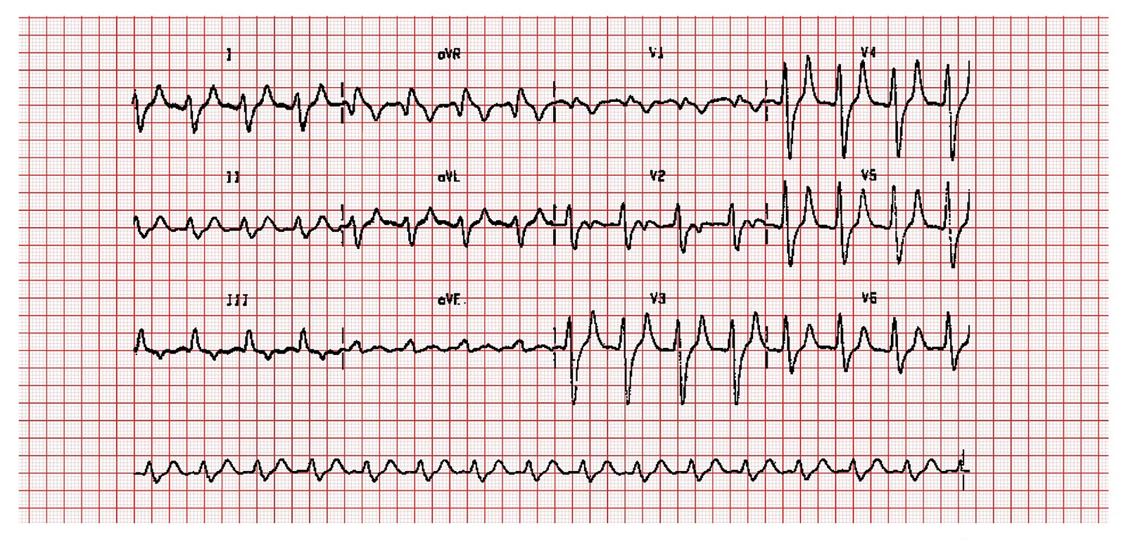
Long description
ECG of a 58-year-old haemodialysis patient with severe hyperkalaemia (serum potassium 9.6 mmol/L), whose file page lists the classic hyperkalaemic changes: small or absent P waves, wide QRS, shortened or absent ST segment, and wide, tall, tented T waves.

- Serum potassium on a lithium heparin sample, confirmed against a serum sample when pseudohyperkalaemia is possible. [1]
- Venous or arterial blood gas in an emergency for a rapid potassium result, plus pH and bicarbonate to identify acidosis. [1]
- Urea, creatinine and GFR to distinguish acute from chronic kidney disease. [3]
- Full blood count for haemolysis, sepsis, thrombocytosis or leucocytosis. [3]
- Blood glucose and glycosylated haemoglobin for diabetes and insulin deficiency. [3]
- Creatine kinase for rhabdomyolysis, and lactate dehydrogenase or alanine aminotransferase for haemolysis or tumour lysis. [3]
- Serum bicarbonate, plasma osmolality and renin, angiotensin and aldosterone levels when the cause is not obvious, to separate aldosterone deficiency from resistance. [3]
- Urine potassium, sodium and creatinine, or a transtubular potassium gradient (TTKG), to judge distal secretion (TTKG is expected to exceed 10 in normal renal response; an inappropriately low value suggests hypoaldosteronism or a tubular defect). [3]
- ECG and continuous cardiac monitoring, as above. [1]
Diagnosis is clinical and biochemical: a raised serum potassium above the reference range, confirmed on a non-haemolysed sample, with an assessment of severity and cause. [1][3] Severity bands are given in the Definition table above. [1] In practice the trigger for urgent treatment is a potassium of 6.5 mmol/L or more, or any ECG abnormality with a rising potassium, because the risk increases sharply from that level. [1]
Admit and assess using an early warning (NEWS) score. [1] Measure potassium urgently, get a 12-lead ECG for a serum potassium of 6.0 mmol/L or more, and place at least continuous 3-lead monitoring on patients with potassium of 6.5 mmol/L or more, with ECG changes, or with moderate disease who are unwell. [1] Manage on an acute medical, renal or coronary care unit with cardiac monitoring. [1] Stop all nephrotoxic and potassium-retaining drugs and any potassium supplement. [3]
The UK Kidney Association's five steps are: assess cardiac risk, shift potassium into cells, remove it from the body, monitor blood results, and prevent recurrence. [1] The typical emergency sequence is: [1][2]
| Step (minutes) | Action | Dose and practical points |
|---|---|---|
| 0 to 5 | Protect the heart (only if ECG changes are present) | 30 mL 10% calcium gluconate over 10 minutes, or 10 mL 10% calcium chloride over 5 minutes (equivalent 6.8 mmol). Prefer calcium gluconate except in resuscitation, where calcium chloride is preferred. Onset 1 to 3 minutes, duration 30 to 60 minutes; can be repeated if ECG changes persist. [1][2] |
| 0 to 15 | Shift potassium into cells | 10 units soluble insulin with 25 g glucose by intravenous infusion. Recommended for severe hyperkalaemia (potassium 6.5 mmol/L or more) and suggested for moderate disease (6.0 to 6.4 mmol/L). Serum potassium falls from 15 minutes and peaks at 30 to 60 minutes, with an effect lasting up to 4 to 6 hours. [1][2] |
| Prevent hypoglycaemia | If the pre-treatment blood glucose is below 7.0 mmol/L, add 10% glucose at 50 mL/h for 5 hours after the insulin. [1] [2] | |
| 0 to 30 | Shift (adjunct) | Nebulised salbutamol 10 to 20 mg as an adjunct to insulin-glucose, not as monotherapy. Peak effect in 30 to 90 minutes. [1][2] |
| As needed | Treat acidosis | Sodium bicarbonate is not used routinely. It may be considered for acidosis under senior advice; UK protocols often use a slow infusion. [1] |
| Within first hour | Remove potassium | Loop diuretic such as furosemide intravenously, adjusted to volume status and residual urine. Sodium zirconium cyclosilicate 10 g orally in emergency severe disease. Patiromer is an additional option. Calcium resonium is no longer routine. Add dialysis for refractory severe hyperkalaemia or anuria. [1][2] |
| Cardiac arrest | Special circumstances | 10 mL 10% calcium chloride intravenously plus 50 mmol sodium bicarbonate intravenously, through separate lines or with a flush between, plus standard resuscitation and early dialysis. [2] |
Salbutamol lower doses are used where there is ischaemic heart disease; its main side effect is tachycardia. [1] Avoid glucose-insulin therapy as the sole measure when haemodialysis is immediately available, but give standard medical therapy if dialysis will be delayed. [1]
- Re-check serum potassium at 1 and 2 hours to see whether treatment has worked, then at 4 and 6 hours for rebound as the insulin and salbutamol wear off, and again at 24 hours. [1]
- Aim for a serum potassium below 6.0 mmol/L within 2 hours. [1]
- Monitor capillary glucose at 0, 30, 60, 90, 120, 180, 240, 300 and 360 minutes after insulin-glucose; hypoglycaemia is defined as glucose below 4.0 mmol/L. [1]
- Hypoglycaemia most often occurs in the first 1 to 3 hours and can occur as late as 6 hours, especially with repeated insulin doses. [1]
- Repeat the ECG after calcium. [1]
In haemodialysis patients with severe hyperkalaemia (potassium 6.5 mmol/L or more), dialysis is the definitive treatment and should be given urgently; intravenous calcium is still given first if there are toxic ECG changes. [1] Standard medical therapy is used if dialysis is not immediately available. [1] Continuous renal replacement therapy is more suitable for haemodynamically unstable patients, although its potassium clearance is lower. [3]
- Identify and remove the potassium source: stop oral or parenteral potassium supplements, review potassium-containing foods and salt substitutes. [3]
- Treat dehydration, acidosis and poor glycaemic control. [3]
- Use dietitian input for patients with chronic kidney disease; potassium restriction is usually introduced only when serum potassium is persistently 5.5 mmol/L or more. [1][3]
- Prevent constipation, which can raise potassium in advanced renal failure. [1][3]
- Refer to renal or critical care services for persistent or severe hyperkalaemia, particularly with advanced or oliguric kidney failure. [1]
- Review the need for potassium-retaining drugs on admission and discharge, and re-introduce them cautiously after an episode. [1]
- In chronic kidney disease, avoid combining RAAS blockers, and do not start or continue RAAS inhibition routinely when serum potassium is 5.0 mmol/L or more; discontinue it if potassium reaches 6.0 mmol/L. [1]
- Trimethoprim can raise potassium by 0.36 to 1.21 mmol/L within days, so prescribe it cautiously with kidney disease or RAAS blockers. [1][3]
- Give sick day rules for drugs that predispose to acute kidney injury and hyperkalaemia, including RAAS blockers, NSAIDs, diuretics, metformin and SGLT2 inhibitors, during dehydrating illness. [1]
- A potassium binder can be used to keep potassium below the danger level while maintaining RAAS inhibitor therapy for cardiac or renal protection: sodium zirconium cyclosilicate and patiromer are started in secondary care, and patiromer is stopped if RAAS therapy is stopped. [1]
- Monitor serum potassium weekly for a month after starting or changing a binder dose, then monthly, and re-check within a week of stopping it. [1]
- Arrange community blood monitoring on discharge for anyone who has needed hospital treatment. [1]
Children. Hyperkalaemia in a child is usually a paediatric nephrology or critical care problem; renal and gut potassium excretion are limited and management principles are the same, with body-weight-based drug doses. [3]
Haemodialysis. Dialysis patients with severe hyperkalaemia need urgent dialysis; medical therapy bridges the gap. [1]
Pregnancy. No specific hyperkalaemia management changes are asserted here beyond standard obstetric and renal advice; no evidence-based pregnancy-specific protocol is available in the sources used. [3]
Cardiac pacemakers and BRASH. Bradycardia from hyperkalaemia can be resistant to atropine; treat the potassium and involve critical care early. [1]
- Life-threatening arrhythmia (ventricular tachycardia, ventricular fibrillation, asystole) and sudden death from untreated severe hyperkalaemia. [1][3]
- Hypoglycaemia after insulin-glucose therapy, with its own risk of brain injury. [1]
- Recurrent rebound hyperkalaemia as the effect of insulin and salbutamol wears off. [1]
- Undertreatment of pain, heart failure or kidney protection if protective drugs are stopped and not restarted. [1]
- Drug interactions and constipation with older cation exchange resins; calcium resonium is no longer routine for acute disease because evidence of benefit is limited. [1]
Mortality increases with hyperkalaemia severity in hospital, but hyperkalaemia is partly a marker of underlying kidney, heart and diabetic disease rather than a single cause of death. [1] Even severe ECG abnormalities are reversible with prompt membrane stabilisation and potassium lowering, which is why no patient with suspected severe hyperkalaemia should wait for a formal laboratory result before treatment when the clinical picture is clear. [1][3]
- A point-of-care blood gas analyser gives a rapid potassium in minutes and should be used in emergencies while a formal sample is processed. [1]
- When ECG is unavailable, do not delay treatment: protect the airway and circulation, stop potassium sources, give glucose-insulin if severe hyperkalaemia is clinically suspected, and arrange the fastest possible transfer. [1][2]
- If calcium salts are unavailable, none of the other drugs protects the myocardial membrane in the same immediate way; prioritise transfer to a facility with IV calcium. [1][2]
- If salbutamol or insulin are scarce, give whichever is available plus hydration if there is urine output; benzodiazepines are not relevant to potassium. [2][3]
- If sodium zirconium cyclosilicate or patiromer are unavailable, fall back on older cation exchange resins with awareness of their limited evidence, along with loop diuretics if urine is produced. [1][3]
- If dialysis is unavailable, transfer early; meanwhile control the intake and shift the potassium with insulin-glucose, and correct acidosis under senior advice. [1][2]
- Prevention is cheap: drug review, avoiding over-the-counter potassium supplements, treating constipation and arranging basic blood monitoring. [1]
- Hyperkalaemia is a serum potassium above 5.0 mmol/L on many reference ranges and 5.5 mmol/L or more on the UK Kidney Association/European Resuscitation Council definition, with mild 5.5 to 5.9, moderate 6.0 to 6.4 and severe 6.5 mmol/L or more. [1]
- Reduced renal excretion is the root problem, especially once GFR falls below 15 mL/min/1.73 m2. [3]
- Drugs that suppress aldosterone or block its effect (ACE inhibitors, ARBs, spironolactone, amiloride, NSAIDs, trimethoprim, heparin, calcineurin inhibitors) are the commonest triggers. [1][3]
- Tissue breakdown, acidosis and insulin deficiency redistribute potassium and can raise it within hours. [3]
- Pseudohyperkalaemia must be excluded before treating a well patient; repeat on a lithium heparin sample from a large vein. [3]
- ECG changes run from peaked T waves through flat P and wide QRS to sine wave, but a normal ECG never excludes severe disease. [1]
- Emergency sequence: IV calcium if ECG changes, then 10 units soluble insulin in 25 g glucose, nebulised salbutamol 10 to 20 mg, consider sodium bicarbonate for acidosis, then remove potassium with diuretics, binders or dialysis. [1][2]
- Monitor serum potassium at 1, 2, 4, 6 and 24 hours and glucose at 0, 30, 60, 90, 120, 180, 240, 300 and 360 minutes. [1]
- Aim for potassium below 6.0 mmol/L within 2 hours. [1]
- Never start or continue RAAS blockade routinely when potassium is 5.0 mmol/L or more; discontinue at 6.0 mmol/L. [1]
- Potassium binders (sodium zirconium cyclosilicate, patiromer) allow continued use of heart and kidney protective drugs. [1]
- Sieve for hyperkalaemia before stopping protective therapy, and restart therapy after recovery. [1]
Answer each one in your head first, then open the answer.
A 72-year-old woman with chronic kidney disease (eGFR 22) takes ramipril, spironolactone and trimethoprim for a urinary tract infection. Her laboratory potassium is 6.6 mmol/L and her ECG shows peaked T waves only. What do you do in the first 15 minutes?
Show answerHide answer
Admit to a monitored area, stop the ramipril, spironolactone and trimethoprim and any potassium supplement, and give intravenous calcium because of the ECG changes: 30 mL 10% calcium gluconate over 10 minutes (or 10 mL 10% calcium chloride over 5 minutes) followed by 10 units soluble insulin in 25 g glucose intravenously and nebulised salbutamol 10 to 20 mg as an adjunct. Send urgent repeat electrolytes and start 10% glucose at 50 mL/h for 5 hours if her pre-treatment glucose is below 7 mmol/L. Do not use calcium resonium as routine treatment. [1][2]
Why can severe hyperkalaemia coexist with a normal ECG, and what does that imply for management?
Show answerHide answer
ECGs have low sensitivity for hyperkalaemia: sensitivity was only 0.19 in one report, and roughly half of patients with potassium 6.5 mmol/L or more can have no typical ECG change. Abnormalities also cluster or skip steps rather than appear in sequence. Therefore management is driven by the potassium level, the rate of rise and the clinical state as well as the ECG; a normal ECG never justifies deferring treatment of severe or rapidly rising hyperkalaemia. [1]
How would you distinguish a true hyperkalaemia from a pseudohyperkalaemia in a healthy man with a potassium of 6.2 mmol/L found on a routine check?
Show answerHide answer
Pseudohyperkalaemia occurs when potassium leaks from cells after sampling, in haemolysis, prolonged tourniquet or fist clenching, capillary sampling with excess alcohol, or thrombocytosis and leucocytosis. Repeat the test on a free-flowing lithium heparin sample from a large vein with gentle traction and prompt analysis, and use a capillary blood gas to obtain a rapid true plasma potassium. If the repeat is normal, no treatment is needed. [3]
A patient with severe hyperkalaemia receives insulin-glucose and her potassium falls from 6.9 to 5.4 mmol/L in the first hour. What happens next and what do you watch for?
Show answerHide answer
The fall is consistent with the 0.5 to 1 mmol/L drop expected from insulin-glucose, but the effect wears off over 4 to 6 hours, so rebound hyperkalaemia is likely. Re-check potassium at 1, 2, 4, 6 and 24 hours, aim for a value below 6.0 mmol/L by 2 hours, and continue removal with binders or diuretics. At the same time monitor glucose at 0, 30, 60, 90, 120, 180, 240, 300 and 360 minutes, give 10% glucose at 50 mL/h for 5 hours if her pre-treatment glucose was below 7 mmol/L, and be ready to treat hypoglycaemia below 4.0 mmol/L, which is commonest in the first 1 to 3 hours. [1]
During a dehydrating diarrhoeal illness, a patient is taking ramipril and spironolactone. What advice or action reduces the risk of dangerous hyperkalaemia?
Show answerHide answer
Sick day rules: temporarily stop drugs that predispose to acute kidney injury and hyperkalaemia during dehydrating illness, including RAAS blockers, NSAIDs, diuretics and metformin, with a clear plan to restart them. Re-check renal function and re-introduce protective therapy cautiously only after recovery, and avoid repeating the interacting drug combination. [1]
Plain text names only, because the following topics are placeholders until separately written: chronic kidney disease, acute kidney injury, type 4 renal tubular acidosis, diabetic ketoacidosis, rhabdomyolysis. Add formal links once those topics are published.
- Alfonzo A, Harrison A, Baines R, Chu A, Mann S, MacRury M. UK Kidney Association Clinical Practice Guideline: Management of Hyperkalaemia in Adults (updated July 2026). UK Kidney Association; updated July 2026, review date October 2026. Accessed .
- Deakin C, Benson Clarke A, Boulton A, DeBono J, Gosling A, Greig P, Pocock H, Wragg V. Special circumstances guidelines. Resuscitation Council UK; published 27 October 2025. Accessed .
- Lehnhardt A, Kemper MJ. Pathogenesis, diagnosis and management of hyperkalemia. Pediatric Nephrology 2011;26:377-384. doi:10.1007/s00467-010-1699-3. Accessed .
Figure credits
- Figure 1: Original illustration by MedGrasp, CC BY 4.0.
- Figure 2: Michael Rosengarten BEng, MD.McGill, via Wikimedia Commons, CC BY-SA 3.0, CC BY-SA 3.0. resized.
Educational content for medical students and doctors. It is not a substitute for clinical judgement or local guidelines.