Hyponatraemia
Low serum sodium: how to find the cause, judge severity, and correct it safely.
- Hyponatraemia is a serum sodium concentration below 135 mmol/L (under 135 mEq/L). It is the commonest electrolyte disorder in hospital patients and reflects water in excess of solute, not simply sodium deficiency. [4]
- The first job is to ask whether the sodium is really low: high glucose, lipids or proteins can mimic it (translocational and pseudohyponatraemia).
- Once it is real, serum osmolality separates hypotonic from isotonic or hypertonic hyponatraemia, and an assessment of volume status (hypovolaemic, euvolaemic, hypervolaemic) separates the causes.
- The brain suffers when serum becomes suddenly hypotonic (water shifts into brain cells); symptoms range from nausea and headache to confusion, seizures and coma. Symptoms often appear when effective plasma osmolality falls below 240 mOsm/kg, and severe ones are more likely below a serum sodium of 115 mmol/L. [1]
- Correction must never be rushed. The standard ceiling is no more than 8 mmol/L in the first 24 hours; for severe symptoms, small boluses of 3% saline (about 100 mL over 15 minutes, repeated once if needed) are given first. [1]
By the end of this page you should be able to:
- Define hyponatraemia and state its severity cut-offs.
- Explain translocational and pseudohyponatraemia and calculate the glucose correction.
- Classify hypotonic hyponatraemia by serum osmolality and volume status and name the common causes of each.
- Apply the SIADH diagnostic criteria, including urine osmolality and urine sodium.
- Recognise the symptom pattern by severity and the conditions that make osmotic demyelination more likely.
- Plan safe acute correction with hypertonic saline, including rates, ceilings, desmopressin and what to do if overcorrection occurs.
- Choose and monitor the treatments for chronic hyponatraemia (fluid restriction, salt tablets, urea, loop diuretics, vaptans) and contrast European and US guidance.
- Apply a practical approach in low-resource settings.
DefinitionHyponatraemia
Hyponatraemia is a serum sodium concentration below 135 mmol/L (under 135 mEq/L), caused by an excess of water relative to solute. Some references set the upper limit of normal at 136 mmol/L. [1]
Laboratory reference ranges for serum sodium are usually 135 to 145 mmol/L (136 to 145 in the Merck Manual). A low result is not always a water-sodium imbalance: severe hyperglycaemia, hyperlipidaemia or extreme hyperproteinaemia can each produce a spuriously or translocationally low number. The serum sodium concentration is normally a reasonable proxy for serum osmolality, and the two fall together in true hypotonic hyponatraemia. [1]
Hyponatraemia is common in hospital practice. [4]
These must be excluded before any treatment.
- Translocational hyponatraemia. When severe hyperglycaemia (or exogenous mannitol or glycerol) raises plasma osmolality, water shifts out of cells into the extracellular fluid and dilutes the sodium. The serum sodium falls by approximately 1.6 mmol/L for every 100 mg/dL (5.55 mmol/L) rise in serum glucose above normal. [1] This is expected and harmless in itself; correcting the glucose corrects the sodium.
- Pseudohyponatraemia. With severe hypertriglyceridaemia or extreme hyperproteinaemia (as in multiple myeloma), the non-aqueous volume of serum confuses some autoanalysers and the reported sodium is low while the true concentration is unchanged. Direct ion-selective electrode measurement (point-of-care analysers, or by request in some laboratories) excludes it. [1]
Serum osmolality (or the calculated value: 2 x sodium + glucose + urea, in mmol/L) sorts hyponatraemia into three groups.
- Hypertonic hyponatraemia. Effective osmolality is high; water shifts out of cells and the reported sodium is low. Seen with severe hyperglycaemia and, rarely, after mannitol or glycerol. Treat the underlying osmotic load.
- Isotonic hyponatraemia. Serum osmolality is normal. This is almost always pseudohyponatraemia from lipids or proteins. No water correction is needed.
- Hypotonic hyponatraemia. Serum osmolality is below 275 mOsm/kg (below 275 mmol/kg). Water is genuinely in excess. This is the group worked up by volume status. [1]
Once hypotonic hyponatraemia is established, bedside volume assessment separates three broad mechanisms. [1]
Both total body water and total body sodium are reduced, with proportionally more sodium lost. Vasopressin is released non-osmotically to defend volume, so water is retained and the dilution worsens.
- Renal losses. Thiazide (and, less often, loop) diuretics; mineralocorticoid deficiency (primary adrenal insufficiency); osmotic diuresis (glucose, urea, mannitol); salt-losing nephropathy (interstitial nephritis, medullary cystic disease, partial obstruction, polycystic kidney disease). [1][2]
- Cerebral salt wasting. A separate, renal salt-losing state that can occur after brain injury or intracranial surgery or bleeding. Serum and plasma osmolality are low, urine osmolality is high (above 100, often above 300 mOsm/kg), urine sodium is usually above 40 mmol/L, and serum uric acid is low. It tends to resolve with isotonic saline, whereas SIADH does not. [1]
- Extrarenal losses. Severe diarrhoea, protracted vomiting, third-space sequestration (burns, pancreatitis, peritonitis, rhabdomyolysis, obstruction). These are typically accompanied by urine sodium under 10 mmol/L, because the kidneys conserve sodium. [1]
When volume is contracted, urine sodium under 20 mmol/L suggests an extrarenal cause; urine sodium above 20 mmol/L suggests a renal salt-losing cause (mineralocorticoid deficiency, salt-losing nephropathy). With metabolic alkalosis from vomiting, urine chloride (not sodium) is the better discriminator. [1]
Total body sodium is normal; total body water is increased.
- SIADH (see below).
- Hypothyroidism and adrenal (glucocorticoid) deficiency, both of which impair free water excretion.
- Primary polydipsia, where water intake (often with a psychiatric illness or compulsive drinking) overwhelms renal excretion; normal kidneys can clear up to 25 litres in 24 hours, so polydipsia alone only causes hyponatraemia with very large volumes or reduced renal function. [1]
- Low solute (beer potomania, "tea and toast"): when solute intake is so low that the kidneys cannot excrete the ingested water even at maximally dilute urine.
- Drug-induced vasopressin release or potentiation (thiazides, barbiturates, carbamazepine, opioids, several others). [1]
- Other triggers: stress, nausea, pain, the postoperative state, MDMA (ecstasy), and transurethral resection of the prostate with hypotonic irrigation. [1]
Effective circulating volume is reduced despite an expanded extracellular fluid volume, so baroreceptor-driven vasopressin (and angiotensin II) are high.
- Heart failure, cirrhosis, nephrotic syndrome, acute or chronic kidney disease. [1]
- Urine sodium is usually under 10 mmol/L and urine osmolality is high relative to serum osmolality. [1]
Relevant distinction: effective plasma volume is not the same as extracellular fluid volume. Heart failure and cirrhosis, for instance, present with expanded extracellular fluid but decreased effective plasma volume. [1]
SIADH is the commonest cause of euvolaemic hypotonic hyponatraemia. It is defined as urine that is not maximally dilute in the presence of serum hypo-osmolality, in a patient with normal adrenal, thyroid, renal, hepatic and cardiac function and no hypotension, volume depletion or other physiologic cause for vasopressin release. [1][2]
Vasopressin release (or its renal effect) is enhanced or inappropriate in:
- Cancer: lung (especially small cell carcinoma), central nervous system, duodenum, pancreas, lymphoma. [2]
- Lung disease: pneumonia, lung abscess, tuberculosis, aspergillosis. [2]
- Brain disease: meningitis, encephalitis, stroke, subarachnoid or subdural haemorrhage, head trauma, brain abscess, acute psychosis, acute intermittent porphyria, pituitary surgery. [2]
- Drugs: the most often implicated groups are analgesics (opioids, NSAIDs), antidepressants (SSRIs, venlafaxine), antiseizure drugs (carbamazepine), antipsychotics and cytotoxic agents (cyclophosphamide, vincristine). [2]
- Pain, nausea, stress, the postoperative state, and positive-pressure ventilation. [1][2]
A full list appears in the Merck Manual table of disorders associated with SIADH. [2] Note that LGI1-antibody autoimmune encephalitis is an uncommon central cause, in which hyponatraemia may be the first clue. [5]
- Hypotonic hyponatraemia. Serum sodium below 135 mmol/L with serum osmolality below 275 mmol/kg (mOsm/kg).
- Inappropriately high urine osmolality. Should be under 100 mOsm/kg if the kidney were doing its job; in SIADH it is usually 120 to 150 mmol/L, and always above 100. [1][2]
- Euvolaemia. No clinical oedema, no obvious volume depletion.
- Urine sodium usually above 30 mmol/L and fractional excretion of sodium above 1%. [1][2]
- Normal adrenal, thyroid, renal, hepatic and cardiac function. Exclude hypothyroidism and glucocorticoid deficiency with thyroid and cortisol testing. [2]
- No diuretics, no hypotonic fluid excess, no other physiologic vasopressin stimulus that explains it.
A serum uric acid that is low (and a normal BUN and creatinine) supports SIADH. [2] When the cause remains unclear after history and examination, a chest radiograph looks for an infiltrate or mass, and brain imaging is reserved for patients with a suspected neurological cause or no other explanation. [2]
Reset osmostat (a lower but stable set point) produces milder hyponatraemia that corrects incompletely; it is a diagnosis of exclusion. [2] Nephrogenic syndrome of inappropriate antidiuresis (gain-of-function vasopressin receptor mutations) mimics SIADH, especially in infants. [5]
Symptoms are those of water intoxication: brain cell swelling as serum becomes hypotonic. They generally appear when effective plasma osmolality falls below 240 mOsm/kg (below 240 mmol/kg), and are more severe with faster onset. [1][2]
- Mild. Often no symptoms. Nausea is common and may be an early sign; falls are not unusual.
- Moderate. Headache, nausea, disequilibrium, fatigue, lethargy, altered personality, confusion.
- Severe. With a serum sodium below 115 mmol/L: stupor, neuromuscular hyperexcitability, hyperreflexia, seizures, coma, and death. [1][2]
Severe cerebral oedema can occur with acute hyponatraemia, particularly in premenopausal women; a rapid fall matters more than the absolute number, and even a slow decline to a low value may be surprisingly well tolerated. [1] Established chronic hyponatraemia, by contrast, can present with minimal symptoms at very low serum sodium values.
The practical question is not "what is the sodium?" but "why is water in excess?". The algorithm is:
- Is it true hypotonic hyponatraemia? (Exclude pseudohyponatraemia and translocational.)
- What is the volume status? (Hypovolaemic, euvolaemic, hypervolaemic.)
- For euvolaemic (or equivocal) cases, is it SIADH or one of its mimics (hypothyroidism, glucocorticoid deficiency, polydipsia, low solute intake, reset osmostat)?
Hyponatraemia associated with brain pathology can be either SIADH or cerebral salt wasting; the two differ in volume status and response to saline (section above). [1]
- First-line. Serum and urine sodium and osmolality, glucose, urea, creatinine, blood gas if unwell, thyroid function, cortisol (or a short synacthen test where adrenal insufficiency is possible), full blood count, liver function tests. [1][2]
- ECG for moderate to severe, or earlier with heart disease, digoxin use or symptoms.
- Chest radiograph for suspected SIADH (infiltrate or mass); brain imaging only if clinically suspected or no other explanation found. [2]
- Fractional excretion of sodium (above 1% supports SIADH or another renal loss). [1][2]
- In metabolic alkalosis from vomiting, urine chloride is more informative than urine sodium. [1]
Osmolality can be estimated or measured; the normal serum range is 275 to 295 mOsm/kg. [1][6]
| Volume status | Mechanism | Common causes |
|---|---|---|
| Hypovolaemic | Salt lost more than water | Thiazide diuretics (especially older adults); primary adrenal insufficiency; osmotic diuresis; salt-losing nephropathy; cerebral salt wasting; vomiting, diarrhoea, third-space loss [1][2] |
| Euvolaemic | Water retained, sodium near normal | SIADH (cancer, lung, brain, drugs, pain, surgery); hypothyroidism; glucocorticoid deficiency; primary polydipsia; low solute intake; drug-induced vasopressin release [1][2] |
| Hypervolaemic | Sodium and water both increased, water more | Heart failure; cirrhosis; nephrotic syndrome; acute or chronic kidney disease [1] |
Approach to hyponatraemia
- Low serum sodium (under 135 mmol/L)First confirm on the analyser; note the sample and avoid haemolysis
- Exclude pseudohyponatraemia and translocational causesHigh glucose: subtract ~1.6 mmol/L per 100 mg/dL above normal; use direct ion-selective electrode if lipids or proteins are high
- Question: Serum osmolality?
High (> 295)
- Hypertonic hyponatraemiaTreat the osmotic load (hyperglycaemia, mannitol); sodium recovers as glucose falls
Normal (275-295)
- PseudohyponatraemiaRe-measure with direct ion-selective electrode; no water correction needed
Low (below 275)
- Hypotonic hyponatraemia: assess volume statusBedside examination plus urine sodium and osmolality
- Question: Volume status?
Hypovolaemic
- Renal vs extrarenal lossUrine Na under 10 = extrarenal; above 20 = renal salt wasting
- Treat hypovolaemia0.9% saline; fix the cause
Euvolaemic
- SIADH or mimicUrine Na usually > 30, urine osmolality > 100; check thyroid and cortisol
- Treat the cause plus water restriction250 to 500 mL/24 h; loop diuretic +/- saline; urea or tolvaptan for resistant cases
Hypervolaemic
- Edematous disorderHeart failure, cirrhosis, nephrotic syndrome, kidney disease; diuretic and treat the underlying disease
- Correction ceiling checkNo more than 8 mmol/L rise in the first 24 h; slower is safer
Doses and rates in the flowchart are taken from the Merck Manual (reference 1). [1]
Reduce continuing losses, replenish interacting electrolytes (especially potassium and magnesium, which track with many of the same causes), identify and treat the cause, and correct the sodium only as fast as the patient needs but never faster than is safe. [1][2]
Give 0.9% saline to restore intravascular volume. As volume is replaced, the vasopressin stimulus falls and the kidneys excrete the excess water, so the sodium can correct quickly on its own: watch the levels. [1] If the serum sodium was below 120 mmol/L, or the patient remains symptomatic, add free-water restriction (500 to 1000 mL/24 h). [1]
Treat the underlying disorder (heart failure, cirrhosis, kidney disease). Restrict fluids. A loop diuretic (with escalating doses) is often needed; combine with 0.9% saline where there is an electrolyte deficit, and replace potassium (and magnesium) lost in the urine. For heart failure, an ACE inhibitor with a loop diuretic can resolve refractory hyponatraemia. Severe or resistant cases may need haemofiltration. [1]
- Stop any offending drug, treat infection, water restriction 250 to 500 mL/24 h. [2]
- A loop diuretic may be combined with intravenous 0.9% saline, as in hypervolaemic hyponatraemia. [2]
- Where the cause is not correctable (for example metastatic cancer) and water restriction is intolerable, demeclocycline (300 to 600 mg orally every 12 hours) induces a renal concentrating defect, but it risks acute kidney injury and is little used. [2]
- Oral urea is very effective and safe (about 30 g once or twice daily, flavoured to improve taste): it acts as an osmotic diuretic and is well supported as a second-line treatment. [2][3]
- Salt tablets (oral NaCl) or fludrocortisone are used for mild to moderate chronic hyponatraemia, with dose adjusted to symptoms; salt tablets are limited by hypertension and heart failure. [1][2]
The goal is to reverse cerebral oedema without overcorrecting.
- Correction ceiling (both European and US sources). Raise the serum sodium by no more than 8 mmol/L in the first 24 hours; in general correct no faster than 0.5 mmol/L per hour, although up to 2 mmol/L per hour for the first 2 to 3 hours is used with seizures or severely altered consciousness. [1][3] The European 2024 standard stresses that gradual correction with clinical assessment is preferred over rapid normalisation towards the reference range. [3]
- Severe neurologic symptoms (seizures, coma). Give no more than 100 mL per hour of 3% saline over 4 to 6 hours, enough to raise the serum sodium by 4 to 6 mmol/L. [1] Alternatively, 100 mL of 3% saline intravenously over 15 minutes, repeated once if symptoms persist, is used for rapid-onset (under 24 hours) cases or when neurologic symptoms are severe; this 4 to 6 mmol/L is the target initial rise. [1] The 3% solution contains 513 mmol of sodium per litre. [1]
- Desmopressin (1 to 2 mcg every 8 hours), given concurrently with hypertonic saline, prevents an unpredictable water diuresis that can follow abrupt normalisation of endogenous vasopressin as the underlying disorder is corrected. Stop it once the sodium has been corrected at the appropriate rate for 24 hours. [1]
- If a clear rapidly progressive symptom develops during correction (for example after a rise of more than 14 mmol/L in 8 hours), stop the hypertonic fluids, induce hyponatraemia with hypotonic fluid if needed, and re-lower the serum sodium to limit demyelination. [1]
European (European Society of Intensive Care Medicine, European Society of Endocrinology and ERA-EDTA ERBP 2014, and the 2024 treatment standard) and US (expert panel 2013 and the Merck Manual) recommendations differ in detail rather than principle. [3][4][5] The 2024 European standard favours rapid intermittent boluses of 100 to 150 mL of 3% saline, 500 mL per day initial fluid restriction for asymptomatic mild cases, and urea and tolvaptan as the most effective second-line agents for SIADH. [3] US sources have historically emphasised 100 mL boluses for neurologic symptoms, a controlled rise of 1 to 2 mmol/L per hour for the first 2 to 3 hours in seizures, and treatment by volume status, with capping at 8 mmol/L in 24 hours. [1][5] Where they disagree, the local guideline and senior advice should govern; the shared rule is to avoid overcorrection.
- Fluid restriction is first line, usually 500 mL per day to start, titrated to the serum sodium; many patients find severe restriction (250 to 500 mL) unacceptable. [2][3]
- Vaptans (vasopressin V2 receptor antagonists) are options for severe or resistant euvolaemic or hypervolaemic hyponatraemia.
- Conivaptan (intravenous): loading dose then a continuous infusion for up to 4 days; not for advanced chronic kidney disease (eGFR under 30 mL/min) or anuria, and caution in cirrhosis. [1]
- Tolvaptan (oral): once daily, limited to 30 days because of liver toxicity, and not for liver disease or advanced kidney disease. [1][2]
- Both require close monitoring (aiming at no more than an 8 mmol/L rise in 24 hours), both are strong CYP3A inhibitors with many drug interactions, and neither is for hypovolaemic hyponatraemia. [1]
- Both European and US sources agree that almost half of SIADH patients do not respond to fluid restriction, which is where urea and tolvaptan earn their place. [3]
As with any salt-wasting or salt-retaining plan, check that potassium and other electrolytes are maintained.
When hypokalaemia coexists (often with diuretic use or vomiting), the same disorder may be driving SIADH or renal salt loss; correct potassium and (where possible) magnesium as part of the plan, using 0.9% saline and not glucose as the diluent. [1]
Figure 1CPM3
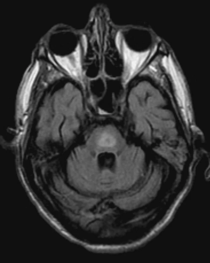
Long description
A single MRI FLAIR image whose own file description reads 'Central pontine myelinolysis, MRI FLAIR'.

- Osmotic demyelination syndrome (central pontine myelinolysis). Classically follows too-rapid correction. More common in patients with alcohol use disorder, undernutrition or other chronic debilitating illness. It may produce flaccid paralysis, dysarthria, dysphagia, and locked-in syndrome, and damage is often permanent. [1]
- Seizures, cerebral oedema, herniation and death in acute severe. [1]
- Longest survival problem: recurrence, especially in uncorrectable SIADH (malignancy). [2]
- Treatment-related: aquaretic-induced (vaptan) overly rapid correction; conivaptan or tolvaptan toxicity.
Outcome depends chiefly on the underlying cause (correctable SIADH vs metastatic cancer, heart failure, cirrhosis) and on avoiding overcorrection, which is the one iatrogenic complication that does permanent harm. Chronic, mildly low sodium in older or inpatient populations is associated with more symptoms (gait, cognition, falls) than a single number implies. [1][3]
- Older adults are particularly prone to thiazide-induced hyponatraemia, sometimes severe and rapid within weeks of starting a thiazide. [1]
- Premenopausal women are at higher risk of severe cerebral oedema with acute hyponatraemia, possibly because sex hormones reduce solute extrusion from brain cells. [1]
- Children are outside the scope of most of these sources; adapt ranges and drugs by weight.
- Pregnancy and breastfeeding are not covered by the sources used here and are left to specialist guidance.
- Work from the bedside: volume status (postural pulse, skin turgor, oedema, JVP) plus basic chemistry (sodium, glucose, urea, creatinine) is usually enough to classify.
- Urine sodium is still the cheapest discriminating test available in most district labs; urine osmolality and serum osmolality are helpful where available. Where they are unavailable, history plus urine dipstick and fractional sodium estimation help.
- Fluid restriction costs nothing and is the backbone for euvolaemic cases; use 500 mL per day initially and titrate. [2][3]
- Avoid rapid correction even where monitoring is sparse: prefer modest, evenly spaced interventions, with sodium checked at least daily (twice daily if low and severe).
- 3% saline may be unavailable. The same sodium can be delivered by careful dilution of 10% saline in 0.9% saline, made up under senior supervision with frequent rechecking; the same 4 to 6 mmol/L target rise applies.
- Treat the cause first: oral rehydration, antiemetics, stopping the offending thiazide, treating infection, and managing heart failure or cirrhosis.
- Refer recurring, severe or treatment-resistant cases, and any patient with coma, seizures or acute overcorrection, to a centre with nephrology and intensive care.
- Hyponatraemia is a serum sodium below 135 mmol/L, most often from an excess of water rather than a lack of sodium.
- Pseudohyponatraemia and translocational (hyperglycaemic) hyponatraemia must be excluded before treating: sodium falls about 1.6 mmol/L per 100 mg/dL rise in glucose above normal. [1]
- Real (hypotonic) hyponatraemia has serum osmolality under 275 mmol/kg; classify it as hypovolaemic, euvolaemic or hypervolaemic from the bedside.
- SIADH requires hypotonic hyponatraemia with inappropriately high urine osmolality (above 100, typically 120 to 150 mmol/L), urine sodium usually above 30 mmol/L, euvolaemia and normal adrenal, thyroid, renal, hepatic and cardiac function. [2]
- Causes of SIADH: cancer (especially small-cell lung cancer), lung disease, brain and meningeal disease, drugs (analgesics, SSRIs, antiseizure, antipsychotic, cytotoxic agents), pain, surgery, stress.
- Symptoms follow falling effective plasma osmolality: nausea first, then headache and confusion, with seizures, coma and death when sodium is below 115 mmol/L (especially with rapid onset).
- Correction ceiling: no more than 8 mmol/L rise in the first 24 hours; 0.5 mmol/L per hour normally, up to 2 mmol/L per hour for the first 2 to 3 hours in severe neurologic cases; give 100 mL of 3% saline over 15 minutes (repeated once) for abrupt neurologic symptoms, or no more than 100 mL per hour over 4 to 6 hours otherwise. [1]
- Use desmopressin 1 to 2 mcg every 8 hours with hypertonic saline to prevent unpredictable diuresis; if overcorrection exceeds 14 mmol/L in 8 hours, stop hypertonic fluids and re-lower the sodium.
- Chronic: fluid restriction, oral salt (limited by hypertension and heart failure), oral urea (effective and safe), loop diuretics, and vaptans (conivaptan IV up to 4 days, not eGFR under 30 or anuria; tolvaptan oral, 30 days maximum, not liver or kidney disease), with close monitoring.
- Low solute intake ("beer potomania", "tea and toast") and primary polydipsia are recognised causes.
- Merck and current conservative sources use an 8 mmol/L 24-hour ceiling; the 2013 US expert panel and the 2014 European guideline set 10 to 12 mmol/L in the first 24 hours for low-risk patients. Both families agree on avoiding overcorrection, and they differ in emphasis on 3% saline bolus size and on second-line drugs.
Answer each one in your head first, then open the answer.
A 70-year-old man had a serum sodium of 124 mmol/L. His serum osmolality is 288 mOsm/kg, urine osmolality 40 mmol/kg and spot urine sodium 8 mmol/L. He is clinically dry. What is the most likely explanation and the urine finding?
Show answerHide answer
Pseudohyponatraemia or a labelling error is more likely than true hypotonic hyponatraemia, because serum osmolality is normal (275 to 295 mOsm/kg). True hyponatraemia with dilute urine suggests primary polydipsia or low solute intake, in which urine should be maximally dilute. Given the dry volume status, hypovolaemia with an extrarenal loss would give urine sodium under 10 mmol/L, but serum osmolality would be low. Recheck the sample. [1][6]
A 60-year-old woman has euvolaemic hypotonic hyponatraemia (serum sodium 128 mmol/L). Urine sodium is 55 mmol/L and urine osmolality 480 mOsm/kg; thyroid and cortisol are normal. Name the diagnosis and its three common pulmonary causes.
Show answerHide answer
Syndrome of inappropriate ADH secretion (SIADH). Common pulmonary causes include pneumonia, lung abscess and tuberculosis (and aspergillosis). The brain and cancer categories (especially small cell lung cancer) and drugs should also be considered, with a chest radiograph.
How much does serum sodium rise with an acute hyperglycaemia of 450 mg/dL above a normal fasting glucose?
Show answerHide answer
About 7.2 mmol/L of apparent (translocational) hyponatraemia: 1.6 mmol/L per 100 mg/dL (450 / 100 = 4.5; 4.5 x 1.6 = 7.2). The serum sodium will rise as the glucose is treated; no specific sodium therapy is usually needed.
A patient with euvolaemic hyponatraemia has been restricting fluids to 500 mL per day for four days, yet serum sodium remains 126 mmol/L. What are the two treatment options the European 2024 standard lists as the most effective second-line agents?
Show answerHide answer
Urea (oral) and tolvaptan. Urea is also described as very effective and safe. Fluid restriction fails in roughly half of SIADH patients.
A postmenopausal patient with chemotherapy-induced SIADH has a serum sodium of 118 mmol/L and is lethargic with one seizure. Outline the safe first 24 hours.
Show answerHide answer
Give no more than 100 mL per hour of 3% saline over 4 to 6 hours (or one 100 mL bolus of 3% saline over 15 minutes, repeated once if symptoms persist) aiming to raise serum sodium by 4 to 6 mmol/L initially, then at no more than 0.5 mmol/L per hour, with a 24-hour ceiling of 8 mmol/L. Add desmopressin 1 to 2 mcg every 8 hours to prevent an unpredictable water diuresis. Stop the hypertonic infusion and re-lower the sodium if the rise exceeds 14 mmol/L over 8 hours.
Which patients are at highest risk of permanent neurologic damage from overcorrection, and what is the speed threshold quoted as dangerous?
Show answerHide answer
Patients with alcohol use disorder, undernutrition or another chronic debilitating illness. A rise of more than 14 mmol/L in 8 hours is quoted as the speed at which osmotic demyelination may follow; the 24-hour ceiling is 8 mmol/L.
Name the conditions that make tolvaptan an unsafe choice.
Show answerHide answer
Liver disease, advanced kidney disease, hypovolaemia and treatment beyond 30 days (because of liver toxicity). Both vaptans are also CYP3A inhibitors with many drug interactions, require close monitoring and are not for hypovolaemic hyponatraemia.
A patient with subarachnoid haemorrhage is hyponatraemic. How does the response to isotonic saline help to distinguish SIADH from cerebral salt wasting?
Show answerHide answer
Cerebral salt wasting (rare, renal salt loss after brain pathology) is expected to resolve with isotonic saline; SIADH is not, and may worsen with hypotonic fluids. Cerebral salt wasting is also suggested by a fractional sodium excretion pattern and a urine sodium above 40 mmol/L, whereas SIADH usually gives a urine sodium above 30 mmol/L.
Related topics (plain text, linked once the discipline is complete): SIADH, cerebral salt wasting, primary adrenal insufficiency (Addison disease), hypothyroidism, hyperglycaemia, acute kidney injury, chronic kidney disease, heart failure, cirrhosis, nephrotic syndrome, acid-base disorders, hyponatraemia/hypernatraemia corrections.
- Lewis JL III. Hyponatremia. Merck Manual Professional Version, full review June 2025, last updated December 2025. Accessed .
- Lewis JL III. Syndrome of inappropriate ADH secretion (SIADH). Merck Manual Professional Version, full review June 2025. Accessed .
- Spasovski G. Hyponatraemia-treatment standard 2024. Nephrology Dialysis Transplantation 2024;39(10):1583-1592. doi:10.1093/ndt/gfae162. Accessed .
- Spasovski G, Vanholder R, Allolio B, et al. Clinical practice guideline on diagnosis and treatment of hyponatraemia. European Journal of Endocrinology 2014;170(3):G1-G47. doi:10.1530/EJE-13-1020. Accessed .
- Verbalis JG, Goldsmith SR, Greenberg A, et al. Diagnosis, evaluation, and treatment of hyponatremia: expert panel recommendations. American Journal of Medicine 2013;126(10 Suppl 1):S1-S42. doi:10.1016/j.amjmed.2013.07.006. Accessed .
- UCSF Health (A.D.A.M.). Osmolality blood test (normal range 275 to 295 mOsm/kg). Medical test reference, reviewed July 13, 2025. Accessed .
Figure credits
Educational content for medical students and doctors. It is not a substitute for clinical judgement or local guidelines.