Nephrolithiasis
How kidney stones form, present and are treated: types, diagnosis, pain relief, drainage, surgery, and prevention.
- A kidney stone is a solid crystal mass that forms when the urine becomes supersaturated with salts and minerals; the most common stones are calcium-based.
- It usually announces itself with sudden, severe renal colic: waves of pain from the loin to the groin, often with nausea, restlessness and sometimes blood in the urine.
- Most small stones pass on their own, but fever or rigors with obstruction is a urological emergency because of infected urine behind the stone.
- Non-contrast CT is the most accurate way to confirm and localise a stone; ultrasound is the safer first test in pregnancy and in children. [2]
- Prevention is the real treatment: enough fluid to produce more than 2.5 L of urine daily, normal calcium intake, limited salt, and targeted drug therapy guided by stone and urine analysis.
By the end of this page you should be able to:
- Explain why kidney stones form, and name the five common stone types with their frequency, urine pH, X-ray appearance and treatment.
- Take a history and examination that separates renal colic from pyelonephritis, a leak of an abdominal aortic aneurysm and other causes of loin pain.
- Choose and interpret the imaging and laboratory tests for an acute stone, including the special considerations for pregnancy and children.
- Manage acute renal colic, including drug treatment, medical expulsive therapy, urgent drainage and the main definitive treatments.
- Plan prevention: lifestyle and diet, metabolic evaluation, and the drugs used for each stone type.
- Discuss the recommendations of the EAU and AUA guidelines and their application in a low-resource setting.
Nephrolithiasis means stones located within the kidney itself (renal calyces and pelvis) or passing through the ureter. The wider term urolithiasis adds stones of the bladder and prostate. This page deals mainly with renal and ureteric stones. Stone disease is common, often recurrent, and usually manageable, but an obstructed infected kidney is an emergency. Stone care has two jobs: getting the present stone out safely, and stopping the next one forming. This is a large subject and this page is written to be read once for understanding and returned to as a reference. It is educational material and not a substitute for clinical judgement or local guidelines.
DefinitionNephrolithiasis
The formation of solid crystalline aggregates, called renal calculi or kidney stones, anywhere in the renal collecting system or ureter. A "staghorn" stone is one that branches through the renal pelvis and calyces and can fill much of the collecting system.
Stones are classified first by composition, because composition decides everything else: which pH favours them, whether they show on a plain X-ray, how likely they are to recur, and how to prevent them. They can also be classified by size (the guideline categories are under 5 mm, 5 to 10 mm, 10 to 20 mm and over 20 mm), by location (calyx, pelvis, ureter) and by aetiology (infectious, metabolic, genetic or drug-related).[2]
Kidney stone disease is the most common chronic disease of the urinary tract. A recent narrative review gives a global prevalence of 7 to 13%, and notes that the figure has risen over the last three decades in parallel with obesity, diabetes, diet change and warming climates.[1] Prevalence exceeds 10% in some economically developed countries; tropical and subtropical regions report 5 to 10% compared with 1 to 5% in cooler zones, although detection of asymptomatic stones complicates the comparison.[1] Incidence peaks between the fourth and sixth decades of life; the highest rates in series are in men aged 20 to 49 years, but the sex gap is narrowing as stones become more common in women.[1] High ambient temperature, low fluid intake, a sedentary job and obesity all increase risk.[1][2]
Recurrence is the rule rather than the exception. A review of first-time stone formers calculates a five-year recurrence of about 26%,[2] while broader cohorts give 30 to 50% within 5 to 10 years and 75% within 20 years.[1] About half of people who recur have a single further episode in life; a little over 10% have highly recurrent disease.[2] The European Association of Urology (EAU) Urolithiasis Guidelines note a possible link between stone disease and chronic kidney disease, and flag stones in a solitary kidney, infection stones, brushite, uric acid and cystine as carrying particular renal risk.[2]
Clinical pearlStones as a public health problem
Because nephrolithiasis is most common in working-age adults, a single stone can mean time off work, repeated attendance and, in countries that charge, significant bills. Prevention is cheap and permanent; surgery is neither.
The EAU classifies stones into four broad groups by aetiology: non-infection stones (calcium oxalate, calcium phosphate, uric acid, ammonium urate), infection stones (magnesium ammonium phosphate, that is struvite, and carbonate apatite), genetically determined stones (cystine, xanthine, 2,8-dihydroxyadenine) and drug stones.[2] Most stones have a mixed origin and more than one factor contributes.
Risk factors, from the literature and the guidelines, include:
- Diet and fluid. Low fluid intake is the single commonest and most modifiable factor. High salt intake raises urinary calcium and lowers citrate; high animal protein lowers urine pH and raises uric acid and oxalate; excessive oxalate intake adds to calcium oxalate risk; over-restriction of dietary calcium paradoxically increases oxalate absorption because calcium no longer binds oxalate in the gut.[1][2]
- Metabolic and medical. Hypercalciuria, hyperoxaluria, hyperuricosuria, hypocitraturia, gout, primary hyperparathyroidism, distal renal tubular acidosis, medullary sponge kidney, inflammatory bowel disease or bariatric surgery, exocrine pancreatic insufficiency.
- Infection. Urease-producing organisms, which make alkaline urine, predispose to struvite stones.
- Genetic. Cystinuria, primary hyperoxaluria, Dent disease, renal tubular acidosis and monogenic stones occur in a small minority of adults but in a substantial minority of children and young adults.
- Anatomical. Medullary sponge kidney, pelvi-ureteric junction obstruction, horseshoe kidney, calyceal diverticulum, strictures.
- Environmental. Heat, dehydration, geography and climate, and some occupations. Monogenic disease accounts for 1 to 11% of adults and 12 to 21% of children and young adults with stone disease.[2]
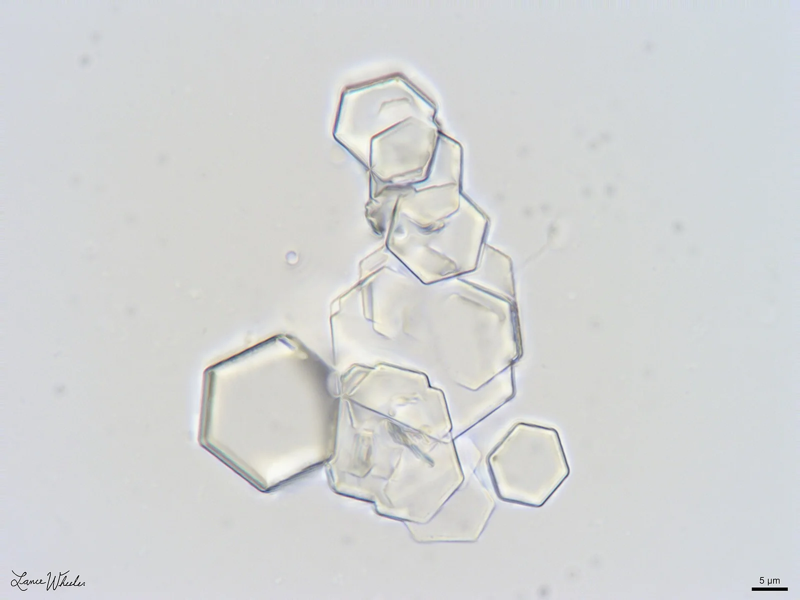
Figure 1Cystine Crystals in Canine Urine Sediment
Long description
Microscopic image of cystine crystals in the urine sediment of a male dog at 1000x magnification with a 5 um scale bar, focus-stacked from 12 exposures.

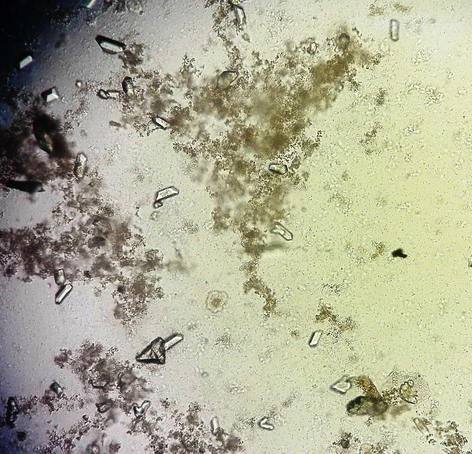
Figure 2Кристаллы трипельфосфата в форме гробовых крышек и призм на фоне аморфных фосфатов. Осадок мочи. Нативный препарат. x400
Long description
Urine microscopy showing struvite (triple phosphate) crystals with their coffin-lid shape among amorphous debris.

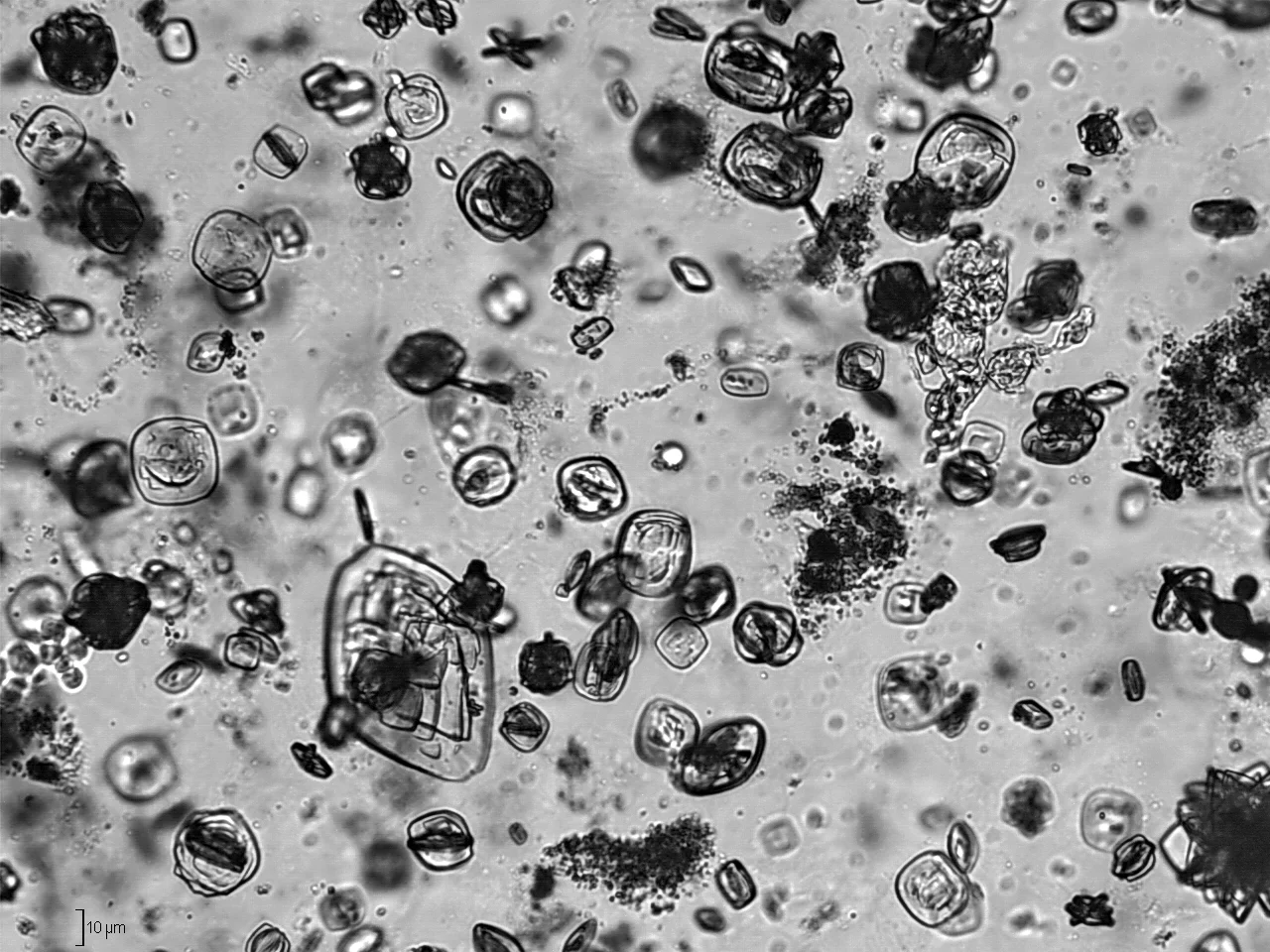
Figure 3Uric acid crystals (urine) - Ürik asit kristalleri (idrar) - 01
Long description
Abundant uric acid crystals in a human urine sample with a pH of 5, as detected by an automated urinalysis system, with some amorphous urate crystal clumps.

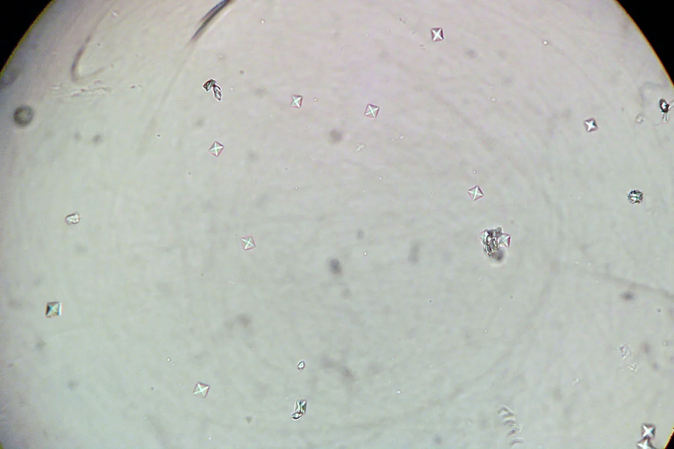
Figure 4OxalicAcidDihidrate
Long description
Urine crystals of oxalic acid in dihydrate form whose description identifies the characteristic mail-envelope appearance with two bright lines crossing in the center, distinguished from struvite crystals.

The table below gives the common types, their frequency, the urine pH that favours them, their appearance on a plain X-ray (kidney-ureter-bladder, KUB) film and the broad direction of prevention and treatment. Frequencies are approximate and vary by region and hospital series.[1][2][3]
| Stone type | Approximate frequency | Urine pH and setting | Plain X-ray appearance | Prevention and treatment notes |
|---|---|---|---|---|
| Calcium oxalate (mono- and dihydrate) | Commonest single type; makes up most of the 70 to 80% of stones that are calcium-based[1] | Forms across a wide pH range; a persistently acidic urine (under 5.5) favours co-crystallisation with uric acid | Radiopaque | High fluid intake; normal dietary calcium; low salt; restriction of oxalate-rich foods if hyperoxaluria; thiazide for hypercalciuria; alkaline citrate for hypocitraturia [2] |
| Calcium phosphate (hydroxyapatite, brushite) | A minority of calcium stones | Alkaline urine (crystallises above pH 6.5 to 6.8); associated with infection, hyperparathyroidism or renal tubular acidosis | Radiopaque | Treat the underlying cause (parathyroid, acidosis, infection); thiazide for hypercalciuria over 8 mmol per 24 hours in the EAU scheme [2] |
| Struvite (magnesium ammonium phosphate) | 2 to 15% of stones analysed[2] | Alkaline urine above pH 7.2, produced by urease-splitting bacteria such as Proteus mirabilis | Poor radiopacity; often forms a staghorn in the collecting system | Complete surgical removal; antibiotics; where indicated, urine acidification with ammonium chloride or methionine |
| Uric acid | About 10% of renal stones[2] | Acidic urine, classically under 5.5; linked to gout, high purine intake, insulin resistance | Radiolucent (invisible on plain film) | Alkalinise to pH 7.0 to 7.2; allopurinol if hyperuricosuric; reduce purines; increased fluids |
| Cystine | 1 to 2% in adults, 6 to 8% in children[3] | Limited solubility at physiological pH; solubility roughly doubles by pH 7.5 | Poor radiopacity (moderate density, about 400 to 700 Hounsfield units on CT) | High fluid intake (over 3 L in adults); potassium citrate to pH 7.5 to 8.0; tiopronin (thiol drug) if needed; surgery if stones form [2] |
| Ammonium urate | Under 1% (rare) | Alkaline urine above pH 6.5 with ammonium; linked to infection, malnutrition or laxative abuse | Radiolucent | Treat infection and malnutrition; address the cause [2] |
Stone formation is usually explained by three simultaneous factors: supersaturation of the urine with stone-forming salts, a deficiency of the small molecules and proteins that inhibit crystallisation (citrate, magnesium, glycosaminoglycans, osteopontin), and a surface on which crystals can anchor and grow. Supersaturation is driven by low urine volume, high excretion of the offending substance and, for salts whose solubility depends on pH, the wrong urine pH. The change in urine pH through the day is why a single 24-hour urine collection is a snapshot, not a verdict. Calcium oxalate, the very common stone, can anchor on Randall's plaques, which are tiny calcium phosphate deposits in the renal papilla that are present in many stone formers and in some people without stones. Cystine precipitates because the dibasic amino acid transporter fails in cystinuria and the urine becomes oversaturated. Uric acid comes out of solution in acid urine because at low pH uric acid is protonated and poorly soluble. Struvite grows quickly in alkaline infected urine because urease breaks urea into ammonia and carbon dioxide, raising pH and providing ammonium. Prevention works because it reverses these steps: more water dilutes, citrate binds calcium and alkalinises, thiazides cut urinary calcium and allopurinol cuts uric acid.[1][2]
Stones often cause no symptoms while they sit in the kidney. They declare themselves in four main ways.
- Renal colic. Sudden, severe, colicky pain starting in the loin and radiating to the groin, testis or labium. It comes in waves and is often described as the worst pain the patient has known. The patient is restless and cannot get comfortable; they pace and shift, which is the opposite of peritonitis, where movement hurts. Nausea and vomiting are common and follow the intensity of the pain rather than preceding it. Haematuria, visible or found on dipstick, is present in most but not all cases.[2][1]
- Lower urinary tract symptoms. Frequency, urgency, dysuria or "gravel" passed once the stone nears the bladder.
- Asymptomatic stones. Found incidentally on imaging done for another reason.
- Infection. Fever, rigors or feeling systemically unwell with an obstructed collecting system is an emergency, because pus accumulates behind the stone.
- Atypical presentations. Older or immunosuppressed patients may present only with fever or confusion and no classic pain.
Examination is often normal. Look for:
- Tenderness over the loin and costovertebral angle; the abdomen may be uncomfortable but there is usually no peritoneal guarding.
- Signs of sepsis: fever, tachycardia, hypotension.
- A distended or palpable kidney if obstruction is high grade.
- A pulsatile abdominal mass or bruit in an older smoker, in whom a leak of an abdominal aortic aneurysm must be excluded first.
Red flagObstructed and infected kidney
Fever or rigors in a patient with a known or suspected stone, or with classic renal colic, is a urological emergency. Drainage must not wait for a definitive scan if the diagnosis is strongly suggested. Delay risks septicaemia and death.
The common confusion is between renal colic and the following:
- Pyelonephritis. Fever, rigors, malaise and a tender kidney with usually a dull, steady ache rather than waves of colic; restlessness is absent. Urine culture is positive.
- Musculoskeletal back pain. Localised, worsened by movement and not radiating to the groin; no haematuria.
- Abdominal aortic aneurysm. Constant pain, often in an older smoker, possibly with a pulsatile mass; hypotension is an emergency.
- Other abdominal emergencies. Appendicitis, diverticulitis, ectopic pregnancy, ovarian torsion or renal infarction can all mimic colic.
- Renal tumour or cyst. Usually painless haematuria without colic.
A small minority of patients have a second condition found while investigating the loin pain; an alternative diagnosis should be sought when the picture does not fit.
The EAU recommends a succinct work-up for every patient with suspected urolithiasis, regardless of risk:[2]
- Urine dipstick for red cells, white cells, nitrites and approximate pH, plus urine microscopy and culture.
- Blood: creatinine, uric acid, (ionised) calcium, sodium, potassium, full blood count and C-reactive protein.
- If intervention is likely, partial thromboplastin time and international normalised ratio.
A passed stone, if it can be retrieved by filtering the urine, should be sent for analysis by infrared spectroscopy or X-ray diffraction rather than wet chemistry.[2]
Figure 5Non-contrast CT of multiple bilateral renal calculi
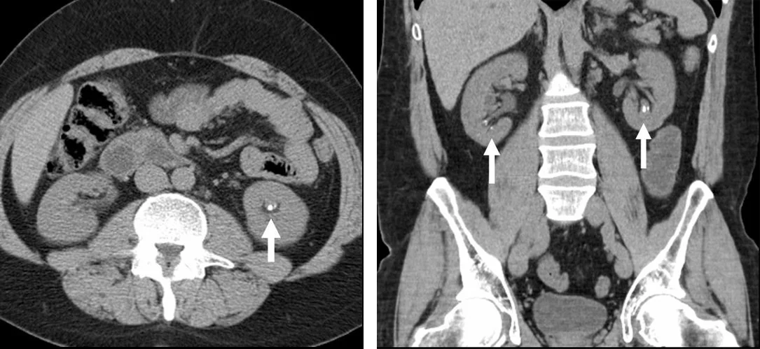
Long description
Non-contrast CT of the abdomen/pelvis demonstrating multiple bilateral renal calculi, presented as an axial view on the left and a coronal reformat on the right (the corresponding Wikipedia figure caption marks the calculi with arrows).

- Adults. The EAU recommends ultrasound as the first-line imaging step, followed by non-contrast CT to confirm the diagnosis, locate the stone and plan treatment. Non-contrast CT is the more accurate test: it detects radiolucent stones (such as uric acid) as well as radiopaque ones, measures stone density and skin-to-stone distance, and guides the choice between shock wave lithotripsy, ureteroscopy and percutaneous surgery.[2] A plain KUB radiograph is useful to separate radiolucent from radiopaque stones and for later comparison during follow-up. A contrast study is added only when the collecting system anatomy must be defined for planned surgery.[2]
- Low-dose CT protocols reduce radiation exposure and have a pooled sensitivity of 93.1% and a specificity of 96.6% for urolithiasis.[2]
- Pregnancy. Ultrasound first; magnetic resonance imaging second; low-dose CT as a last line after justification. In pregnancy the goal is to avoid irradiating the fetus while not missing an obstructed infected kidney.[2]
- Children. Ultrasound first (it needs no anaesthesia and uses no radiation); add a KUB radiograph or low-dose non-contrast CT if ultrasound does not answer the question. Children with stones are usually high-risk patients and need a full metabolic evaluation.[2]
Non-steroidal anti-inflammatory drugs (NSAIDs) are the first drug of choice. They are more effective than opioids for stone colic and patients need less additional analgesia. Opioids, especially pethidine, cause more vomiting and are reserved as second line along with ketamine or ketorolac in the acute setting. Adding an antispasmodic to an NSAID does not improve pain control. NSAIDs can affect renal function in patients with already reduced renal function, and long-term oral diclofenac and ibuprofen increase cardiovascular risk; for the short duration of acute colic these are usually acceptable, but kidney disease, dehydration and cardiovascular disease should be considered beforehand and dosed according to local guidance. If pain cannot be controlled medically, drainage or stone removal is indicated.[2]
Encourage oral fluid intake if the patient is not vomiting. If vomiting or dehydration is present, give intravenous isotonic saline while monitoring for volume overload, especially in older adults or anyone with heart or kidney disease. Fluids are supportive care, not the cure: they will not flush a stone through an obstruction and should never delay analgesia or imaging.
For ureteric stones that are suitable for conservative management, the EAU recommends offering an alpha-blocker for distal ureteric stones of 5 to 10 mm. Tamsulosin is the most familiar agent; silodosin appears more effective than tamsulosin in pooled analyses. These drugs are used off-label for this purpose, relax ureteric smooth muscle, and should be given with analgesia and increased fluids for up to several weeks with planned re-imaging.[2] The greatest benefit appears to be in stones above 5 mm. Combination therapy (for example an alpha-blocker with a phosphodiesterase-5 inhibitor) has been studied but the evidence is mixed.[2]
Drainage is indicated for:
- Infected obstruction: fever or rigors with an obstructing stone. Use a percutaneous nephrostomy or a ureteric stent; both are similarly effective, and nephrostomy may be preferred when retrograde access is difficult. Give antibiotics immediately after sending blood and urine cultures, and delay definitive stone removal until the infection has cleared.[2]
- Analgesia-refractory colic.
- Anuria or bilateral obstruction, or obstruction in a solitary kidney.
- Stones with significant comorbidity where pain cannot be safely controlled at home.
Definitive stone treatment should wait until sepsis has resolved.[2]
The choice among shock wave lithotripsy (SWL), ureteroscopy (URS) and percutaneous nephrolithotomy (PCNL) depends on stone size, number, position, density and composition, on the patient's body habitus and on local expertise.[2]
- Observation is appropriate for many small ureteric stones that are likely to pass, with analgesia, hydration and expulsive therapy where suitable. A plain film or ultrasound can confirm passage.
- Extracorporeal shock wave lithotripsy (SWL) is non-invasive and effective for many renal and ureteric stones. Success depends on stone hardness, position and size, skin-to-stone distance and technique. Good practice includes adequate analgesia, correct coupling (air bubbles in the coupling gel deflect up to 99% of the shock wave) and a frequency of 1.0 to 1.5 Hz. Routine stenting before SWL does not improve stone-free rates. Routine antibiotic prophylaxis is not needed when the urine is sterile. Complications include Steinstrasse in about 4%, macroscopic haematuria in 17.2%, pain in 12.1%, an auxiliary procedure in 6.9% and sepsis in about 0.15%.[2]
- Ureteroscopy (rigid or flexible) is the standard for many ureteric stones and for renal stones, with a modern tip diameter under 8 French. Digital scopes and flexible instruments offer good stone-free rates and are the treatment of choice for many stones that are not suitable for SWL.
- Percutaneous nephrolithotomy (PCNL) is used for larger, harder or more complex stones, including staghorn calculi, and for stones not amenable to retrograde treatment.
- Chemolysis (alkalinisation) can dissolve uric acid stones at a urine pH of about 7.0 to 7.2, either alone or combined with tamsulosin; struvite may be dissolved by irrigation with hemiacidrin in selected cases. Percutaneous chemolysis is rarely used today.[2]
- Open or laparoscopic stone surgery is reserved for rare situations where none of the above is possible or safe.
Acute flank pain with suspected renal colic
- History and examinationColicky loin-to-groin pain, restlessness, nausea, haematuria; fever is a red flag
- Clinical red flagsFever or rigors, solitary kidney, anuria, vomiting, pregnancy, older age with vascular risk
- Question: Any red flag or diagnostic uncertainty?
Yes
- Urgent imaging and blood testsUltrasound first; add non-contrast CT as needed; creatinine, CRP, urine culture
- If infected obstruction is suspectedAntibiotics and decompression (stent or nephrostomy); delay stone removal
No
- AnalgesiaNSAID first line if no contraindication; opioid, ketamine or ketorolac as second line
- Confirm the stoneUltrasound first, then non-contrast CT to confirm and localise
- Small distal stoneObserve with analgesia and increased fluids; consider an alpha-blocker for distal ureteric stones of 5 to 10 mm
- Persistent pain, infection, obstruction or failure to passDrain or remove the stone: SWL, ureteroscopy or percutaneous nephrolithotomy
The EAU gives a single set of general measures that apply to every stone former, regardless of stone type:[2]
- Fluids: 2.5 to 3.0 L per day, aiming for a urine volume above 2.5 L per day; water is the preferred drink. More is needed in hot weather, heavy exercise or gastrointestinal losses.
- Salt (sodium chloride): 4 to 5 g per day or less.
- Dietary calcium: 1.0 to 1.2 g per day, taken with meals rather than between them, so it binds oxalate in the gut.
- Animal protein: 0.8 to 1.0 g per kilogram per day.
- Fruit and vegetables: encouraged for their fibre and water content; excessive oxalate-rich foods should be limited when hyperoxaluria is present.
- Lifestyle: normal body mass index, adequate physical activity, control of diabetes and metabolic syndrome, limitation of sugar-sweetened drinks and alcohol.
Every stone former should have a basic work-up. High-risk patients, that is those with recurrence, stones in childhood or young adulthood, a family history, solitary kidney, brushite or cystine or infection stones, renal tubular acidosis, hyperparathyroidism, inflammatory bowel disease or known metabolic abnormality, should have a full evaluation: two consecutive 24-hour urine collections for volume, pH, calcium, oxalate, uric acid, citrate, sodium and magnesium, taken about three months after the acute episode and after the patient is stable, uninfected and on their usual diet. Stone analysis by infrared spectroscopy or X-ray diffraction is the anchor that turns generic advice into specific treatment.[2]
Drug treatment follows the stone type and the urine abnormality, not a universal recipe:
- Thiazide (hydrochlorothiazide 25 to 50 mg daily) for hypercalciuria in calcium oxalate and calcium phosphate stones. Warn patients taking hydrochlorothiazide of the increased risk of non-melanoma skin cancer and to have their skin checked regularly.[2]
- Alkaline citrate (potassium citrate, 3.25 to 9.75 g per day, divided into doses) for hypocitraturia, calcium oxalate, uric acid and cystine stones, alkalinising the urine. Dose is titrated to urine pH; too high a pH risks calcium phosphate formation. For cystinuria the target is usually pH 7.5 to 8.0. [2][3]
- Allopurinol (100 to 300 mg daily) for hyperuricosuria, including calcium oxalate stones driven by hyperuricosuria; febuxostat is second line. [2]
- Adjuncts for calcium oxalate stones: magnesium supplements for hypomagnesuria, pyridoxine for primary hyperoxaluria, calcium with meals for enteric hyperoxaluria.
- Uric acid stones: potassium citrate to pH 7.0 to 7.2, with allopurinol when hyperuricosuric.
- Cystine stones: hyperhydration (urine volume over 3 L daily), potassium citrate to pH 7.5 to 8.0, sodium restriction under 100 mmol daily, animal protein restriction, with tiopronin (800 to 2000 mg daily) or D-penicillamine as thiol drugs if conservative measures are insufficient.[3]
- Struvite (infection) stones: complete removal where possible, appropriate antibiotics for persistent bacteriuria, and urine acidification with ammonium chloride 1 g two or three times daily or methionine 200 to 500 mg one to three times daily. Urease inhibitors such as acetohydroxamic acid exist but are not available in all regions.[2]
- Primary hyperoxaluria and enteric hyperoxaluria are managed according to their own pathways (fluid intake of 3.5 to 4 L daily, pyridoxine, lumasiran for genetic type 1, calcium with meals and lower fat and oxalate diet respectively).[2]
The EAU Guidelines on Urolithiasis (2026) are organised as classification, diagnosis, treatment, and metabolic evaluation and recurrence prevention. They recommend ultrasound as the initial imaging step with non-contrast CT to confirm; NSAIDs as first-line analgesia; alpha-blockers (off-label) for distal ureteric stones of 5 to 10 mm; urgent decompression for infected obstruction or anuria; fluid intake sufficient to produce above 2.5 L of urine daily; salt 4 to 5 g daily; calcium 1.0 to 1.2 g daily; and the drug therapies above for high-risk stone formers. They stress stone analysis and individualised risk assessment before pharmacological prevention.[2]
The AUA Medical Management of Kidney Stones Guideline (2026), published in two parts in The Journal of Urology, updates the evaluation and dietary management of stone formers in Part I and treatment and follow-up in Part II, including medical rather than surgical management. It is directed at personalised long-term stone prevention, with emphasis on stone and urine analysis, diet and 24-hour urine testing in recurrent and high-risk formers.[4] It and the EAU guideline are the two most commonly referenced guidelines for kidney stone management; local practice and the patient's preferences should guide the final choice.
- Pregnancy. Ultrasound first, then MRI, then low-dose CT as a last resort; avoid NSAIDs late in pregnancy; involve obstetrics, urology and anaesthetics. Most stones are observed if uncomplicated. If infection and obstruction are present, decompress promptly.[2]
- Children. They are almost always high-risk patients, so a full metabolic evaluation and stone analysis matter more than in adults. Ultrasound is the preferred imaging modality. Monogenic causes are much more common than in adults. Conservative surgery (ureteroscopy, miniaturised percutaneous approaches) and careful radiation sparing are priorities.[2][3]
- Older adults. Consider abdominal aortic aneurysm in the differential; use NSAIDs cautiously with chronic kidney disease and cardiovascular disease; hydrate carefully.
- Chronic kidney disease and solitary kidney. Higher risk of CKD and stones; NSAIDs may worsen function; stones and their treatment must be individually planned.
When advanced tests or drugs are unavailable:
- Diagnosis: ultrasound plus a plain KUB radiograph will pick up most stones if CT is unavailable; if the clinical picture is classic and ultrasound shows hydronephrosis, treat the colic while arranging transfer rather than chasing the exact localisation.
- Analgesia: oral or injectable NSAIDs are usually the cheapest effective option; opioids are second line. Avoid repeated NSAID dosing if renal function is impaired.
- Drainage: if no urology service exists, transfer promptly for infected obstruction. A percutaneous nephrostomy can sometimes be placed under ultrasound at a district level.
- Prevention: hydration, low salt, normal dietary calcium and moderate animal protein cost little and work. Thiazides, allopurinol and potassium citrate are inexpensive where available; if stone analysis is impossible, urine pH and dipstick findings can guide pH-directed therapy. Heat means patients need to drink more than textbook volumes suggest.
- Obstruction and hydronephrosis, with flank pain, infection and impaired renal function.
- Urosepsis from infected obstructed urine, the most dangerous complication.
- Renal impairment in recurrent or high-burden stone disease, particularly struvite, cystine, xanthine, 2,8-dihydroxyadenine and anatomical disease.
- Procedural complications: SWL (Steinstrasse, haematuria, sepsis); URS or PCNL (bleeding, infection, ureteric perforation, stricture).
- Staghorn stones may destroy renal parenchyma silently.
The outlook for an uncomplicated calcium oxalate stone is excellent once the stone is passed or removed and the patient hydrates and reduces salt. The main long-term issue is recurrence, which is substantially reduced by the general measures and, where indicated, targeted drug therapy. Prognosis worsens with struvite and cystine stones, recurrent infection, obstruction and chronic kidney disease; these patients need specialist follow-up.
- Nephrolithiasis is common, often recurrent and usually preventable.
- The stone type decides the urine pH, X-ray appearance, recurrence risk and prevention plan; analyse any retrieved stone.
- Common types: calcium oxalate (most of the 70 to 80% that are calcium-based), calcium phosphate, struvite (2 to 15%), uric acid (about 10%), cystine (1 to 2% of adults, 6 to 8% of children) and rare ammonium urate.
- Renal colic presents with severe waves of loin-to-groin pain, restlessness, nausea and often haematuria; infection with obstruction is an emergency.
- Ultrasound first, then non-contrast CT, is the usual imaging sequence; CT is most accurate and sees radiolucent stones.
- NSAIDs are first-line analgesia; alpha-blockers are used off-label for distal ureteric stones of 5 to 10 mm.
- Treat fever with obstruction urgently: antibiotics plus nephrostomy or stent, delaying definitive stone removal.
- SWL, ureteroscopy and PCNL are chosen by stone size, site, density and patient factors.
- All stone formers need fluids producing above 2.5 L of urine daily, 4 to 5 g of salt or less, normal calcium with meals, moderate animal protein and weight control.
- Targeted therapy: thiazide for hypercalciuria; alkaline citrate for uric acid, cystine and hypocitraturia; allopurinol for hyperuricosuria; antibiotics and acidification for struvite.
- Use EAU and AUA guidance, but adapt to the patient and the resources available.
Answer each one in your head first, then open the answer.
A 35-year-old man has sudden severe left loin pain radiating to the groin, is pacing the room and vomits before the peak of the pain. Dipstick shows blood 2+. What is the most likely diagnosis, and why does he pace rather than lie still?
Show answerHide answer
Renal colic from a left ureteric stone. The waves of colic are produced by ureteric spasm against an obstruction; because movement does not worsen visceral pain the patient cannot find a comfortable position and paces. In peritonitis the pain is parietal and worsens with movement, so the patient lies still. Haematuria supports a calculus but is not diagnostic.
Which stone type is radiolucent and dissolves when the urine is alkalinised to about pH 7.0 to 7.2?
Show answerHide answer
Uric acid. It becomes more soluble as pH rises. Confirming the diagnosis on imaging may need non-contrast CT because plain film can miss it; definitive treatment combines alkalinisation, fluids and sometimes allopurinol for hyperuricosuria.
A 28-year-old woman at 22 weeks of pregnancy has loin pain and fever. What is the appropriate imaging sequence and the immediate management priority?
Show answerHide answer
Ultrasound first, then MRI if needed, and low-dose CT only as a last justified resort. The emergency is an obstructed infected kidney: give analgesia, fluids and antibiotics promptly and decompress (nephrostomy or stent) without waiting for perfect imaging if sepsis is suspected.
A patient with recurrent calcium oxalate stones has low 24-hour urine citrate and high urine calcium. Which drugs would you consider, and what lifestyle advice stands for every stone former?
Show answerHide answer
Consider an alkaline citrate for the hypocitraturia and a thiazide for the hypercalciuria. For everyone: fluids producing more than 2.5 L of urine daily, salt under 4 to 5 g daily, dietary calcium of 1.0 to 1.2 g taken with meals, animal protein of 0.8 to 1.0 g per kilogram daily, and weight control. [2]
A 6-year-old has a cystine stone. What does that imply about stone frequency and management?
Show answerHide answer
Cystine makes up 6 to 8% of paediatric stones. Management is aggressive prevention: urine volume over 3 L (or 40 to 50 mL per kilogram) daily in divided doses including overnight, potassium citrate to pH 7.5 to 8.0, sodium under 100 mmol daily, protein restriction, and a thiol drug such as tiopronin if targets are not met. Stones often need ureteroscopy, especially for stones up to 20 mm, or percutaneous nephrolithotomy for larger ones. [2][3]
Acute kidney injury; chronic kidney disease; haematuria; hypercalcaemia; hyperuricaemia and gout; urinary tract infection; renal tubular acidosis; cystinuria; primary hyperoxaluria; metabolic syndrome; thiazide diuretics; calcium and vitamin D metabolism.
- Leonova, A., Sinisi, R., Alba, S., et al. Personalised prevention and management of kidney stones: a narrative review on the importance of integrating environmental, social and genetic factors. Public Health Reviews 2026;47:1609208.. Accessed .
- European Association of Urology Guidelines Panel on Urolithiasis. EAU Guidelines on Urolithiasis (2026 edition). European Association of Urology, 2026. Online guideline chapters: Guidelines; Metabolic Evaluation and Recurrence Prevention.. Accessed .
- Zambudio Munuera, A., Rodriguez Parras, P., Galan Llopis, J.A., et al. Cystinuria: diagnosis, medical and surgical management, and emerging therapies. A narrative review. Research and Reports in Urology 2026;18:629402.. Accessed .
- Pearle, M.S., Matlaga, B.R., Antonelli, J.A., et al. Medical Management of Kidney Stones: AUA Guideline (2026). The Journal of Urology 2026;216(5):613-631. American Urological Association, 2026 (Part I: evaluation and dietary management; Part II: treatment and follow-up).. Accessed .
Figure credits
- Figure 1: Lance Wheeler, via Wikimedia Commons, CC BY-SA 4.0, CC BY-SA 4.0. resized.
- Figure 2: Vladimir064, via Wikimedia Commons, CC BY 4.0, CC BY 4.0. resized.
- Figure 3: Doruk Salancı, via Wikimedia Commons, CC BY-SA 3.0, CC BY-SA 3.0.
- Figure 4: J3D3, via Wikimedia Commons, CC BY-SA 3.0, CC BY-SA 3.0.
- Figure 5: Kristie Guite, Louis Hinshaw and Fred Lee, via Wikimedia Commons, CC BY 3.0, CC BY 3.0.
Educational content for medical students and doctors. It is not a substitute for clinical judgement or local guidelines.