Polycystic kidney disease
Autosomal dominant and recessive polycystic kidney disease: cyst growth, complications, diagnosis, blood pressure control, tolvaptan, and low-resource care.
- Polycystic kidney disease is a group of inherited disorders in which fluid-filled cysts grow in the kidneys from tubules that should stay microscopic.
- The common, adult form is autosomal dominant polycystic kidney disease (ADPKD), usually from variants in PKD1 or PKD2; it often stays silent until early adulthood.
- The recessive form (ARPKD) presents before or soon after birth, is more severe, and always involves the liver.
- Early control of blood pressure and identifying rapidly progressive disease (Mayo imaging class and PROPKD score) are the two management decisions that change the long-term outcome.
- Tolvaptan is the only drug shown to slow cyst growth in rapidly progressing ADPKD, but it needs liver enzyme monitoring and a high water intake. [1]
By the end of this page you should be able to:
- Contrast ADPKD and ARPKD in inheritance, gene, age of onset, kidney and liver features.
- Explain how loss of polycystin function leads to cysts, hypertension and kidney failure.
- Recognise the renal and extrarenal features of ADPKD and know which are common.
- Use ultrasound criteria by age, alongside family history, genetics and MRI total kidney volume, to diagnose and risk-stratify.
- Outline management: blood pressure targets and first-line drugs, water intake, tolvaptan and its safety, cyst complications, cerebral aneurysm screening, and kidney replacement.
- Describe the main points for genetic counselling and care in low-resource settings.
- Distinguish simple cysts, medullary sponge kidney and tuberous sclerosis from PKD.
DefinitionPolycystic kidney disease
The two principal forms are compared in the table below. "PKD" is sometimes used loosely for either; the distinction matters because prognosis and management differ.
| Feature | ADPKD (adult-type) | ARPKD (infantile/childhood) |
|---|---|---|
| Inheritance | Autosomal dominant; 50% risk to each child of an affected parent | Autosomal recessive; 25% recurrence risk for siblings [1][3] |
| Main gene | PKD1 (chromosome 16) in about 78% of cases; PKD2 (chromosome 4) in about 15%; minor genes in under 7% | PKHD1 (chromosome 6); rare DZIP1L and other variants [1] |
| Usual onset | Cysts from birth, often silent until the second to third decade | Most often detected in utero or at birth; occasionally later childhood |
| Kidneys | Bilateral, variably sized cysts, heavy cystic burden, large kidneys | Bilaterally enlarged, uniformly hyperechoic kidneys with tiny radially arranged cysts; retained reniform outline |
| Blood pressure | Hypertension in 50 to 70% by age 30, often before kidney function falls | Hypertension in up to 80% of children [1] |
| Kidney failure | Median age about 54 years with PKD1 and about 78 years with PKD2; wide family variation | Variable; severe perinatal cases die; survivors can maintain function into adulthood |
| Liver | Liver cysts in over 80% by age 30; severe polycystic liver disease under 5% | Obligatory ductal plate malformation: biliary ectasia and congenital hepatic fibrosis, with portal hypertension |
| Common extrarenal | Intracranial aneurysms, mitral valve prolapse, stones, hernias, diverticular disease | Pulmonary hypoplasia at birth, cholangitis, portal hypertension, combined liver-kidney transplant [1] |
| Specific treatment | Tolvaptan for rapidly progressing disease; ACE inhibitor or ARB for blood pressure | Blood pressure control; manage portal hypertension; combined transplant when needed [1] |
These figures come from the large open reviews used for this page. Most patients who develop kidney cysts in adult life and have a family history turn out to have one of the PKD1 or PKD2 variants; about 10% of clinically diagnosed patients are negative on PKD1 and PKD2 testing.
ADPKD is the most common single-gene kidney disease: it affects up to 12 million people worldwide, with an estimated annual incidence of 2.5 per 100,000. Prevalence estimates based on imaging are around 1 in 2000, while studies using large genetic databases suggest the true figure may be closer to 1 in 1072 because mild cases are easily missed. The disease occurs in all ancestries and both sexes, although in one US series rates of kidney failure were higher in men than women (8.2 versus 6.8 per million people). ADPKD accounts for 5 to 10% of all kidney failure, making it the fourth leading cause after diabetes, hypertension and glomerulonephritis; in the United States it produces about 5% of new dialysis cases each year. Around 98% of men and 77% of women with ADPKD reach kidney failure by age 80, though two-thirds of patients with typical ADPKD are rapid progressors.[1] [2][3]
ARPKD is far rarer: about 1 in 20,000 live births in European ancestry populations, with a carrier frequency of roughly 1 in 70. The exact incidence is uncertain because the most severe cases die perinatally without a diagnosis. Consanguineous populations can have much higher rates. [3]
ADPKD is autosomal dominant with high penetrance but variable expression. Every child of an affected parent has a 50% chance of inheriting the disease; males and females are equally affected. Presentation and speed of decline vary strikingly even within one family: identical twins differ by an average of about 2 years in the age at kidney failure, siblings with the same variant by about 7 years, and relatives overall by about 13 years. This intrafamilial variability reflects genetic background (modifier genes), epigenetic and environmental factors. [1][2]
Pathogenic variants in PKD1 are usually truncating or in critical domains and produce a heavier burden of cysts; these patients fail earlier. Non-truncating PKD1 variants are milder; PKD2 variants are milder still. A number of other genes (IFT140, GANAB, DNAJB11, ALG5, ALG8, ALG9 and others) cause ADPKD-like disease with smaller cystic burden but can still lead to chronic kidney disease or kidney failure later in life.
ARPKD is autosomal recessive. Mutations in PKHD1 are found in about 80% of patients; homozygous truncating mutations usually cause severe or lethal perinatal disease, while at least one missense variant tends to be milder. Monoallelic PKHD1 variants can cause a mild dominant cystic kidney disease, and early ADPKD can mimic ARPKD. DZIP1L and other ciliopathy genes can also phenocopy ARPKD. [3]
Other recognised risk factors are not "modifiable" in the usual sense: a positive family history; being male (slightly faster decline); and, in several studied populations, Black race reaching kidney failure about 1.5 years earlier. Hypertension and obesity are repeatedly associated with faster progression.
Figure 1Polycystic kidneys, gross pathology CDC PHIL
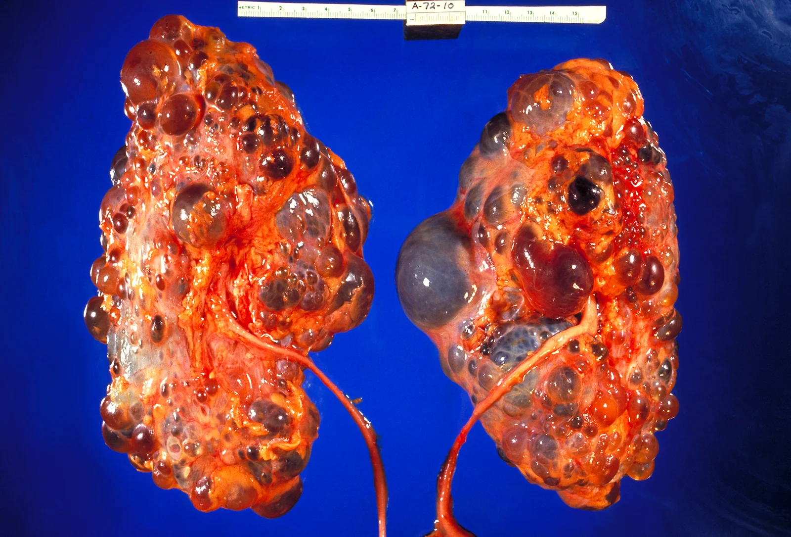
Long description
Gross specimen of both kidneys in polycystic kidney disease, enlarged and studded with cysts of varying size.

The polycystin complex sits on the membrane and in the primary cilia of renal tubular cells. PC1 (from PKD1) and PC2 (from PKD2) normally act together as a calcium-permeable channel, regulating intracellular calcium, cyclic AMP and cell homeostasis. When either protein is deficient, two things happen. First, intracellular calcium falls and cAMP rises, activating protein kinase A; this drives epithelial proliferation and chloride-driven fluid secretion into the lumen through CFTR, so cysts grow. Second, tubular cells lose their normal planar polarity and differentiate abnormally.
Arginine vasopressin reinforces this: by binding V2 receptors it raises cAMP further, which is why blocking the V2 receptor (tolvaptan) slows cyst growth and why hydration that lowers vasopressin is encouraged. As cysts enlarge, they compress intrarenal vessels, causing ischaemia and chronic renin-angiotensin-aldosterone activation; this explains the early onset of hypertension, often before kidney function visibly declines.
Most patients are asymptomatic until early adulthood; symptoms usually appear in the third decade. The classic triad is flank or abdominal pain, haematuria, and hypertension. Patients describe a dragging or heavy feeling from nephromegaly, sharp pain from a ruptured or bleeding cyst, and renal colic from stones.
- Flank or abdominal pain is reported by up to 60% at some point and by about half as their main symptom; pain can be acute (cyst haemorrhage, infection, stone) or chronic from capsular stretch. Chronic pain is a major driver of quality of life loss and does not correlate with kidney size. [1]
- Gross haematuria occurs in up to 40% over a lifetime, especially with larger kidneys and younger age of onset; it is usually self-limiting within a week. [1]
- Hypertension is often found incidentally and is present in 50 to 70% by age 30 despite preserved kidney function; it affects up to 75 to 82% as chronic kidney disease progresses and up to 20% of children and adolescents. [1]
- Urinary tract infection and cyst infection (or pyelonephritis) affect up to about 30 to 50% of patients depending on the series; women are more often affected. [1]
- Kidney stones occur in roughly 3 to 59% of patients; uric acid stones are disproportionately common (about half of stones, versus under 10% in the general population), because of lower urine pH, hypocitraturia and distortion of the collecting system. [1]
- Cyst rupture can produce retroperitoneal haemorrhage, but this is uncommon.
Red flags requiring urgent review: severe unremitting flank pain with fever, gross haematuria with clots or anaemia, thunderclap headache, sudden neurological deficit, acute abdominal pain with peritonism, or an infected cyst not responding to oral antibiotics.
The kidneys may be palpable and ballotable as large, smooth, lobulated masses; hepatomegaly suggests polycystic liver disease. Measure blood pressure carefully. Check for cardiac murmurs (mitral valve prolapse) and signs of dehydration or fluid overload. In ARPKD in the newborn, the Potter facies, contracted limbs, club feet and pulmonary hypoplasia dominate, and the abdomen is massively distended by kidneys.
- Liver cysts are the commonest extrarenal manifestation: present in over 80% by age 30 and in about 80 to 90% after age 35. Severe, symptomatic polycystic liver disease is uncommon (under 5%). Risk factors for a heavier liver burden are oestrogen exposure and multiple pregnancies; the liver may compress the stomach or diaphragm and cause early satiety, dyspnoea and weight loss. Oestrogen-containing contraceptives and hormone replacement should be avoided or limited when moderate or severe liver disease is present. [1]
- Intracranial aneurysms occur in roughly 8 to 18% of patients depending on the series, versus about 3% in the general population, and more often with a family history of aneurysm or subarachnoid haemorrhage. Most are small, saccular and in the anterior circulation. Rupture carries high morbidity and mortality. [1]
- Mitral valve prolapse is seen in 3 to 26% and is usually silent; echocardiography is needed when there is a murmur or symptoms. [1]
- Pericardial effusion in up to 20%, usually asymptomatic. [1]
- Cardiomyopathy in up to 8%, more often associated with PKD2. [1]
- Abdominal wall hernias in about 45% of those who have reached kidney failure. [1]
- Diverticular disease, pancreatic cysts (about 19%) and seminal vesicle cysts (about 40% of men) also occur. [1]
- Renal cell carcinoma is rare (under 1%), but when it occurs in PKD it tends to be earlier, bilateral and multicentric; routine screening is not recommended. [1]
| Condition | Key distinguishing feature |
|---|---|
| Simple (acquired) renal cysts | Usually solitary or few, increase with age, no family history, kidneys are not massively enlarged |
| Autosomal recessive PKD | Perinatal or early childhood onset, enlarged echogenic kidneys, congenital hepatic fibrosis and portal hypertension |
| Tuberous sclerosis complex (TSC1/TSC2) | Autosomal dominant; kidney cysts plus angiomyolipomas, often with skin, neurological or cardiac findings; contiguous TSC2/PKD1 deletion causes severe early cystic disease |
| Von Hippel-Lindau disease | Renal cysts and clear cell carcinoma, central nervous system and retinal haemangioblastomas |
| HNF1B-related nephropathy | Small kidneys with cortical cysts, diabetes, genital tract abnormalities, elevated liver enzymes |
| Medullary sponge kidney | Cystic dilatation of medullary collecting ducts, nephrocalcinosis, stone disease, usually preserved kidney function |
| Nephronophthisis | Small kidneys with corticomedullary cysts, autosomal recessive, onset usually before age 25 |
| Acquired cystic kidney disease of dialysis | Cysts appear after long-standing dialysis; kidneys are usually small |
Baseline tests include serum creatinine and estimated GFR (eGFR), a full blood count (anaemia worsens as kidney function falls), urea and electrolytes, urine dipstick for blood and protein (proteinuria is usually low-grade), a 24-hour urine collection when stones are suspected, and liver function tests. Urine pH and stone risk factors matter in patients with nephrolithiasis. Blood cultures and inflammatory markers are taken when a cyst infection is suspected. [1]
Figure 2CT scan autosomal dominant polycystic kidney disease
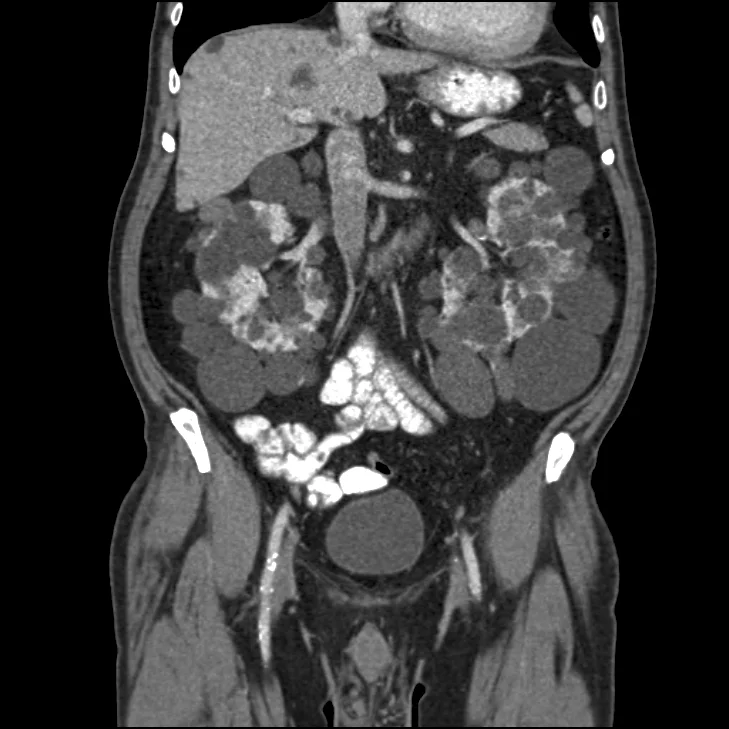
Long description
Coronal contrast CT of autosomal dominant polycystic kidney disease with both kidneys enlarged and replaced by cysts.

Kidney ultrasound is the first-line screening test for relatives and the usual first test when PKD is suspected: cheap, radiation-free and widely available. The modified Pei-Ravine ultrasound criteria, which vary with age, are summarised below for families with a known PKD1 or PKD2 variant. Magnetic resonance imaging (MRI) or computed tomography (CT) is more sensitive for small cysts, for measuring total kidney volume and for risk stratification; in people under 40, MRI or contrast CT is preferred because ultrasound may miss early disease. [4]
| Age band | Criterion (Pei 2009; adult and older adult) | More sensitive Pei/Gabow criteria used in PKD2 and younger people |
|---|---|---|
| 5 to 14 years | — | At least 1 cyst total |
| 15 to 19 years | 3 or more cysts in total (15 to 39) | At least 1 cyst in each kidney (or 2 in one kidney and none in the other) |
| 15 to 39 years | 3 or more cysts in total | 2 in one kidney and 1 in the other (cysts with 3 or more bilaterally) |
| 40 to 59 years | At least 2 cysts in each kidney | At least 2 cysts in each kidney |
| 60 years and over | At least 4 cysts in each kidney | At least 4 cysts in each kidney |
Notes that follow from the same reviews: in an at-risk individual, no cysts over the age of 40 on ultrasound effectively rules out ADPKD regardless of genotype; and the presence of four or fewer cysts in someone under 40 is usually enough to exclude it. In people with an atypical or unclear picture, high-resolution MRI or CT, or genetic testing, is needed. [1]
Testing is not needed where the clinical and imaging picture is clear and the family is already known to carry a pathogenic PKD1 or PKD2 variant. It is essential or highly useful when:
- there is no family history, or an atypical or asymmetric picture;
- the patient is a child with very early onset disease;
- a young living kidney donor is being assessed;
- there is a discrepancy between kidney size and the rate of eGFR decline;
- reproductive planning or pre-implantation genetic testing is being considered;
- extrarenal findings suggest an alternative syndrome (tuberous sclerosis, von Hippel-Lindau).
A next-generation sequencing panel covering PKD1, PKD2 and the rarer cystic kidney disease genes is the standard first test. PKD1 is difficult to sequence because of its large pseudogenes nearby, so interpretation must be done by an experienced genetics laboratory. Variants are reported using standard ACMG classes (pathogenic, likely pathogenic, uncertain significance, likely benign, benign). Mosaicism occurs and can make testing falsely negative. Genetic information affects not only the patient but family members; counselling should cover insurance and employment implications, which vary by country.
World Health Organization
Total kidney volume, usually height-adjusted (htTKV), is the strongest non-invasive predictor of future kidney function. Two points are well established: an htTKV above 600 mL/m predicts stage 3 chronic kidney disease within about 8 years, and a baseline TKV above 1500 mL was associated with a GFR decline of 4.33 mL/min/year. GFR falls by 2 to 5 mL/min/year once decline has begun, and half of patients reach kidney failure by age 60. [1]
The Mayo imaging classification (MIC) divides typical ADPKD into five classes (1A to 1E) by htTKV and age. Classes 1C to 1E predict kidney failure before age 65 and are the groups in which tolvaptan is considered. Because small errors in TKV measurement can move a patient between classes, planimetry rather than the simpler ellipsoid method is preferred in people under 25.
The PROPKD score estimates the risk of kidney failure before age 60 from genetics and clinical history: male sex (1 point), hypertension before age 35 (2 points), a first urological event before age 35 (2 points), and variant type (PKD2 = 0, non-truncating PKD1 = 2, truncating PKD1 = 4). A score of 3 or less has a low risk (about 81% negative predictive value for reaching kidney failure before 60), and a score above 6 a high risk (positive predictive value about 91%). Its use is limited by the need for genetic testing and its intermediate zone (4 to 6). [1]
ADPKD can present before adulthood (early-onset, before 15, or very early onset, in utero to 18 months). Criteria for very early onset disease in utero include oligohydramnios and hyperechoic enlarged kidneys; up to 18 months the diagnosis requires enlarged palpable kidneys plus at least one of hypertension, GFR under 90 mL/min/1.73 m2, or persistent proteinuria. From 18 months to 15 years, the diagnosis requires at least one of enlarged palpable kidneys, elevated blood pressure, decreased GFR or persistent proteinuria. Ultrasound (one or more cysts in a child with a positive family history) is the preferred screening test, but the medical, legal and psychological consequences of labelling a child need careful discussion with the family. Genetic testing is recommended for atypical, early-onset or apparently sporadic cases. Deciding whether to screen children under 18 remains contentious; early detection helps blood pressure and lifestyle, but there is psychosocial burden. [1]
Approach to a patient at risk of or with polycystic kidney disease
- Family history or incidental cysts on imagingTake a careful three-generation pedigree; measure blood pressure
- First test: renal ultrasoundUse age-specific criteria; MRI or CT if under 40 or if ultrasound is equivocal
- Question: Cysts match age criteria and family history positive?
Yes: typical ADPKD likely
- Measure blood pressure, creatinine, eGFRAnnual or as indicated
- MRI or CT to measure height-adjusted total kidney volumeFor risk stratification and tolvaptan eligibility
- Stratify by Mayo imaging class and PROPKD scoreDecide whether disease is likely rapidly progressive
- Start ACE inhibitor or ARB and target BP (under 120 mmHg systolic if aged 50 or over; 110/75 mmHg if aged 18 to 50 with eGFR above 60)
- Consider tolvaptan if MIC 1C to 1E or proven rapid progressionStart early; give high water intake advice; monitor liver enzymes
- Screen for cerebral aneurysm if family history of aneurysm, subarachnoid haemorrhage or unexplained sudden death, high-risk occupation, or before major surgeryTime-of-flight MRA; repeat every 5 years in high-risk or every 10 years otherwise
- Manage complicationsCyst infection, stones, haematuria, pain; dialysis and transplant when kidney failure arrives
No: atypical or negative
- Repeat or upgrade imaging (MRI or CT)Four or fewer cysts under 40 usually excludes ADPKD
- Genetic testing panel (PKD1, PKD2 and related genes)Essential with no family history, early onset, donor assessment or extrarenal syndromes
- If all tests negative, consider simple cysts, ARPKD, TSC, VHL, HNF1B, medullary sponge kidneyRefer to nephrology and genetics
Every patient benefits from: stop smoking (especially important because of aneurysm risk), avoid nephrotoxins such as NSAIDs, keep a normal weight, limit salt, treat pain, and drink enough fluid. Advise up to about 3 litres of water per day to suppress vasopressin and reduce stone risk; trials show this lowers urine osmolality but has not consistently slowed GFR or kidney volume decline over three years. Counsel about insurance and employment implications of the diagnosis where relevant. [1]
Hypertension is usually the earliest treatable feature and cardiovascular disease is the leading cause of death in ADPKD. Targets in rapidly cited guidance are:
- under 50 years with preserved kidney function (GFR above 60 mL/min/1.73 m2): target 110/75 mmHg or lower;
- 50 years and over, or any stage of kidney disease: systolic blood pressure under 120 mmHg (some national groups adopt under 130 mmHg after shared decision making). [1]
First-line drugs are an ACE inhibitor or, if that is not tolerated, an angiotensin receptor blocker (ARB); either alone is adequate in most patients. Combining two renin-angiotensin blockers is avoided, because it increases hyperkalaemia without added renal benefit. Blood pressure should be measured with a validated device, including home and ambulatory readings where possible.
Cyst infection presents with fever, flank or abdominal pain and raised inflammatory markers; it is often indistinguishable from pyelonephritis, so treat long. Obtain urine and blood cultures before antibiotics, and image (CT, MRI, PET-CT or indium-labelled white-cell scan) to find the infected cyst. Preferred antibiotics are lipid-soluble agents with good cyst penetration, such as fluoroquinolones or trimethoprim-sulfamethoxazole; beta-lactams are less reliable for cysts. A cyst infection needs 4 to 6 weeks of treatment; if there is no clinical response, CT-guided aspiration or drainage is considered. Drainage procedures are best done in a centre with PKD expertise. [1][2]
Management starts with hydration, analgesia and treatment of obstruction. For stones that need intervention, flexible ureteroscopy is often preferred over shock-wave lithotripsy or percutaneous nephrolithotomy because cystic kidneys distort the anatomy. Prevention relies on high fluid intake (over 3 L per day aiming at urine output over 2.5 L), a low-salt, low-animal-protein diet and correction of metabolic defects found on 24-hour urine; potassium citrate for hypocitraturia, and allopurinol or febuxostat may be considered when uric acid stones recur.
Most cyst bleeds settle with rest and hydration. In severe or persistent bleeding, suspending ACE inhibitors or ARBs may reduce the risk of acute kidney injury; blood transfusion should be used cautiously to avoid sensitisation before transplantation; interventional radiology embolisation is indicated for persistent or unstable bleeding; and unilateral nephrectomy is a last resort.
Non-drug measures (heat, ice, physical therapy) come first. Paracetamol is the usual first-line analgesic; adjuvant agents such as gabapentin may help; NSAIDs may be used cautiously for short periods when kidney function is preserved; opioids are reserved for severe pain. Larger symptomatic cysts can be treated by aspiration with or without sclerotherapy, and refractory cases may need nerve block, ablation or, rarely, nephrectomy or partial hepatectomy.
Tolvaptan, a vasopressin V2 receptor antagonist, is the only disease-modifying drug approved for ADPKD. It slows cyst growth by lowering cAMP. In the TEMPO 3:4 trial it reduced kidney function decline by about 30% and kidney growth by about 49% over three years in patients with total kidney volume over 750 mL and creatinine clearance over 60 mL/min/1.73 m2. In REPRISE it slowed eGFR decline by about 35% over one year in patients with more advanced chronic kidney disease (creatinine clearance 25 to 65 mL/min/1.73 m2). It remains effective down to an eGFR of about 15 mL/min/1.73 m2 and is continued until kidney replacement therapy is needed. Best results require early use in confirmed rapid progressors (Mayo class 1C to 1E, PROPKD above 6, or documented eGFR decline above 3 mL/min/year). [1]
Practice points:
- Individualise the discussion; older patients (over 55) with slower decline have a smaller benefit-to-risk ratio.
- Up-titrate to the maximum tolerated dose, 90 mg on waking and 30 mg 8 hours later. [2]
- Expect aquaretic side effects: polyuria, nocturia, thirst. Tolerability, not urine osmolality, drives dosing; some clinicians measure urine osmolality as an adherence marker.
- Monitor liver enzymes before starting and on treatment because of a risk of reversible drug-induced liver injury; tolvaptan is contraindicated in pregnancy, lactation, a history of significant liver injury, hypovolaemia and urinary tract obstruction.
- Somatostatin analogues can shrink liver volume and symptoms but are not recommended to protect kidney function; their use is usually restricted to polycystic liver disease and hepatology care.
Screening with time-of-flight non-contrast magnetic resonance angiography (or CT angiography if MRI is unsuitable) is recommended for:
- a family history of intracranial aneurysm, subarachnoid haemorrhage or unexplained sudden death;
- a high-risk occupation (for example pilots or professional drivers);
- before major elective surgery such as kidney or liver surgery.
If an aneurysm is found, management is by a multidisciplinary team and depends on size, symptoms and age: smaller aneurysms under 7 mm in older patients are usually observed with serial imaging, while larger or symptomatic ones in younger patients need intervention. If no aneurysm is found, repeat screening every 5 years in high-risk patients and every 10 years otherwise, after discussing benefits and risks. Management of ruptured aneurysm and subarachnoid haemorrhage follows standard neurocritical care. [1]
When kidney failure develops, living-donor kidney transplantation gives the best long-term outcomes and is preferred. Haemodialysis and peritoneal dialysis are both appropriate bridges or alternatives; peritoneal dialysis is not contraindicated in ADPKD, although patients carry a higher risk of abdominal hernias and renal volumes can cause technical difficulty. Nephrectomy is considered for recurrent cyst infection or bleeding, severe refractory pain, suspected cancer, or to create space for a transplant in selected cases. Kidney and liver transplantation may be combined in patients with severe polycystic liver disease.
Counselling covers inheritance (50% risk to children in ADPKD, 25% in ARPKD), prognosis which varies more than genotype alone predicts, testing options, family screening, and insurance or employment implications. Pre-implantation genetic testing for monogenic disorders is an option for some couples; its availability and cost vary. During pregnancy, women with ADPKD should avoid ACE inhibitors and ARBs (use labetalol, nifedipine or alpha-methyldopa), monitor blood pressure closely, and be aware of risks of pregnancy-induced hypertension and pre-eclampsia. Multiple pregnancies have been associated with faster GFR decline, and oestrogen-containing contraception can accelerate liver cyst growth, so liver volume should be assessed before starting hormonal contraception or hormone replacement. [3]
Prognosis is driven by genotype and imaging. Patients with PKD1 variants reach kidney failure at a median of about 54 years (range 53 to 60); PKD1 truncating variants around 55.6 years versus 67.9 years for non-truncating; PKD2 variants around 77 to 80 years. Identical twins differ by a mean of about 2 years, siblings by about 7 and relatives overall by about 13 years. Rapid progression is defined by an annual eGFR decline of 3 mL/min/year or more (ERA-EDTA) and is seen in about two-thirds of typical patients. Other adverse factors include male sex, hypertension onset before 35, early urological events, proteinuria, obesity and enlarged total kidney volume. [1][3]
- Simple (acquired) cysts: common age-related findings, often solitary, not inherited; kidneys are not massively enlarged. One cyst in an over-40 adult essentially rules out ADPKD; under 40, four or fewer cysts usually excludes it.
- Medullary sponge kidney: congenital ectasia of medullary collecting ducts with nephrocalcinosis and stone disease; kidney function usually remains normal. It is not the same as medullary cystic kidney disease (now often called autosomal dominant tubulointerstitial kidney disease).
- Tuberous sclerosis complex: autosomal dominant TSC1 or TSC2 variants, birth incidence about 1 in 6000; renal cysts in about half and angiomyolipomas in about 80%; cystic disease may mimic ADPKD. A deletion spanning both TSC2 and PKD1 causes early severe polycystic disease. [3]
- HNF1B-related, von Hippel-Lindau, nephronophthisis and other ciliopathies: all can present with cystic kidneys and are important mimics in children and in people without a family history.
Children: see the diagnostic criteria above. Asymptomatic children do not need drug treatment. Ambulatory blood pressure monitoring is preferred, blood pressure is controlled with renin-angiotensin blockade, and lifestyle measures (salt, weight, hydration) matter. Tolvaptan in children is still under study. About 20% of children with ADPKD have hypertension. [1]
Pregnancy: see genetic counselling above. Pre-conception counselling should discuss pre-implantation testing where available.
Older adults: patients over 55 with slower decline have less to gain from tolvaptan; decisions should still follow individual goals.
Where ultrasound is the only available test, use age-specific criteria and treat an over-40 adult with no cysts as not having ADPKD. Family history and blood pressure are valuable and cost nothing. Where genetic testing is unavailable, counsel on recurrence risk using the family pattern. Ensure ACE inhibitors or ARBs are available for blood pressure; they are inexpensive and effective. Emphasise hydration for stones, and avoid NSAIDs. If cyst infection occurs, use the best locally available antibiotic with cyst penetration (fluoroquinolone where affordable) for the full 4 to 6 weeks. Refer early if dialysis or transplantation may be needed; peritoneal dialysis can be used. Virtual consultation models can connect community clinicians with PKD specialists.
NoteRegional note on blood pressure targets
Some national hypertensive societies target systolic blood pressure under 130 mmHg rather than under 120 mmHg. Both values appear in current guidance; the choice is made with the patient after shared decision making, and the under-110/75 mmHg target for young adults with good kidney function remains the most aggressive standard target.[2]
- ADPKD is autosomal dominant: children of an affected parent have a 50% risk of inheriting it. PKD1 accounts for about 78% of cases and PKD2 about 15%. [1]
- Its course is highly variable, even within a family: identical twins differ on average by 2 years and relatives by 13 years in the age at kidney failure.
- The kidneys enlarge with bilateral cysts; kidney function falls by about 2 to 5 mL/min/year once decline begins and about half reach kidney failure by age 60.
- Hypertension is common (50 to 70% by age 30) and is an independent driver of progression.
- Common complications: haematuria (up to 40%), kidney infection (up to about 30%), stones (up to 50%), chronic pain (up to 60%), liver cysts (over 80% by age 30), intracranial aneurysms (roughly 8 to 18%, higher with a family history), mitral valve prolapse (3 to 26%).
- ARPKD presents perinatally, kills 30 to 50% of neonates from pulmonary hypoplasia, and always involves congenital hepatic fibrosis.[3]
- Diagnosis: age-specific ultrasound criteria, family history, genetics when needed, MRI or CT for total kidney volume.
- Management: ACE inhibitor or ARB and blood pressure targets (110/75 mmHg in 18 to 50-year-olds with preserved GFR; under 120 mmHg systolic when 50 or older); up to 3 L water daily; tolvaptan 90 mg/30 mg for rapid progressors with liver monitoring. [1]
- Screen for cerebral aneurysms in high-risk patients and operate when larger or symptomatic.
- Living-donor transplantation is preferred for kidney failure; peritoneal dialysis is allowed.
- Other cystic diseases (simple cysts, medullary sponge kidney, tuberous sclerosis) must be distinguished, especially in children and in people without a family history.
Answer each one in your head first, then open the answer.
A 24-year-old man has an affected father with ADPKD. Ultrasound shows 4 cysts across both kidneys. What is the diagnosis and what should be done next?
Show answerHide answer
He meets the modified ultrasound criterion for 15 to 39 years (3 or more cysts in total), so ADPKD is likely. Arrange a baseline blood pressure, serum creatinine and eGFR, a plan for MRI total kidney volume and risk stratification (Mayo class, PROPKD), and blood pressure treatment with an ACE inhibitor or ARB to a target of 110/75 mmHg. Discuss family screening, genetics, aneurysm screening based on family history, and high fluid intake. [2][3][4]
Compare the amount of cystic disease and kidney failure age in PKD1 against PKD2.
Show answerHide answer
PKD1 produces a heavier cystic burden and an earlier median age at kidney failure, about 54 years (range 53 to 60), versus about 77 to 78 years (range 60 to 85) for PKD2. Truncating PKD1 variants are earlier than non-truncating ones. [1]
Why does a V2 receptor antagonist slow ADPKD, and why is high water intake recommended despite mixed trial evidence?
Show answerHide answer
Vasopressin raises cAMP through V2 receptors, which drives cyst epithelial proliferation and CFTR-mediated chloride secretion into cysts. Blocking the receptor (tolvaptan) slows growth. Diluting vasopressin with water lowers urine osmolality and theoretically suppresses vasopressin; hydration also helps stones, although drinking up to 3 L per day has not consistently slowed GFR decline or kidney volume growth over three years.
A newborn has massively enlarged kidneys, oligohydramnios and pulmonary hypoplasia, and dies of respiratory failure. Autopsy shows cystic kidneys and congenital hepatic fibrosis. What is the diagnosis and its recurrence risk?
Show answerHide answer
Autosomal recessive polycystic kidney disease from PKHD1. The recurrence risk for future siblings is 25%. Counseling must also cover the associated portal hypertension and the role of combined liver-kidney transplantation later in childhood.
Which patients with ADPKD should be offered tolvaptan, and what safety step is essential?
Show answerHide answer
Rapid progressors: Mayo imaging class 1C to 1E, PROPKD over 6, or documented eGFR decline over 3 mL/min/year, started early, titrated to 90 mg on waking and 30 mg 8 hours later. Liver enzymes must be monitored because of the risk of reversible drug-induced liver injury. Tolvaptan is contraindicated in pregnancy, lactation, significant liver injury, hypovolaemia and urinary tract obstruction.
A patient with ADPKD has a headache described as 'the worst ever'. What must you consider and how would you screen an asymptomatic relative?
Show answerHide answer
Consider a ruptured intracranial aneurysm with subarachnoid haemorrhage; case fatality is high, reported from about 35% to over 50%. Screen high-risk relatives (family history of aneurysm, haemorrhage or unexplained sudden death, high-risk occupation, before major surgery) with non-contrast time-of-flight MR angiography, repeating every 5 years in high-risk and every 10 years in lower-risk patients if normal. [1]
Closely related renal topics include chronic kidney disease, hypertension, stone disease and imaging of the urinary tract, which are covered on their own pages. Reproductive and genetic counselling overlaps with the genetics topics.
- Borghol AH, Bou Antoun MT, Hanna C, et al. Autosomal dominant polycystic kidney disease: an overview of recent genetic and clinical advances. Renal Failure 2025;47(1):2492374. doi:10.1080/0886022X.2025.2492374. Accessed .
- Lanktree MB, Ashawasega N, Bevilacqua M, et al. Canadian Society of Nephrology Commentary on the 2025 KDIGO Clinical Practice Guidelines for Autosomal Dominant Polycystic Kidney Disease. Canadian Journal of Kidney Health and Disease 2026;13:20543581261455635. doi:10.1177/20543581261455635. Accessed .
- Bergmann C. Genetics of autosomal recessive polycystic kidney disease and its differential diagnoses. Frontiers in Pediatrics 2018;5:221. doi:10.3389/fped.2017.00221. Accessed .
- Harnan S, Gittus M, Falzon L, et al. The diagnostic accuracy of ultrasound and genomic tests for the diagnosis of autosomal-dominant polycystic kidney disease: a systematic mapping review. Clinical Kidney Journal 2025;18(7):sfaf187. doi:10.1093/ckj/sfaf187. Accessed .
Figure credits
Educational content for medical students and doctors. It is not a substitute for clinical judgement or local guidelines.