Anatomical terminology and planes
Standardized anatomical language and positional references for precise clinical communication.
Anatomical terminology relies on a standardized reference position to ensure precision, much like a map oriented with north at the top. The normal anatomical position is defined as the body standing upright, with the feet at shoulder width and parallel, and toes forward. In this position, the upper limbs are held out to each side, and the palms of the hands face forward. This standardisation reduces confusion because terms are used as if the body is always in this position, regardless of its act [1]
Ancient Greek and Latin words are used to build anatomical terms, providing a universal language. Regions of the body are identified using precise terms such as “occipital” for the back of the head, which is more accurate than common phrases. The human body’s numerous regions have specific terms to help increase precision. The term “brachium” or “arm” is reserved for the upper arm, while “antebrachium” or “forearm” is used for the lower arm. [1]
Certain directional anatomical terms appear throughout anatomy textbooks and are essential for describing relative locations. An anatomist might describe one band of tissue as “inferior to” another or a physician might describe a tumor as “superficial to” a deeper body structure. Images of the body’s interior commonly align along one of three planes: the sagittal, frontal, or transverse. The serous membranes have two layers—parietal and visceral—surrounding a fluid filled space. [1] [2]
Because anatomy is a live and active science its terminology can be expected to be at any one time in flux. [2]
DefinitionAnatomical Position
The body standing upright, with the feet at shoulder width and parallel, toes forward, upper limbs held out to each side, and the palms of the hands facing forward.
| Common Term | Anatomical Term | Specific Region |
|---|---|---|
| Arm | Brachium | Upper arm |
| Forearm | Antebrachium | Lower arm |
| Leg | Crus | Between knee and ankle |
| Armpit | Axilla | Axillary region |
| Collarbone | Clavicle | Pectoral region |
Anatomical terminology is not designed to confuse, but to increase precision and reduce medical errors. By using precise terms, ambiguity is eliminated, ensuring that descriptions such as the location of a scar are clear rather than vague. The language derives from ancient Greek and Latin, languages whose meanings remain static because they are no longer used in everyday conversation. [1]
The standardisation of this language has evolved through several key historical milestones. The history began with ad hoc usage until 1895, when the Anatomische Gesellschaft published the Basle Nomina Anatomica (BNA), a list entirely in Latin. The International Federation of Associations of Anatomists (IFAA) was created in 1903 with the objective of selecting a uniform nomenclature. Subsequent updates included the Birmingham Revision (BR) in 1933 and the Jenenser Nomina Anatomica (JNA) in 1935. [2]
In 1989, the Federative Committee on Anatomical Terminology (FCAT) was created to review terminology, starting with NA5 as the base. This committee published TA1, the first edition to include a complete set of English equivalent terms alongside Latin. FCAT later evolved into the Federative International Program on Anatomical Terminology (FIPAT), which updated the terminology to TA2. This edition was published online in 2019 and accepted at the IFAA World Congress in 2020. [2]
DefinitionStandardisation
Standardised terminology is fundamental for effective scientific and clinical communication, strengthening anatomical education and improving research reliability. [3]
To alleviate confusion, the ATI website was developed by Leiden University Medical Center and the Clinical Anatomical Terminology (CAT) Committee of the American Association of Clinical Anatomists (AACA). ATI presents listings of both IFAA official terms and other terms in use, allowing users to look up synonyms. It serves as a conduit between clinicians and anatomists, informing clinicians of standardized terminology in place of eponyms. [2]
| Year | Document/Committee | Key Feature |
|---|---|---|
| 1895 | Basle Nomina Anatomica (BNA) | Entirely in Latin |
| 1903 | IFAA Creation | Objective: uniform nomenclature |
| 1933 | Birmingham Revision (BR) | Updated BNA |
| 1935 | Jenenser Nomina Anatomica (JNA) | Published by AG |
| 1989 | FCAT Creation | Review started with NA5 |
| 2019 | TA2 | Published online |
Inconsistent use of terms complicates student learning and compromises clinical reasoning. Although TA was published to reduce discrepancies, terminological inconsistency persists. The adoption of the officially recognized terminology, such as "recurrent laryngeal nerve," is recommended for scientific use. This consistency ensures that anatomy and medicine speak the same language, allowing both fields to advance effectively. [2] [3]
Figure 1Body regions
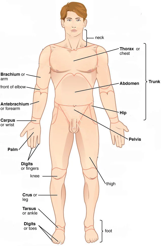
Long description
Body regions. Labelled anatomical illustration from Wikimedia Commons, used under its open licence.

Figure 1: Body regions.
Anatomical terminology serves as the universal language of medicine, yet it is not static. Renaming structures, despite good intent to improve or clarify terminology, also contributes to part of the body of synonyms as previous terms often linger for generations. However, the previous term is still widely used, as is shown from a Pubmed search where over 18,000 articles with the term “common bile duct” have appeared since the publication of Terminologia Anatomica (TA) (https://pubmed.ncbi.nlm.nih.gov/?term=common+bile+duct&filter=years.1998-2024). [2]
Regional and cultural differences also influence the used anatomical terminology. For example, the iliofemoral ligament is commonly known as Bertin’s ligament in France after Exupere Joseph Bertin (1712–1781) and known as Bigelow’s ligament in the UK and the USA after Henry Jacob Bigelow (1819–1890) (Malenfant et al. ; Pećina ). Another example is the proximal deep inguinal lymph node which is known as Cloquet’s node in France and the UK after the French anatomist and surgeon Jules Germain Cloquet (1790–1883) (Loukas et al. ), but it is known as Rosenmüller–Lymphknoten in German-speaking countries (Buttner et al. ). Even in regular, non-eponymous terminology regional differences arise. English-speaking countries, for example, usually omit the word “muscle” in muscle names, sometimes leading to lack of clarity about the intended type of structure. [2]
Divergence in meaning can also arise due to differences between usage in different countries and languages. For example, the concept of anatomical “region” tends to be interpreted as a three-dimensional anatomical volume in English-speaking countries, but as a predominantly two-dimensional area of surface anatomy in mainland Europe, as discussed in FCAT meetings (1989–1998) (Whitmore, personal observation). Also, usage of the term “fascia” has been broadly interpreted by anglophone anatomists as both superficial and deep sheaths or layers of connective tissue in the body, but in mainland Europe preferentially as a term referring more narrowly to the connective tissue layer ensheathing muscles (Singer ; Neumann ). [2]
Other anatomical homonyms are still fully with us. “Os,” for example, can mean either “bone” or “mouth” in Latin. “Cervix” can refer to either the neck (“collum” in Terminologia Anatomica, second edition (TA2)) or to a part of the uterus (cervix uteri). “Ileum,” the distal small intestine, is pronounced the same as “ilium,” a bone of the pelvis. The anatomical malapropism, “ilioc(a)ecal junction,” rather than “ileoc(a)ecal junction” (Bogers and Van Marck ), is a frequently seen result of this ho [2]
However, it is important to recognize that literal accuracy of anatomical terms is an ideal. [2]
Finally, terms may be contentious for different reasons. [2]
To make a definition uniquely defining it may be necessary to focus on specific anatomical details, which require prior knowledge, thus making the definition less accessible. [2]
The correct and internationally accepted term is “recurrent laryngeal nerve”. The consistent use of standardized terminology is crucial for precise communication in education, clinical practice, and research. A thorough understanding of the recurrent laryngeal nerve's anatomical course and variations is essential for minimizing the risk of iatrogenic injury during surgical procedures in the neck, particularly thyroid and parathyroid surgeries. [3]
The final stage involved compiling a detailed description of the anatomical course, functional role, and clinical relevance of the nerve. This was accomplished through a comprehensive review of authoritative anatomy textbooks and peer-reviewed articles, with the aim of supporting students, educators, researchers, and clinicians who rely on precise and standardized terminology. [3]
Analysis of a recent major anatomical textbook further supports the continued predominance of “recurrent laryngeal nerve”. In the 9th edition of Moore ‒ Clinically Oriented Anatomy (2024), Dalley and Agur consistently use “recurrent laryngeal nerve” in their description of laryngeal innervation, with no use of “inferior laryngeal nerve” as a synonym. This finding reinforces the contemporary validity of “recurrent laryngeal nerve” as the standard designation in major anatomical references. [3]
Historically, “inferior laryngeal nerve” was common in 19th- and early 20th-century texts,,,, particularly in European anatomical traditions, whereas “recurrent laryngeal nerve” gradually became preferred because it more accurately reflects the nerve’s retrograde course and offers greater anatomical clarity. Its adoption in modern literature and in the TA reflects this standardization. However, the persistence of “inferior laryngeal nerve” in contemporary literature likely reflects historical continuity and continued practical use. In some surgical and anatomical contexts, particularly in descriptions of branching near the larynx, the term is still used for the terminal portion of the recurrent laryngeal nerve rather than the entire nerve, which perpetuates terminological ambiguity. [3]
For example, in the anatomical position the “base” of the heart is located posterosuperiorly, not inferiorly as expected, but at the present time, this is the convention by which it is described. [2]
The correct and internationally accepted term is “recurrent laryngeal nerve”. The consistent use of standardized terminology is crucial for precise communication in education, clinical practice, and research. A thorough understanding of the recurrent laryngeal nerve's anatomical course and variations is essential for minimizing the risk of iatrogenic injury during surgical procedures in the neck, particularly thyroid and parathyroid surgeries. [3]
Anatomical terminology must be precise to ensure patient safety and effective communication. Eponyms, such as names derived from individuals, are synonyms for structures but are often discouraged by anatomists because they convey no descriptive information about location or characteristics. Despite this, clinicians frequently use them. Confusion arises when terms are homonyms, sharing spelling or pronunciation but having different meanings. For instance, "peroneal" refers to the lateral leg area. Woywodt et al. () argue against the use of eponyms honoring individuals who collaborated with the National Socialists of Germany in World War II. [2]
Practitioners will realistically accept that a number of terms, albeit literally inaccurate, are nevertheless commonly used references or labels to identify a structure or its parts. [2]
A Spigelian hernia occurs through the Spigelian fascia, which is the aponeurotic layer between the rectus abdominis muscle and the semilunar line. These hernias are rare and often difficult to diagnose clinically because they are interparietal, meaning they do not protrude through the anterior abdominal wall fascia but rather lie between the muscle layers. They typically present as a painful mass in the lower abdomen, lateral to the rectus sheath. Diagnosis is often confirmed via ultrasound or CT imaging, which visualises the defect in the aponeurosis. Surgical repair is usually required due to the high risk of incarceration, given the narrow neck of the hernia sac.
A diaphragmatic hernia involves the protrusion of abdominal contents into the thoracic cavity through a defect in the diaphragm. Congenital forms, such as Bochdalek hernias, usually occur posterolaterally due to failure of the pleuroperitoneal membranes to close. Acquired hernias may result from trauma or iatrogenic injury. Symptoms include respiratory distress and gastrointestinal obstruction. In adults, hiatal hernias are the most common type, involving the oesophageal hiatus. Clinical examination may reveal bowel sounds in the chest. Management depends on severity, with surgical repair often indicated for large defects or symptomatic cases to prevent strangulation of the herniated viscera.
Horner syndrome results from disruption of the sympathetic nerve supply to the eye and face. The classic triad consists of ptosis (drooping eyelid), miosis (constricted pupil), and anhidrosis (lack of sweating) on the affected side. The lesion can occur anywhere along the three-neuron pathway: first-order neurons from the hypothalamus to the spinal cord, second-order neurons from the spinal cord to the superior cervical ganglion, and third-order neurons from the ganglion to the eye. Causes include Pancoast tumours, carotid artery dissection, or neck trauma. Diagnosis involves pharmacological testing with apraclonidine and cocaine drops to confirm sympathetic denervation. Identifying the level of the lesion is crucial for determining the underlying aetiology and appropriate management.
The abdominal cavity is commonly subdivided using the quadrants approach, which uses one horizontal and one vertical line intersecting at the patient’s umbilicus. This method is simpler and more commonly used in clinical medicine than the nine-region approach. The nine-region approach utilizes one horizontal line immediately inferior to the ribs and one immediately superior to the pelvis. [1]
Serous membranes, or serosae, cover the walls and organs within the thoracic and abdominopelvic cavities. The parietal layer lines the body cavity walls, while the visceral layer covers the internal organs. A very thin, fluid-filled serous space exists between these two layers. [1]
This creates terminological confusion, which is the converse of synonymy. Such confusion often arises from linguistic evolution or regional differences in terminology. [2]
The Terminologia Anatomica (TA1) first edition favoured "common fibular nerve" over "common peroneal nerve" in 1998. Despite this standardisation, the older terms persist among many clinicians to the present day. [2]
Effective anatomical definitions should be succinct and accessible to reduce student cognitive load. The main purpose of a definition is to provide enough understanding to avoid confusing the concept with related ones. [2]
Left-sided vocal fold palsy is more often linked to these thoracic causes. Preservation of the recurrent laryngeal nerve is critical in cervical surgeries. Transient paralysis rates range from 5% to 8% following such procedures. Permanent paralysis occurs in 0.3%–3% of cases. [3]
Such anomalies profoundly alter the nerve's trajectory. [3]
Exam trapPolysemy Trap
Remember that polysemy means one term has many meanings, not many terms for one structure.
Clinical pearlQuadrant vs Nine Regions
The quadrants approach intersects at the umbilicus and is more common in medicine than the nine-region method.
DefinitionSerous Membrane
A thin membrane covering cavity walls (parietal) and organs (visceral), separated by a fluid-filled space.
| Feature | Statistic / Detail |
|---|---|
| Branching on both sides | 43% (Rustad) |
| Multiple branches given off | 87.5% (Tang) |
| Non-recurrent nerve incidence (Right) | 0.3%‒0.8% |
| Non-recurrent nerve incidence (Left) | 0.004% |
| Transient paralysis rate | 5% to 8% |
| Permanent paralysis rate | 0.3%–3% |
Students frequently confuse anatomical orientation with patient position, leading to errors in clinical communication. A body lying face down is described as prone, while a body lying face up is described as supine. These terms are sometimes used in describing the position of the body during specific physical examinations or surgical procedures. It is critical to remember that 'anterior' and 'posterior' refer to the standard anatomical position, not the patient's current orientation on a bed. [1]
Another significant error involves the literal interpretation of anatomical names. Inaccuracy in the literal meaning of anatomical terms is another reputed cause for confusion and a reason put forward for terms to be changed. For example, in the adult the left atrium is clearly not “left” but “posterior;” the right ventricle not “right,” but “anterior”. Students must learn that historical naming conventions often reflect embryological development or early misconceptions rather than adult spatial [2]
Interpreting medical imaging requires a precise understanding of sectional planes. A section is a two-dimensional surface of a three-dimensional structure that has been cut. Modern medical imaging devices enable clinicians to obtain “virtual sections” of living bodies. Body sections and scans can be correctly interpreted, however, only if the viewer understands the plane along which the section was made. A plane is an imaginary two-dimensional surface that passes through the body. There are three planes commonly referred to in anatomy and medicine, as illustrated in. Confusing axial, sagittal, and coronal planes leads to mislocalisation of pathology. [1]
Exam trapLiteral Misinterpretation
In the adult the left atrium is clearly not “left” but “posterior;” the right ventricle not “right,” but “anterior”. Do not rely on the directional prefix in the name to determine spatial location in the adult thorax.
DefinitionAnatomical Plane
A plane is an imaginary two-dimensional surface that passes through the body. Understanding these planes is essential for interpreting virtual sections obtained by modern imaging devices.
Knowledge of anatomical terminology is indispensable at the bedside, in the operating theatre, and during imaging reviews. The anterior (ventral) cavity has two main subdivisions: the thoracic cavity and the abdominopelvic cavity. The thoracic cavity is the more superior subdivision of the anterior cavity, and it is enclosed by the rib cage. The thoracic cavity contains the lungs and the heart, which is located in the mediastinum. The diaphragm forms the floor of the thoracic cavity and separate [1]
In surgery, particularly thyroidectomy, anatomical variations pose significant risks. The risk of injury to the RLN is ever present during neck surgery[1]. Near the inferior pole of the thyroid gland, the right RLN is intimately related to the inferior thyroid artery and its branches[1]. The RLN may cross anterior or posterior to branches of the artery, or it may pass between them[1]. Due to this close association, the inferior thyroid artery is connected at a certa [3]
RLN Surgical Risk
- Identify Thyroid PoleLocate inferior pole
- Question: Right or Left Side?
Right
- Check Artery RelationRLN crosses anterior, posterior, or between branches of inferior thyroid artery
Left
- Check Vertical AscentNerve ascends from superior mediastinum; close association near inferior pole
NoteSurgical Anatomy
The RLN may cross anterior or posterior to branches of the artery, or it may pass between them[1]. This variability necessitates careful dissection to avoid voice dysfunction.
In examinations, students must define structures uniquely. But which characteristic or combination of characteristics of a structure, for instance a muscle, is most striking, or essential? Is it its function, location, or appearance? And how many characteristics are needed to uniquely define the structure? A definition for biceps brachii such as “two-headed muscle of the anterior arm” is succinct, but it is not yet “uniquely defining” as it may be interpreted to also cover the pectoralis major muscle. Hence, an extra characteristic must be added to ensure that only the biceps brachii is meant. Adding more characteristics of a structure to a definition assures it is “uniquely defining” but makes it less succinct. The goals “uniquely defining” and “succinct” may conflict. [2]
| Concept | Issue | Resolution |
|---|---|---|
| Uniquely Defining | Conflicts with succinctness | Add extra characteristics |
| Succinctness | May lack uniqueness | Replace wording carefully |
| Standardisation | Multiple terms in parallel use | Use professionally endorsed TA |
Despite efforts at standardisation, usage varies. Laitman () and Gobée et al. () noted that the usage of a single set of standard anatomical terms by all researchers, educators, and clinicians may well be an illusory ideal. It is difficult to change a habit. We tend to use terms because we are comfortable and more familiar with them. Natural languages are not controllable. Despite acknowledging that a professionally endorsed TA exists and is actively maintained for this purpose, these authors su [2]
Prone describes a face-down orientation, and supine describes a face up orientation. [1] A section is a two-dimensional surface of a three-dimensional structure that has been cut. Body sections and scans can be correctly interpreted, however, only if the viewer understands the plane along which the section was made. The anterior (ventral) cavity has two main subdivisions: the thoracic cavity and the abdominopelvic cavity. The diaphragm forms the floor of the thoracic cavity and separates it from the more inferior abdominopelvic cavity. [1] In the adult the left atrium is clearly not “left” but “posterior;” the right ventricle not “right,” but “anterior”. The risk of injury to the RLN is ever present during neck surgery[1]. The RLN may cross anterior or posterior to branches of the artery, or it may pass between them[1].
Answer each one in your head first, then open the answer.
What term describes a patient lying face down?
Show answerHide answer
ProneWhat term describes a patient lying face up?
Show answerHide answer
SupineWhat forms the floor of the thoracic cavity?
Show answerHide answer
The diaphragmWhere is the right ventricle located in the adult heart?
Show answerHide answer
AnteriorWhat is the risk associated with the RLN near the inferior thyroid artery?
Show answerHide answer
Injury during neck surgery
- Betts JG, Young KA, Wise JA, et al. Anatomical Terminology. OpenStax, Anatomy and Physiology 2e (CC BY-NC-SA 4.0). Accessed .
- Gobée OP, Sulaiman S, Boaz NT, et al. Can AnatomicalTerms.info with its synonyms and succinct open definitions be a solution to address variations in usage of anatomical terminology?. Anatomical science international 2024;99(4):378-386. doi:10.1007/s12565-024-00761-x. Accessed .
- da Silva JGM, Moretti EC, Lima PO, et al. Speaking the same anatomical language: standardizing laryngeal nerve terminology matters in anatomy and medicine. Brazilian journal of otorhinolaryngology 2026;92(6):101884. doi:10.1016/j.bjorl.2026.101884. Accessed .
Figure credits
Educational content for medical students and doctors. It is not a substitute for clinical judgement or local guidelines.