Anatomical position and directional terms
Standardised reference posture and vocabulary for describing human anatomy.
The anatomical position is the standard reference posture used to ensure precision when describing the human body. Just as maps are oriented with north at the top, this standard body “map” requires the body to be standing upright. The feet are positioned at shoulder width and parallel, with the toes pointing forward. The upper limbs are held out to each side, and the palms of the hands face forward. This standardisation reduces confusion because anatomical terms are applied as if the subject is in this position, regardless of their actual orientation. For example, a scar in the “anterior (front) carpal (wrist) region” is described as being on the palm side of the wrist. The term “anterior” is used even if the hand were placed palm down on a table. [1] [3]
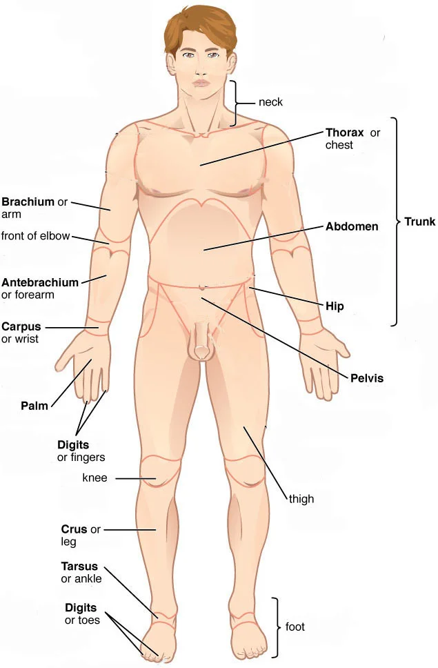
Directional anatomical terms are essential for describing the relative locations of different body structures. These terms allow an anatomist to describe one band of tissue as “inferior to” another. Similarly, a physician might describe a tumor as “superficial to” a deeper body structure. The human body’s numerous regions have specific terms to increase precision. The term “brachium” or “arm” is reserved for the “upper arm”. The term “antebrachium” or “forearm” is used rather than “lower arm”. Similarly, “femur” or “thigh” is correct, and “leg” or “crus” is reserved for the portion of the lower limb between the knee and the ankle. Ancient Greek and Latin words are used to build these anatomical terms. Regions are identified using terms such as “occipital” that are more precise than common phrases such as “the back of the head”. [1] [3]
Abduction and adduction movements are seen at condyloid, and saddle joints. [3]
To describe a structure, one must identify its location relative to these standard terms. The body’s organs are organized in one of two main cavities—dorsal (also referred to posterior) and ventral (also referred to anterior). These cavities are further sub-divided according to the structures present in each area. Images of the body’s interior commonly align along one of three planes: the sagittal, frontal, or transverse. The serous membranes have two layers—parietal and visceral—surrounding a fluid f [1]
Anatomical terminology is not designed to confuse but to eliminate ambiguity in clinical communication. By using precise language derived from ancient Greek and Latin, we ensure that descriptions of location and structure remain consistent, as these languages do not change meaning over time[1]. This precision is critical; for instance, without standard terms, it is unclear if a scar is on the forearm or at the base of the hand, or whether it is on the palm-side or back-side[1].
To understand these terms, one must recognise that they are constructed from roots, prefixes, and suffixes. The root typically refers to an organ, tissue, or condition, while prefixes or suffixes modify the root. For example, in hypertension, the prefix “hyper-” means “high” or “over,” and the root “tension” refers to pressure, resulting in the meaning of abnormally high blood pressure[1].
Body movements and positions are always described relative to a standard reference frame. This reference is the anatomical position, defined as an upright stance with the upper limbs at the side of the body and the palms facing forward[1]. When a patient is lying down, specific terms are used to describe orientation. A body lying face-down is described as prone, whereas a body lying face-up is described as supine[1]. These positional terms are frequently used during physical examinations or surgical procedures to ensure clear communication among healthcare providers[1]. [3]
DefinitionAnatomical Position
An upright stance with upper limbs at the side of the body and palms facing forward.
Movements such as flexion and extension are also defined relative to this position. Flexion of the knee is the bending of the knee to bring the foot toward the posterior thigh, while extension is the straightening of the knee[1]. Note that extension of the thigh beyond the anatomical standing position is greatly limited by the ligaments that support the hip joint[1]. These movements are seen at hinge, condyloid, saddle, and ball-and-socket joints of the limbs[1]. [3]
Directional terms within the nervous system also require careful classification. For convenience, terms like ventral and dorsal are used to reference structures within the spinal cord to underscore relationships between components. Furthermore, spinal nerve systems connecting to the brain are typically contralateral, meaning the right side of the body connects to the left side of the brain and vice versa. [4]
The peripheral nervous system (PNS) is everything else. [2]
A single axon can cross the boundary between these systems. For example, retinal axons are called the optic nerve in the PNS and the optic tract in the CNS. This naming convention clarifies which terms apply to the central or peripheral nervous systems. [2]
Anatomical position serves as the universal reference frame for describing the human body, ensuring that directional terms remain consistent regardless of the patient’s actual posture. In this standard position, the individual stands erect, faces forward, arms are at the sides, and palms face anteriorly. This convention allows clinicians to communicate precise locations without ambiguity.
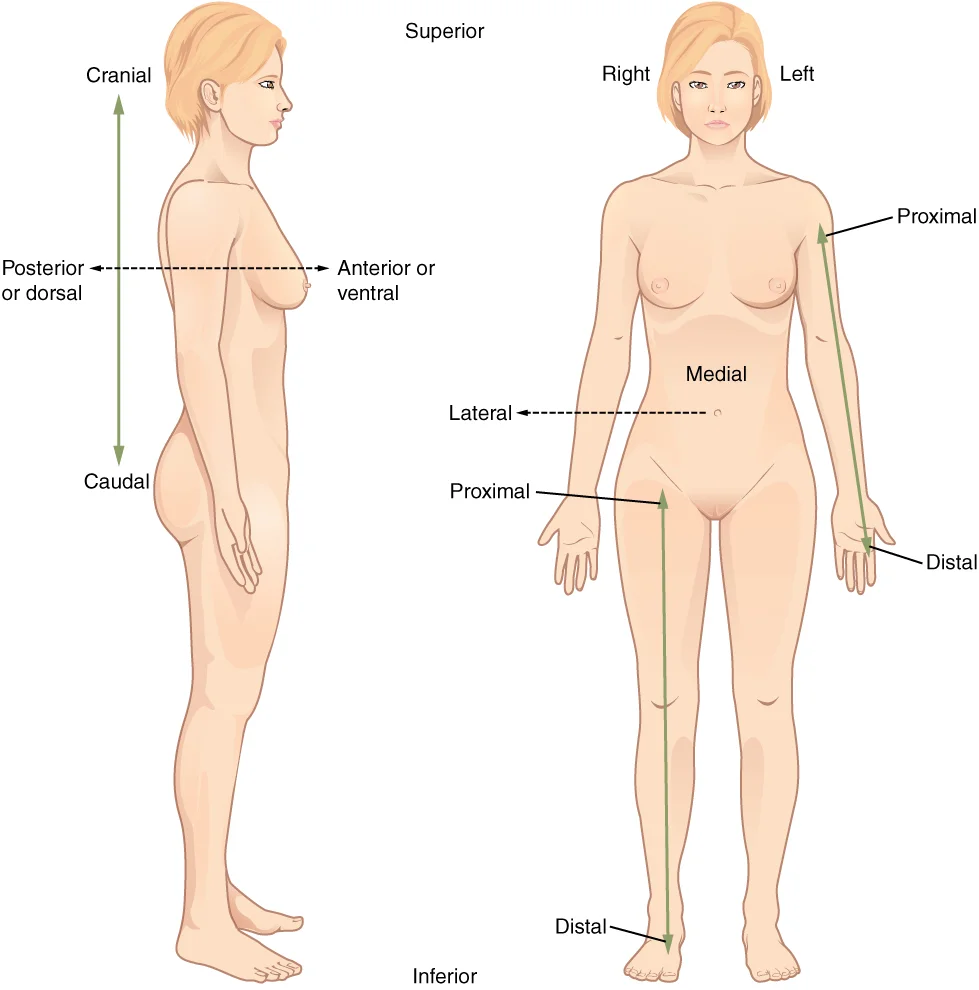
Figure 1Directional Terms
Long description
Paired directional terms are shown as applied to the human body.

Figure 1: Paired directional terms are shown as applied to the human body.
Directional terms describe the relative position of structures. Anterior (ventral) refers to the front of the body, while posterior (dorsal) refers to the back. Superior (cranial) indicates a position toward the head, whereas inferior (caudal) indicates a position toward the feet. Medial describes structures closer to the midline, and lateral describes those farther away. Distal and proximal are used for limbs; distal means farther from the point of attachment, and proximal means closer.
DefinitionAnatomical Position
The standard upright stance with feet together, arms at the sides, and palms facing forward, used as the reference for all directional descriptions.
Figure 2Body regions
Long description
Body regions. Labelled anatomical illustration from Wikimedia Commons, used under its open licence.

Figure 2: Body regions.
The head is superior to the thorax. The thorax is superior to the abdomen. The abdomen is superior to the pelvis. The right arm is lateral to the trunk. The left hand is distal to the left elbow. The right knee is proximal to the right ankle. The sternum is medial to the clavicles. The umbilicus is inferior to the xiphoid process.
The scapulae are posterior to the rib cage. The spine is posterior to the thoracic cavity. The gluteal region is posterior to the hip joint. The popliteal fossa is posterior to the knee.
Movements are described relative to anatomical planes. Flexion and extension typically occur in the sagittal plane. For the vertebral column, flexion is an anterior bending of the neck or trunk. Extension involves a posterior-directed motion, such as straightening from a flexed position. Lateral flexion of the vertebral column occurs in the coronal plane and is defined as the bending of the neck or trunk toward the right or left side. [3] [4]
Abduction and adduction motions occur within the coronal plane. Abduction moves the limb laterally away from the midline of the body. Adduction is the opposing movement that brings the limb toward the body or across the midline. For example, abduction is raising the arm at the shoulder joint, moving it laterally away from the body. Adduction brings the arm down to the side of the body. Spreading the fingers or toes apart is also abduction, while bringing the fingers or toes together is adduction. For the thumb, abduction is the anterior movement that brings the thumb to a 90° perpendicular position, pointing straight out from the palm. [3]
Understanding these terms is essential for localizing pathology. A lesion superior to the diaphragm is in the thorax. A fracture distal to the elbow is in the forearm. Knowing the anatomical position ensures that "left" and "right" are always from the patient’s perspective, not the observer’s.
Understanding the anatomical position and directional terms is fundamental for precise communication in medicine. These standardised descriptors allow clinicians to locate structures unambiguously, regardless of the patient's posture or the observer's perspective. Without a common language, describing the location of a lesion or the pathway of a nerve would be prone to error.
The nervous system is composed of two basic types of cells: neurons and glial cells. Glial cells provide a supportive framework for neurons, while neurons are functionally critical for communication. Each neuron consists of a cell body, or soma, and extensions known as processes. The axon is the specific process that connects a neuron to its target, serving as the primary output fibre. Dendrites are other processes that branch from the soma, typically receiving signals. [2] [4]
Directional terminology relies on the standard anatomical position: the body is erect, facing forward, with arms at the sides and palms facing anteriorly. In this position, 'anterior' refers to the front, and 'posterior' to the back. 'Superior' indicates a position towards the head, while 'inferior' indicates a position towards the feet. 'Medial' describes structures closer to the midline, and 'lateral' describes those further away. 'Proximal' and 'distal' are used for limbs, indicating closeness to or distance from the point of attachment to the trunk.
Terminology for bundles of axons varies by location within the nervous system. A bundle of axons located in the central nervous system (CNS) is termed a tract. Conversely, an equivalent bundle of axons in the peripheral nervous system (PNS) is called a nerve. This distinction is crucial for accurate diagnosis and documentation. The optic nerve provides a clear example of this nomenclature shift. The name changes at the optic chiasm, yet the axons remain continuous. [2]
Clinical pearlNerve vs Tract
Remember that 'nerve' refers to axon bundles in the PNS, while 'tract' refers to axon bundles in the CNS. The optic nerve becomes the optic tract at the chiasm.
Clinical scenarios often involve localising lesions based on these terms. For instance, a patient presenting with weakness in the lateral aspect of the arm may have a lesion affecting a specific nerve root or peripheral nerve. Precise use of 'lateral' versus 'medial' ensures the correct anatomical structure is investigated. Similarly, distinguishing between a tract lesion in the spinal cord and a nerve lesion in the limb guides diagnostic imaging and management.
| Term | Definition | Clinical Example |
|---|---|---|
| Anterior | Towards the front | Anterior abdominal wall incision |
| Posterior | Towards the back | Posterior cranial fossa fracture |
| Superior | Towards the head | Superior mesenteric artery |
| Inferior | Towards the feet | Inferior vena cava |
| Medial | Towards the midline | Medial malleolus fracture |
| Lateral | Away from midline | Lateral collateral ligament |
| Proximal | Closer to trunk | Proximal radius fracture |
| Distal | Further from trunk | Distal phalanx injury |
Accurate application of these terms prevents miscommunication during handovers, surgical planning, and radiological reporting. It ensures that every team member visualises the same anatomical reality.
The anatomical position is the standard reference frame for all directional terminology. In this position, the body is erect, facing forward, with arms at the sides and palms facing anteriorly. This orientation ensures that terms like 'medial' and 'lateral' have consistent meanings regardless of the patient's actual posture. For instance, the thumb is lateral to the little finger in the anatomical position, even if the hand is pronated.
Directional terms describe the relative location of structures. 'Superior' (cranial) indicates a position towards the head, while 'inferior' (caudal) indicates a position towards the feet. 'Anterior' (ventral) refers to the front of the body, and 'posterior' (dorsal) refers to the back. 'Medial' denotes proximity to the midline, whereas 'lateral' denotes distance from it. 'Proximal' and 'distal' are used for limbs, referring to closeness to or distance from the point of attachment to the trunk.
The abdominopelvic cavity is often divided for clinical examination. The quadrants approach uses one horizontal line through the umbilicus and one vertical line intersecting it, creating four regions. This method is simpler and more commonly used in medicine than the nine-region approach. [1][4]
Serous membranes line the thoracic and abdominopelvic cavities. The parietal layer lines the cavity walls, while the visceral layer covers the organs. Between these layers lies a thin, fluid-filled space that reduces friction during movement. [1]
The nervous system is divided into the central and peripheral systems. The central nervous system comprises the brain and spinal cord. The peripheral nervous system includes all neural elements outside these structures. Although some peripheral elements reside within the cranial or vertebral cavities, the system is defined by its location beyond the brain and spinal cord. [2]
The inner ear contains the bony labyrinth, which includes the cochlea for hearing and the vestibule for balance. The cochlea houses the spiral ganglia, whose neurons transmit sound signals. The vestibular system includes three semicircular canals oriented in different planes to detect rotational head movements. The ampulla within each canal contains hair cells that respond to fluid displacement caused by rotation. [4]
Somatosensory signals such as pain and temperature are transduced by free nerve endings. Nociceptors respond to stimuli exceeding a specific threshold, including mechanical, chemical, or thermal damage. Capsaicin, the active component in hot peppers, binds to ion channels in nociceptors sensitive to temperatures above 37°C. [4]
Vision relies on photoreceptors in the retina. Rods contain rhodopsin and are sensitive to low light, while cones contain opsins sensitive to specific wavelengths. The outer segments of these cells undergo chemical changes when light strikes them, initiating neural signals. [4]
A common trap is confusing 'medial' with 'midline'. Medial means towards the midline, not necessarily on it. Another trap is misusing 'proximal' and 'distal' for axial structures; these terms apply only to limbs. Remember that in the anatomical position, the palms face forward, making the thumbs lateral.
Exam trapPalms Orientation
In the anatomical position, palms face anteriorly. This makes the thumb lateral to the little finger, a key distinction for upper limb anatomy.
DefinitionAnatomical Position
The standard reference posture: erect, facing forward, arms at sides, palms forward. All directional terms are defined relative to this position.
| Method | Lines Used | Regions Created | Clinical Use |
|---|---|---|---|
| Quadrants | One horizontal (umbilicus), one vertical | Four | Commonly used in medicine |
| Nine Regions | Two vertical (clavicle midpoints), two horizontal (ribs/pelvis) | Nine | More detailed regional approach |
| Region | Function | Key Structures |
|---|---|---|
| Cochlea | Hearing | Spiral ganglia, oval window |
| Vestibule | Balance | Semicircular canals, ampulla, |
Students frequently confuse the anatomical position with everyday postures, leading to errors in directional terminology. The anatomical position requires the body to be erect, facing forward, with arms at the sides and palms facing anteriorly. A common error is assuming that because the palms face forward in this position, they are also facing the body; in reality, the thumbs point laterally. This distinction is critical when describing the orientation of structures in the upper limb. For insta
Another frequent mistake involves the distinction between central and peripheral nervous tissue characteristics. Students often assume that gray and white matter distinctions apply equally to peripheral nerves. However, the distinction between gray matter and white matter is most often applied to central nervous tissue, which has large regions that can be seen with the unaided eye. Peripheral nerves require microscopic examination and artificial staining to differentiate their components clearly. This is because peripheral structures do not present the same macroscopic colour contrast as the brain or spinal cord. Understanding this difference prevents misinterpretation of histological slides and clinical specimens. [2]
Misunderstanding the function of proprioceptors is also common. Students may confuse interoceptors with proprioceptors, leading to errors in sensory physiology. An exteroceptor is a receptor that is located near a stimulus in the external environment, such as the somatosensory receptors that are located in the skin. In contrast, a proprioceptor is a receptor located near a moving part of the body, such as a muscle, that interprets the positions of the tissues as they move. Interoceptors interpret stimuli from internal organs and tissues, such as the receptors that sense the increase in blood pressure in the aorta or carotid sinus. Confusing these categories results in incorrect localisation of sensory deficits in neurological examinations. [4]
Exam trapPalmar Orientation
Remember that in the anatomical position, the palms face anteriorly, not medially towards the body. The thumbs point laterally.
DefinitionAnatomical Position
The standard reference posture: erect, facing forward, arms at sides, palms forward, thumbs lateral.
Knowledge of anatomical position and directional terms is fundamental at the bedside for accurate clinical communication. When documenting a lesion, precise terminology ensures that any healthcare professional can locate the structure without ambiguity. For example, describing a rash as "distal to the elbow" on the "medial aspect of the forearm" provides a clear map. This precision is vital when coordinating care between different specialties, such as dermatology, orthopaedics, and general practice. Miscommunication can lead to incorrect diagnoses or inappropriate treatments. Therefore, mastering these terms is not merely an academic exercise but a clinical necessity. [3]
In the operating theatre, directional terms guide surgical approaches and incisions. Surgeons must understand the spatial relationships between structures to avoid damaging critical nerves or vessels. For instance, during neck surgery, understanding the rotational capabilities of the cervical spine is important. Rotation can occur within the vertebral column, at a pivot joint, or at a ball-and-socket joint. Specifically, at the atlantoaxial joint, the first cervical (C1) vertebra (atlas) rotates around the dens, the upward projection from the second cervical (C2) vertebra (axis). This allows the head to rotate from side to side as when shaking the head “no”. Awareness of these movements helps in positioning patients safely and anticipating anatomical variations during procedures. [4] [3]
On imaging, directional terms are essential for interpreting radiological scans. Radiologists describe findings relative to the anatomical position, regardless of the patient's position during the scan. For example, in a CT scan of the brain, the frontal lobe is anterior to the occipital lobe. Students must be able to correlate cross-sectional images with standard anatomical descriptions. This skill is particularly important in neurology, where lesions are localised based on their position relative to known landmarks. Understanding the layout of the nervous system helps in predicting the clinical presentation of a lesion. The nervous system is a very complex structure. Within the brain, many different and separate regions are responsible for many different and separate functions. [1] [2]
Directional Terminology Application
- Identify StructureLocate the anatomical feature in question.
- Question: Is it in the limb?
Yes
- Use Proximal/DistalRelative to the trunk.
- Use Medial/LateralRelative to the midline.
No
- Use Anterior/PosteriorRelative to the front/back.
- Use Superior/InferiorRelative to head/feet.
It helps to remember that supination is the motion you use when scooping up soup with a spoon. [3]
NoteClinical Relevance
Accurate terminology prevents errors in patient handovers and surgical planning.
- The anatomical position is the standard reference for all directional terms.
- Palms face anteriorly and thumbs point laterally in the anatomical position.
- Gray and white matter distinctions are primarily macroscopic in the CNS.
- Proprioceptors monitor body position, while interoceptors monitor internal organs.
- The atlantoaxial joint allows for rotation of the head.
- Supination positions the palm anteriorly, like holding soup.
- Precise terminology is essential for clinical communication and imaging.
- Directional terms remain constant regardless of patient position.
Answer each one in your head first, then open the answer.
Which bone rotates around the dens during head rotation?
Show answerHide answer
Axis (C2)What is the primary function of a proprioceptor?
Show answerHide answer
Interpreting positions of moving body partsIn the anatomical position, which way do the palms face?
Show answerHide answer
AnteriorlyWhich joint allows the radius to rotate during pronation and supination?
Show answerHide answer
Proximal radioulnar jointWhat term describes receptors located near external stimuli?
Show answerHide answer
Exteroceptor
- Betts JG, Young KA, Wise JA, et al. Anatomical Terminology. OpenStax, Anatomy and Physiology 2e (CC BY-NC-SA 4.0). Accessed .
- Betts JG, Young KA, Wise JA, et al. Basic Structure and Function of the Nervous System. OpenStax, Anatomy and Physiology 2e (CC BY-NC-SA 4.0). Accessed .
- Betts JG, Young KA, Wise JA, et al. Types of Body Movements. OpenStax, Anatomy and Physiology 2e (CC BY-NC-SA 4.0). Accessed .
- Betts JG, Young KA, Wise JA, et al. Sensory Perception. OpenStax, Anatomy and Physiology 2e (CC BY-NC-SA 4.0). Accessed .
Figure credits
Educational content for medical students and doctors. It is not a substitute for clinical judgement or local guidelines.