Body regions and cavities
Overview of body regions, cavities, and serous membranes.
Surface regions of the body are identified using precise anatomical terminology derived from ancient Greek and Latin, which is more accurate than common descriptive phrases. For instance, the term "occipital" specifically denotes the back of the head. To map these structures consistently, the normal anatomical position serves as the standard reference. Directional terms such as anterior and posterior are essential for describing the relative locations of body structures. Internal images typically align along one of three planes: the sagittal, frontal, or transverse. The body’s organs are organised into two main cavities: the dorsal (posterior) cavity and the ventral (anterior) cavity. These major divisions are further sub-divided according to the specific structures present within each area. [1]
DefinitionSerous Membrane
A serous membrane, or serosa, is a thin membrane that covers the walls and organs in the thoracic and abdominopelvic cavities. [1]
There are three primary serous cavities and their associated membranes. The pleura is the serous membrane that encloses the pleural cavity, which surrounds the lungs. The pericardium is the serous membrane that encloses the pericardial cavity, surrounding the heart. The peritoneum is the serous membrane that encloses the peritoneal cavity, surrounding several organs in the abdominopelvic cavity. These membranes form fluid-filled sacs designed to cushion and reduce friction on internal organs during movement, such as lung inflation or heart contraction. Both parietal and visceral serosa secrete thin, slippery serous fluid located within these cavities. The pleural cavity reduces friction between the lungs and the body wall. Therefore, serous membranes provide additional protection to the viscera by reducing friction that could lead to inflammation. [1] [2]
DefinitionViscera
The visceral layer of the membrane covers the organs, known as the viscera. [1]
A tissue membrane is a thin layer or sheet of cells that covers the outside of the body, the organs, internal passageways leading to the exterior, and the lining of moveable joint cavities. There are two basic types: connective tissue and epithelial membranes. A serous membrane is an epithelial membrane composed of mesodermally derived epithelium called the mesothelium, supported by connective tissue. These membranes line the coelomic cavities, which are cavities that do not open to the outside. They are essentially membranous bags, with mesothelium lining the inside and connective tissue on the outside. Serous fluid secreted by the cells of the thin squamous mesothelium lubricates the membrane and reduces abrasion and friction between organs.
The membrane directly surrounding the heart is the pericardium or pericardial sac. It also surrounds the roots of the major vessels. The pericardium consists of two distinct sublayers: the sturdy outer fibrous pericardium and the inner serous pericardium. The fibrous pericardium is made of tough, dense connective tissue that protects the heart and maintains its position in the thorax. The more delicate serous pericardium consists of two layers: the parietal pericardium, fused to the fibrous pericardium, and an inner visceral pericardium, or epicardium, fused to the heart. The pericardial cavity, filled with lubricating serous fluid, lies between the epicardium and the parietal pericardium. In most organs, visceral serous membranes are microscopic. However, in the heart, it is a macroscopic layer consisting of simple squamous epithelium called a mesothelium, reinforced with loose, irregular, or areolar connective tissue. This mesothelium secretes the lubricating serous fluid that fills the pericardial cavity and reduces friction as the heart contracts. [1] [2]
DefinitionParietal Layer
The parietal layer of the membrane lines the walls of the body cavity. [1]
Between the parietal and visceral layers is a very thin, fluid-filled serous space. This potential space allows for movement without tissue damage. It initially travels within the mediastinum to the left of the vertebral column. As it passes through the thoracic region, it gives rise to visceral branches and parietal branches. The visceral branches include the bronchial arteries, per [1][4]
Anatomical precision relies on a standardized reference framework known as the anatomical position. In this position, the body stands upright with feet at shoulder width and parallel, toes forward, and upper limbs held to each side with palms facing forward. This orientation ensures that directional terms remain consistent regardless of the subject’s actual posture. For instance, the term “anterior” refers to the front surface, such as the palm side of the wrist, even if the hand is placed palm down. When a body is lying down, it is described as prone if face-down or supine if face-up. These positional terms are critical for describing physical examinations and surgical procedures accurately. [1]
The body is divided into specific regions to facilitate precise communication. The term “arm” (brachium) is reserved for the upper arm, while “forearm” (antebrachium) refers to the lower segment. Directional terms such as “inferior” and “superficial” are essential for locating structures relative to one another. [1]
Internally, the body is organized into major compartments separated by membranes. The dorsal (posterior) and ventral (anterior) cavities are the largest of these compartments. The ventral cavity accommodates organs that change size and shape, such as the lungs, heart, stomach, and intestines, allowing them to expand without distorting adjacent tissues. [1] [4]
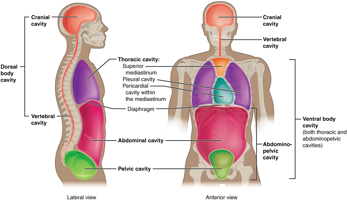
The dorsal cavity is subdivided into the cranial cavity, which houses the brain, and the spinal (vertebral) cavity, which encloses the spinal cord. These two cavities are continuous, reflecting the uninterrupted nature of the central nervous system. [1]
Figure 1Dorsal Ventral Body Cavities
Long description
The ventral cavity includes the thoracic and abdominopelvic cavities and their subdivisions. The dorsal cavity includes the cranial and spinal cavities.

Figure 1: The ventral cavity includes the thoracic and abdominopelvic cavities and their subdivisions.
At the microscopic level, cells are organized into four broad tissue categories: epithelial, connective, muscle, and nervous[1]. Epithelial tissue covers exterior surfaces, lines internal cavities, and forms glands[1]. Connective tissue binds cells and organs, providing support and protection[1]. Muscle tissue is excitable and contracts to provide movement, occurring as skeletal, smooth, or cardiac types[1]. Nervous tissue propagates electrochemical signals to communicate between body regions[1]. These tissues derive from three embryonic germ layers: ectoderm, mesoderm, and endoderm[1]. [3]
In the limbs and neck, vessels are organized into superficial and deeper levels. Deep veins generally correspond to complementary arteries, while superficial veins lack direct arterial counterparts[1]. Superficial veins contribute to thermoregulation; in warm conditions, blood is diverted to these veins to dissipate heat, whereas in cold weather, constriction diverts blood deeper to retain heat[1]. [4]
| Cavity | Subdivisions | Primary Contents |
|---|---|---|
| Dorsal (Posterior) | Cranial, Spinal | Brain, Spinal Cord |
| Ventral (Anterior) | Thoracic, Abdominopelvic | Heart, Lungs, Stomach, Intestines |
DefinitionAnatomical Position
The standard reference posture: standing upright, feet parallel, arms at sides, palms facing forward. All directional terms are defined relative to this position.
Exam trapRegional Terminology
The term “leg” refers only to the segment between the knee and ankle, not the entire lower limb. The “arm” refers only to the upper arm.
To interpret medical imaging accurately, one must understand the geometric planes used to section the body. A section is a two-dimensional surface of a three-dimensional structure that has been cut. Modern medical imaging devices enable clinicians to obtain “virtual sections” of living bodies. We call these scans. Body sections and scans can be correctly interpreted, however, only if the viewer understands the plane along which the section was made. A plane is an imaginary two-dimensional surfac [1]
The sagittal plane divides the body into left and right portions. The coronal (frontal) plane divides the body into anterior and posterior sections. The transverse (axial) plane divides the body into superior and inferior parts.
DefinitionAnatomical Planes
A plane is an imaginary two-dimensional surface that passes through the body. There are three planes commonly referred to in anatomy and medicine. [1]
For clinical communication, such as locating abdominal pain, providers divide the abdominal cavity into specific regions. To promote clear communication, for instance about the location of a patient’s abdominal pain or a suspicious mass, health care providers typically divide up the cavity into either nine regions or four quadrants. This standardisation ensures that a "right upper quadrant" pain is universally understood. [1]
NoteClinical Relevance
To promote clear communication, for instance about the location of a patient’s abdominal pain or a suspicious mass, health care providers typically divide up the cavity into either nine regions or four quadrants.
The body is constructed from tissues, which are groups of cells sharing a common embryonic origin. The cells within a tissue share a common embryonic origin. Microscopic observation reveals that the cells in a tissue share morphological features and are arranged in an orderly pattern that achieves the tissue’s functions. From the evolutionary perspective, tissues appear in more complex organisms. [1][4]
DefinitionTissue
The term tissue is used to describe a group of cells found together in the body. The cells within a tissue share a common embryonic origin. [1]
Membranes line these cavities. The epithelial membrane is composed of epithelium attached to a layer of connective tissue, for example, your skin. The mucous membrane is also a composite of connective and epithelial tissues. Mucus, produced by the epithelial exocrine glands, covers the epithelial layer. The underlying connective tissue, called the lamina propr [3]
DefinitionMucous Membrane
Sometimes called mucosae, these epithelial membranes line the body cavities and hollow passageways that open to the external environment. [3]
The intercostal muscles lie between the ribs, providing structural support. The mediastinum, containing the heart and great vessels, sits centrally. [4]
Clinical pearlAortic Anatomy
After exiting the heart, the ascending aorta moves in a superior direction for approximately 5 cm and ends at the sternal angle. [4]
NoteCirculatory Function
This includes the generalized and more specialized functions of transport of materials, capillary exchange, maintaining health by transporting white blood cells and various immunoglobulins (antibodies), hemostasis, regulation of body temperature, and helping to maintain acid-base balance.
Systemic veins return blood to the right atrium. Since the blood has already passed through the systemic capillaries, it will be relatively low in oxygen concentration. [4]
Exam trapVenous Variability
However, there is a great deal more variability in the venous circulation than normally occurs in the arteries. [4]
As it passes through the body wall and enters the thorax, the axillary vein becomes the subclavian vein. [4]
DefinitionSubclavian Vein Formation
As it passes through the body wall and enters the thorax, the axillary vein becomes the subclavian vein.
Understanding the topographical organisation of the body is fundamental for accurate physical examination and procedural intervention. Clinicians utilise specific anatomical landmarks to divide the abdominal cavity into manageable sections. The more detailed regional approach subdivides the cavity with one horizontal line immediately inferior to the ribs and one immediately superior to the pelvis, and two vertical lines drawn as if dropped from the midpoint of each clavicle (collarbone). There are nine resulting regions. The simpler quadrants approach, which is more commonly used in medicine, subdivides the cavity with one horizontal and one vertical line that intersect at the patient’s umbilicus (navel). This division allows for precise localisation of pain or pathology, such as distinguishing between appendicitis and renal colic. [1]
DefinitionAbdominal Regions
The abdomen is divided into nine regions using two horizontal and two vertical lines, or four quadrants using a single cross through the umbilicus.
The cardiovascular system’s regional anatomy dictates the flow of blood and the presentation of disease. The human heart consists of four chambers: The left side and the right side each have one atrium and one ventricle. Each of the upper chambers, the right atrium (plural = atria) and the left atrium, acts as a receiving chamber and contracts to push blood into the lower chambers, the right ventricle and the left ventricle. The ventricles serve as the primary pumping chambers of the heart, propelling blood to the lungs or to the rest of the body. The right atrium serves as the receiving chamber for blood returning to the heart from the systemic circulation. The two major systemic veins, the superior and inferior venae cavae, and the large coronary vein called the coronary sinus that drains the heart myocardium empty into the right atrium. [2] [4]
| Vein | Drainage Area |
|---|---|
| Superior Vena Cava | Head, neck, upper limbs, and thoracic region |
| Inferior Vena Cava | Lower limbs and abdominopelvic region |
The pulmonary circuit is unique in its oxygenation status. The right ventricle pumps deoxygenated blood into the pulmonary trunk, which leads toward the lungs and bifurcates into the left and right pulmonary arteries. These vessels in turn branch many times before reaching the pulmonary capillaries, where gas exchange occurs: Carbon dioxide exits the blood and oxygen enters. The pulmonary trunk arteries and their branches are the only arteries in the post-natal body that carry relatively deoxygenated blood. Highly oxygenated blood returning from the pulmonary capillaries in the lungs passes through a series of vessels that join together to form the pulmonary veins—the only post-natal veins in the body that carry highly oxygenated blood. [2] [4]
Clinical pearlPulmonary Vessels
Pulmonary arteries carry deoxygenated blood; pulmonary veins carry oxygenated blood. This is the reverse of systemic circulation.
The intercostal muscles lie between the ribs, providing structural support. The mediastinum, containing the heart and great vessels, sits centrally.
The integumentary system provides a protective barrier. The skin is an epithelial membrane also called the cutaneous membrane. It is a stratified squamous epithelial membrane resting on top of connective tissue. The apical surface of this membrane is exposed to the external environment and is covered with dead, keratinized cells that help protect the body from desiccation and pathogens. [3]
Venous return from the head and neck involves complex anastomoses. The superior vena cava drains most of the body superior to the diaphragm. On both the left and right sides, the subclavian vein forms when the axillary vein passes through the body wall from the axillary region. It fuses with the external and internal jugular veins from the head and neck to form the brachiocephalic vein. Each vertebral vein also flows into the brachiocephalic vein close to this fusion. These veins arise from the [4]
Lower limb venous drainage is critical for assessing deep vein thrombosis. It is palpable in patients without excessive adipose tissue. [4]
The heart’s mechanical workload is immense, contracting approximately 108,000 times daily. In a resting adult, each major pumping chamber ejects approximately 70 mL of blood per contraction. This volume equates to 5.25 liters of fluid per minute. Over a full day, the heart pumps approximately 14,000 liters of blood. The organ’s shape resembles a pinecone, broad at the superior surface and tapering to the apex. A typical heart measures 12 cm in length, 8 cm in width, and 6 cm in thickness. Female hearts weigh approximately 250–300 grams, while male hearts weigh approximately 300–350 grams. During diastole, the atrioventricular valves are open and the semilunar valves are closed. During systole, the atrioventricular valves close while the semilunar valves open to eject blood. The left subclavian artery becomes the axillary artery as it passes into the axillary region. The aortic arch gives rise to three major branches: the brachiocephalic artery, the left common carotid artery, and the left subclavian artery. The common carotid artery divides into internal and external branches. The celiac trunk divides into the left gastric, splenic, and common hepatic arteries. The common iliac veins unite at the level of L5 to form the inferior vena cava. [2] [4]
The heart’s vital importance is underscored by its relentless activity. Assuming an average rate of contraction of 75 contractions per minute, a human heart would contract approximately 108,000 times in one day. This activity scales to more than 39 million times in one year and nearly 3 billion times during a 75-year lifespan. Each of the major pumping chambers of the heart ejects approximately 70 mL blood per contraction in a resting adult. This output results in 5.25 liters of fluid per minute. Daily output reaches approximately 14,000 liters. Over one year, that would equal 10,000,000 liters or 2.6 million gallons of blood sent through roughly 60,000 miles of vessels. [2]
The shape of the heart is similar to a pinecone, rather broad at the superior surface and tapering to the apex. A typical heart is approximately the size of your fist: 12 cm (5 in) in length, 8 cm (3.5 in) wide, and 6 cm (2.5 in) in thickness. Given the size difference between most members of the sexes, the weight of a female heart is approximately 250–300 grams (9 to 11 ounces). The weight of a male heart is approximately 300–350 grams (11 to 12 ounces). Cardiac muscle responds to exercise via hypertrophy, where exercise results in the addition of protein myofilaments that increase the size of the individual cells without increasing their numbers. [2]
Valvular status defines the cardiac cycle phases. In diastole, the two atrioventricular valves are open and the two semilunar valves are closed. This occurs when both atria and ventricles are relaxed and when the atria contract to pump blood into the ventricles. In systole, the atrioventricular valves are closed while the two semilunar valves are open. This occurs when the ventricles contract to eject blood into the pulmonary trunk and aorta. Closure of the two atrioventricular valves prevents blood from being forced back into the atria.
Arteries change names as they pass anatomical landmarks. The left subclavian artery becomes the axillary artery as it passes through the body wall and into the axillary region. It then becomes the brachial artery as it flows from the axillary region into the upper arm. The aortic arch has three major branches: the brachiocephalic artery, the left common carotid artery, and the left subclavian artery. Each is classified as an elastic artery. The common carotid artery divides into internal and external carotid arteries. The right common carotid artery arises from the brachiocephalic artery and the left common carotid artery arises directly from the aortic arch. [4]
The celiac trunk divides into the left gastric artery to supply blood to the stomach and esophagus, the splenic artery to supply blood to the spleen, and the common hepatic artery. The external and internal iliac veins combine near the inferior portion of the sacroiliac joint to form the common iliac vein. The common iliac veins come together at the level of L5 to form the inferior vena cava. [4]
Students frequently struggle with the precision of anatomical language, leading to ambiguity in clinical communication. A common error is describing a scar as "above the wrist" without specifying whether it is on the forearm or at the base of the hand, or on the palmar or dorsal surface. Anatomical terms derive from ancient Greek and Latin words, which are no longer used in everyday conversation, ensuring their meaning does not change over time. Using precise terminology eliminates this ambiguity and reduces medical errors. [1]
Another frequent mistake involves the subdivisions of the anterior cavity. Students often assume a physical membrane divides the abdominal and pelvic cavities. In reality, although no membrane physically divides the abdominopelvic cavity, it is useful to distinguish between the abdominal cavity, which houses digestive organs, and the pelvic cavity, which houses organs of reproduction. The diaphragm forms the floor of the thoracic cavity and separates it from the more inferior abdominopelvic cavity. [1]
Misunderstanding the coronary circulation pattern is also prevalent. Many assume blood flow to the heart is continuous. However, coronary circulation is not continuous; rather, it cycles, reaching a peak when the heart muscle is relaxed and nearly ceasing while it is contracting. This distinction is critical when interpreting ischaemic events. [2]
Confusion often arises regarding the branches of the aorta. Students may misidentify the origin of the inferior mesenteric artery. The inferior mesenteric artery supplies blood to the distal segment of the large intestine, including the rectum, and arises approximately 5 cm superior to the common iliac arteries. It does not arise directly from the iliacs.
The termination of the abdominal aorta is another area of error. The abdominal aorta formally ends at approximately the level of vertebra L4, where it bifurcates to form the common iliac arteries. It does not terminate at the diaphragm; that is the end of the thoracic aorta.
Regarding brain blood supply, students sometimes underestimate the constancy of flow. Blood flow to the brain is remarkably constant, with approximately 20 percent of blood flow directed to this organ at any given time. Interruption for even a few seconds can cause a transient ischemic attack.
Damage timelines are often misremembered. Loss of blood flow for longer periods, typically between 3 and 4 minutes, will likely produce irreversible brain damage or a stroke. This is distinct from the temporary loss of neurological function seen in TIAs. [4]
Finally, the structure of the heart muscle is often oversimplified. The muscle pattern is elegant and complex, as the muscle cells swirl and spiral around the chambers of the heart. They form a figure 8 pattern around the atria and around the bases of the great vessels. This complex swirling pattern allows the heart to pump blood more effectively than a simple linear pattern would. [4]
At the bedside, precise anatomical knowledge guides physical examination and diagnosis. When assessing a patient with chest pain, understanding that the heart is located in the mediastinum within the thoracic cavity is essential. The anterior cavity has two main subdivisions: the thoracic cavity and the abdominopelvic cavity. Knowing that the diaphragm separates these cavities helps in interpreting breath sounds and abdominal tenderness.
The intercostal muscles lie between the ribs, providing structural support. The mediastinum, containing the heart and great vessels, sits centrally.
Imaging interpretation depends on knowing vertebral levels. The abdominal aorta terminates when it bifurcates into the two common iliac arteries at the level of the fourth lumbar vertebra. Radiologists use this landmark to identify the end of the abdominal aorta on CT scans. Similarly, the inferior mesenteric artery arises approximately 5 cm superior to the common iliac arteries, a key landmark for identifying vascular anomalies. [4]
In examinations, students must distinguish between valve types. The valves between the atria and ventricles are known generically as atrioventricular valves. The valves at the openings that lead to the pulmonary trunk and aorta are known generically as semilunar valves. The pulmonary semilunar valve prevents backflow from the pulmonary trunk.
Understanding the blood supply to the brain is critical for neurological assessments. The internal carotid arteries along with the vertebral arteries are the two primary suppliers of blood to the human brain. When blood flow is interrupted, even for just a few seconds, a transient ischemic attack (TIA), or mini-stroke, may occur. Recognising these timelines aids in triage.
The structure of the myocardium is relevant in cardiology. The middle and thickest layer is the myocardium, made largely of cardiac muscle cells. It is built upon a framework of collagenous fibers, plus the blood vessels that supply the myocardium and the nerve fibers that help regulate the heart. This knowledge explains the mechanics of contraction.
The septum between the atria and ventricles is known as the atrioventricular septum. It is marked by the presence of four openings that allow blood to move from the atria into the ventricles and from the ventricles into the pulmonary trunk and aorta. This anatomical detail is fundamental for understanding congenital heart defects.
The right ventricle ejects blood into the pulmonary trunk, which branches into the left and right pulmonary arteries that carry it to each lung. The superior surface of the right ventricle begins to taper as it approaches the pulmonary trunk. This anatomical relationship is visible on echocardiography.
Aortic Branching and Termination
- Start: Thoracic AortaDescends in thorax
- Question: Reaches Diaphragm?
Yes
- Passes through Aortic HiatusBecomes Abdominal Aorta
No
- Continue DescentGive off branches
- Abdominal AortaLeft of vertebral column
- Question: Level L4?
Yes
- BifurcationForms Common Iliac Arteries
No
- Give off Branchese.g. Inferior Mesenteric Artery
Anatomical terms derive from ancient Greek and Latin words, ensuring consistent meaning. The anterior cavity has two main subdivisions: the thoracic cavity and the abdominopelvic cavity. The diaphragm forms the floor of the thoracic cavity and separates it from the abdominopelvic cavity. Coronary circulation is not continuous; it cycles, peaking during relaxation. The aortic arch ends at the level of the intervertebral disk between T4 and T5. The abdominal aorta terminates at the level of the fourth lumbar vertebra. The inferior mesenteric artery arises approximately 5 cm superior to the common iliac arteries. Blood flow to the brain is remarkably constant, with approximately 20 percent directed to it.
Answer each one in your head first, then open the answer.
What ensures the meaning of anatomical terms does not change?
Show answerHide answer
Anatomical terms derive from ancient Greek and Latin words, which are no longer used in everyday conversationWhat separates the thoracic cavity from the abdominopelvic cavity?
Show answerHide answer
The diaphragm forms the floor of the thoracic cavity and separates it from the more inferior abdominopelvic cavityWhen does coronary circulation reach its peak?
Show answerHide answer
Coronary circulation is not continuous; rather, it cycles, reaching a peak when the heart muscle is relaxedAt what vertebral level does the abdominal aorta bifurcate?
Show answerHide answer
The abdominal aorta terminates when it bifurcates into the two common iliac arteries at the level of the fourth lumbar vertebraHow long can blood flow be interrupted before irreversible brain damage occurs?
Show answerHide answer
Loss of blood flow for longer periods, typically between 3 and 4 minutes, will likely produce irreversible brain damage
- Betts JG, Young KA, Wise JA, et al. Anatomical Terminology. OpenStax, Anatomy and Physiology 2e (CC BY-NC-SA 4.0). Accessed .
- Betts JG, Young KA, Wise JA, et al. Heart Anatomy. OpenStax, Anatomy and Physiology 2e (CC BY-NC-SA 4.0). Accessed .
- Betts JG, Young KA, Wise JA, et al. Types of Tissues. OpenStax, Anatomy and Physiology 2e (CC BY-NC-SA 4.0). Accessed .
- Betts JG, Young KA, Wise JA, et al. Circulatory Pathways. OpenStax, Anatomy and Physiology 2e (CC BY-NC-SA 4.0). Accessed .
Figure credits
Educational content for medical students and doctors. It is not a substitute for clinical judgement or local guidelines.