Ankle joint
The ankle joint is a uniaxial hinge joint formed by the tibia, fibula, and talus.
Figure 1919 Ankle Feet Joints
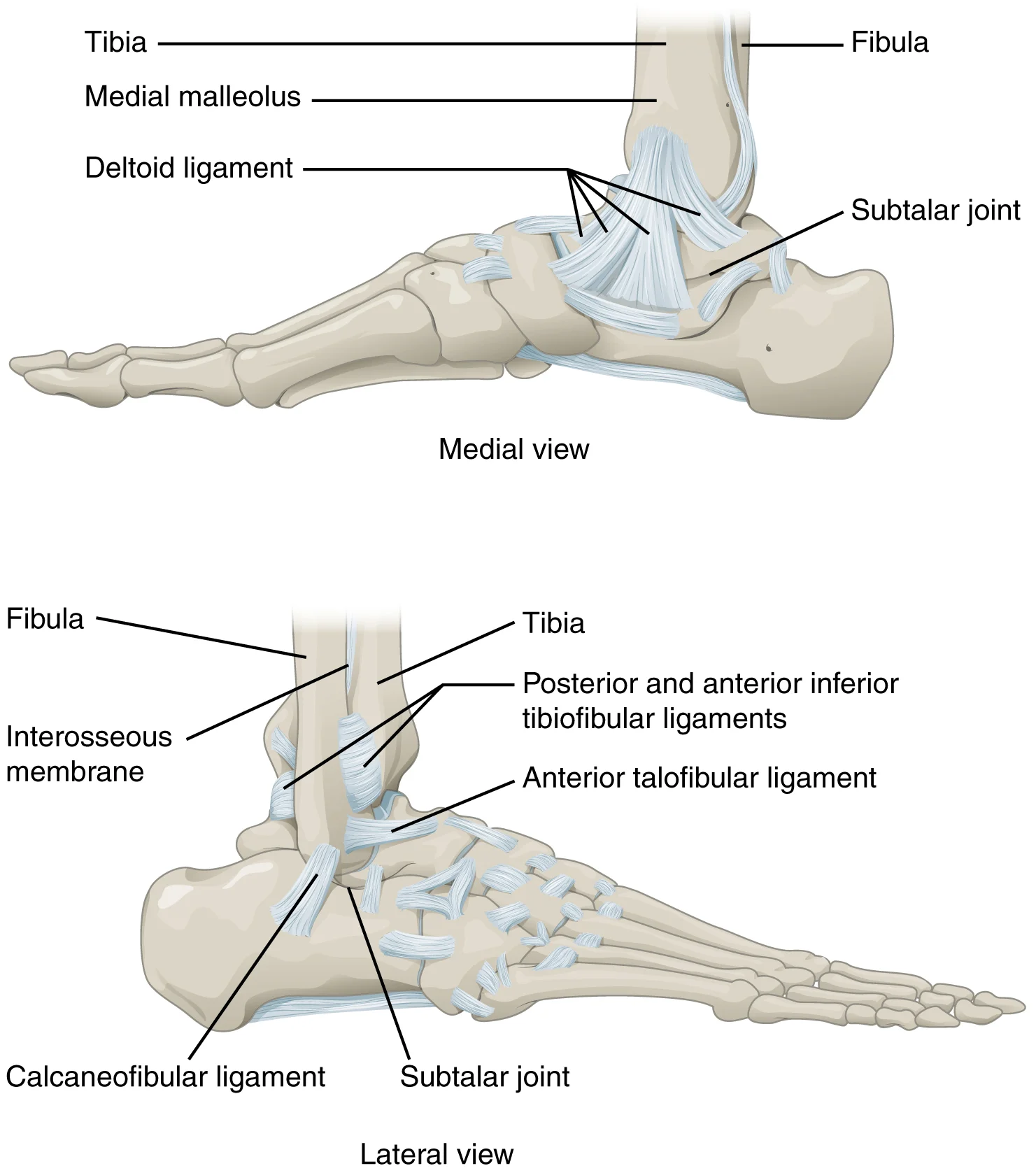
Long description
The talocrural (ankle) joint is a uniaxial hinge joint that only allows for dorsiflexion or plantar flexion of the foot. Movements at the subtalar joint, between the talus and calcaneus bones, combined with motions at other intertarsal joints, enables eversion/inversion movements of the foot. Ligame

Figure 1: The talocrural (ankle) joint is a uniaxial hinge joint that only allows for dorsiflexion or plantar flexion of the foot.
The ankle joint, technically known as the talocrural joint, is a synovial joint that is functionally classified as a diarthrosis. It is structurally a hinge joint, which allows movement primarily in one plane. This specific structural classification dictates that the joint permits dorsiflexion and plantar flexion of the foot. The joint is formed by the articulation between the trochlea of the talus and the distal ends of the tibia and fibula. The medial ma [1][3]
On the medial side is the broad deltoid ligament. The deltoid ligament supports the ankle joint and also resists excessive eversion of the foot. [1]
Injuries to the ankle are common and often involve these supporting structures. An inversion ankle sprain is a common injury that results in injury to one or more of the lateral ankle ligaments. Fractures of the malleoli can also disrupt the integrity of the ankle mortise. While the talocrural joint allows only dorsiflexion and plantar flexion, gliding motions at the subtalar and intertarsal joints of the foot allow for inversion and eversion of the foot. The ability of the bones to move smoothly against each other within the joint cavity provides freedom of joint movement. Synovial joints are strengthened by the presence of ligaments, which hold the bones together and resist excessive or abnormal movements of the joint. Muscles and their tendons acting across a joint can also increase their contractile strength when needed, thus providing indirect support for the joint. [1] [3]
The ankle joint, formally known as the talocrural joint, is a uniaxial hinge joint that permits only dorsiflexion and plantar flexion of the foot. This specific range of motion is dictated by the shape of the articulating surfaces, which are covered by articular cartilage to ensure smooth movement. The joint cavity is enclosed by an articular capsule, and the bones are not directly connected by fibrous tissue or cartilage within the cavity, allowing for free movement.
The superior aspect of the talus bone is square-shaped and possesses three distinct areas of articulation. The top surface of the talus articulates with the inferior tibia, forming the portion of the ankle joint that carries body weight between the leg and the foot. This superior surface is covered by a thin layer of hyaline cartilage, which acts like a Teflon® coating to prevent friction and damage to the underlying bone tissue. This cartilage is not continuous with the cartilage on the opposing bone; instead, it remains separate to allow smooth sliding. [1] [3]
The sides of the talus are firmly held in position by articulations with the medial malleolus of the tibia and the lateral malleolus of the fibula. These lateral and medial bony prominences prevent any side-to-side motion of the talus, thereby restricting the joint to a single axis of movement. Outside of these articulating surfaces, the bones are connected by ligaments, which are strong bands of fibrous connective tissue that anchor the bones together and prevent separation. These ligaments limit the range of motion, preventing excessive or abnormal joint movements. [1]
Lining the inner surface of the articular capsule is a thin synovial membrane. The cells of this membrane secrete synovial fluid, a thick, slimy fluid that provides lubrication to further reduce friction between the bones. This fluid also provides nourishment to the articular cartilage, which lacks blood vessels. The combination of articular cartilage and synovial fluid ensures that the articulating bones move smoothly against each other without damaging the underlying tissue. [3]
The articular cartilage covers these surfaces, and the synovial membrane lines the inner surface of the surrounding articular capsule.
Figure 2Gray356
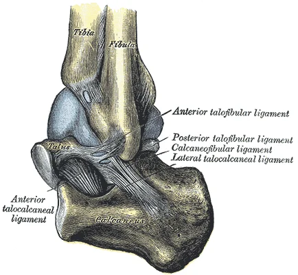
Long description
Capsule of left talocrura articulation (distended). Lateral aspect.

Figure 2: Capsule of left talocrura articulation (distended) (lateral aspect).
The ankle joint, or talocrural joint, is a synovial hinge joint formed by the articulation of the talus with the tibia and fibula. Synovial joints are characterised by the presence of a joint cavity, the walls of which are formed by the articular capsule. This capsule is a fibrous connective tissue structure attached to each bone just outside the area of the articulating surface. [3]
The articular capsule encloses this space. The tibia is the larger, weight-bearing bone located on the medial side of the leg.
Additional support is provided by the muscles and their tendons that act across the joint. A tendon is the dense connective tissue structure that attaches a muscle to bone. As forces acting on a joint increase, the body will automatically increase the overall strength of contraction of the muscles crossing that joint, thus allowing the muscle and its tendon to serve as a “dynamic ligament” to resist forces and support the joint. This type of indirect support by muscles is very important at the shoulder joint, for example, where the ligaments are relatively weak. [3]
DefinitionAnkle Joint
A synovial hinge joint formed by the articulation of the talus with the tibia and fibula.
| Bone | Location |
|---|---|
| Tibia | Medial side of the leg |
| Fibula | Lateral side of the leg |
| Talus | Tarsal bone in the foot |
The thin shaft of the fibula has the interosseous border of the fibula, a narrow ridge running down its medial side for the attachment of the interosseous membrane that spans the fibula and tibia.
| Ligament | Attachments | Function |
|---|---|---|
| Medial (Deltoid) | Medial malleolus to talus, calcaneus, navicular | Resists eversion; stabilises medial ankle |
| Lateral Complex | Lateral malleolus to talus, calcaneus | Resists inversion; stabilises lateral ankle |
The talocrural joint, commonly referred to as the ankle joint, is structurally classified as a uniaxial hinge joint. This classification indicates that it permits motion primarily in a single plane, specifically dorsiflexion and plantar flexion of the foot. The stability of this joint is heavily dependent on its ligamentous architecture, which serves to hold the bones together and resist excessive or abnormal movements. These ligaments are located on the sides of the joint, extending from the malleoli of the tibia and fibula to anchor onto the talus and calcaneus bones. By positioning themselves laterally and medially, these structures allow for the necessary hinge motion while preventing abnormal side-to-side and twisting movements of the talus and calcaneus during eversion and inversion. [5]
Figure 3Gray355
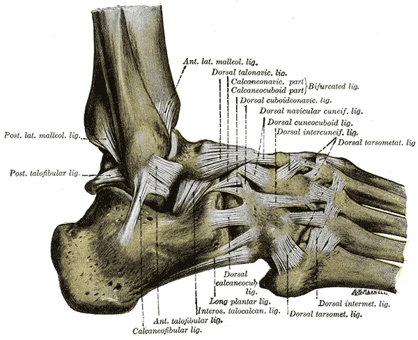
Long description
The ligaments of the foot from the lateral aspect. (Quain.)

Figure 3: The ligaments of the foot from the lateral aspect (Quain.).
The medial aspect of the ankle is reinforced by the deltoid ligament. This structure is described as broad and is situated on the medial side of the ankle joint. Its primary function is to support the ankle joint and resist excessive eversion of the foot. The deltoid ligament also plays a crucial role in preventing side-to-side motions of the talus at the talocrural joint. Because it is a strong, broad band, it provides significant stability against forces that would otherwise pull the foot outw
In contrast, the lateral side of the ankle is supported by a group of smaller, distinct ligaments rather than a single broad band. These include the anterior talofibular ligament, the posterior talofibular ligament, and the calcaneofibular ligament. Collectively, these lateral ligaments support the ankle joint and are specifically tasked with resisting excess inversion of the foot. The difference in structure between the medial and lateral sides reflects the biomechanical demands placed on the joint; the lateral ligaments are more susceptible to injury during inversion sprains, whereas the deltoid ligament is rarely injured due to its strength and breadth. [5]
DefinitionLigament Classification
Ligaments are classified as extrinsic if outside the articular capsule, intrinsic if fused to the capsule wall, or intracapsular if inside the capsule. [6]
The ligaments of the ankle can be understood through the lens of synovial joint strengthening. Synovial joints are strengthened by the presence of ligaments, which hold the bones together and resist excessive or abnormal movements of the joint. While some joints rely on articular discs or menisci for padding and smooth movement, the ankle relies on robust collateral ligaments. Muscles and their tendons acting across the joint also provide indirect support, increasing contractile
| Ligament | Location | Attachments | Primary Function |
|---|---|---|---|
| Deltoid Ligament | Medial | Medial malleolus of tibia to talus and calcaneus | Supports ankle; resists excessive eversion; prevents side-to-side motion of talus |
| Anterior Talofibular Ligament | Lateral | Lateral malleolus of fibula to talus | Supports ankle; resists excess inversion |
| Posterior Talofibular Ligament | Lateral | Lateral malleolus of fibula to talus | Supports ankle; resists excess inversion |
| Calcaneofibular Ligament | Lateral | Lateral malleolus of fibula to calcaneus | Supports ankle; resists excess inversion |
The dome of the talus sits centrally between these bony prominences. Beneath the talus lies the calcaneus. Surrounding these structures are the bursae, which are thin connective tissue sacs filled with lubricating liquid. They are located in regions where skin, ligaments, muscles, or muscle tendons can rub against each other, usually near a body joint. Bursae reduce friction by separating the adjacent structures, preventing them from rubbing directly against each other. [3] [1][4]
Bursae are classified by their location. A subcutaneous bursa is located between the skin and an underlying bone. It allows skin to move smoothly over the bone. Examples include the prepatellar bursa located over the kneecap and the olecranon bursa at the tip of the elbow. A submuscular bursa is found between a muscle and an underlying bone, or between adjacent muscles. In the ankle, subtendinous bursae prevent friction between bone and a muscle tendon. These bursae contain a lubricating fluid that serves to reduce friction between structures. [3]
Figure 4Gray1242
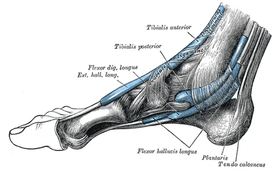
Long description
The mucous sheaths of the tendons around the ankle. Medial aspect.

Figure 4: The mucous sheaths of the tendons around the ankle (medial aspect).
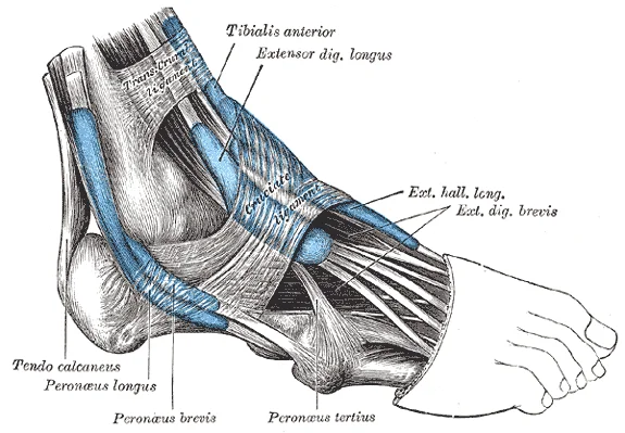
Figure 5Gray1241
Long description
The mucous sheaths of the tendons around the ankle. Lateral aspect.

Figure 5: The mucous sheaths of the tendons around the ankle (lateral aspect).
The ankle joint is a synovial hinge joint, structurally designed to permit movement primarily in one plane. This specialization contrasts with the multiaxial mobility of the shoulder or the weight-bearing stability of the hip, reflecting the lower limb's role in supporting body mass during standing, walking, and running. The joint is formed by the distal ends of the tibia and fibula, which articulate with the dome-shaped trochlea of the talus. This arrangement creates a mortise-and-tenon mechanism, where the talus fits between the malleoli. [1]
DefinitionSyndesmosis
A syndesmosis is a fibrous joint found between parallel bones, such as the tibia and fibula, where the gap is filled with a fibrous interosseous membrane or ligaments.
| Muscle Group | Primary Action | Nerve Supply |
|---|---|---|
| Gastrocnemius and Soleus | Plantarflexion | Tibial nerve |
| Tibialis Anterior | Dorsiflexion | Deep fibular nerve |
| Tibialis Posterior | Inversion and Plantarflexion | Tibial nerve |
| Peroneus Longus and Brevis | Eversion | Superficial fibular nerve |
Stability is further reinforced by strong ligaments and retinacula. Retinacula, such as the superior and inferior extensor retinacula, hold the tendons of the anterior compartment muscles in place against the ankle joint during movement, preventing bowstringing.
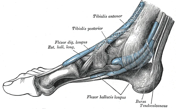
Figure 6Gray442
Long description
The mucous sheaths of the tendons around the ankle. Medial aspect.

Figure 6: The mucous sheaths of the tendons around the ankle (medial aspect).
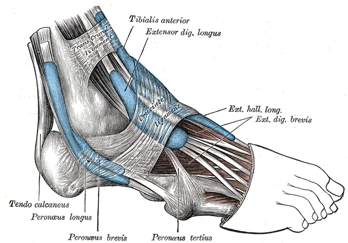
Figure 7Gray441
Long description
The mucous sheaths of the tendons around the ankle. Lateral aspect.

Figure 7: The mucous sheaths of the tendons around the ankle (lateral aspect).
The talus sits inferiorly, its trochlea articulating with the tibia.
NoteClinical Pearl
The ankle is more stable in dorsiflexion because the wider anterior part of the talus is wedged into the mortise, tightening the ligaments.
Inversion and eversion occur primarily at the subtalar and transverse tarsal joints, not the ankle joint itself. The subtalar joint, located between the talus and calcaneus, is the most important contributor to these motions. These plane joints allow slight sliding and rotation, contributing to the overall adaptability of the foot on uneven ground. [1]
The stability of the ankle joint is determined by a combination of bony architecture, ligamentous support, and the functional classification of the joint itself. As a uniaxial hinge joint, the talocrural joint allows only dorsiflexion and plantar flexion. This restriction to a single plane of motion inherently limits the degrees of freedom available for movement, thereby enhancing stability compared to multiaxial joints. A uniaxial joint only allows for motion in a single plane around a single a
The bony structure of the ankle contributes significantly to its stability. The talocrural joint consists of the articulation between the talus bone and the medial malleolus of the tibia, the distal end of the tibia, and the lateral malleolus of the fibula. This mortise-like configuration, where the talus is held between the two malleoli, prevents side-to-side motion of the talus. The strong ligaments located on the sides of the joint further restrict movement to the allowed planes. Since they a
It is important to distinguish the stability of the talocrural joint from the mobility of the subtalar and intertarsal joints. Gliding motions at the subtalar and intertarsal joints of the foot allow for inversion and eversion of the foot. These are plane joints, where the articulating surfaces are flat or slightly curved and of approximately the same size. The motion at plane joints is usually small and tightly constrained by surrounding ligaments. While the talocrural joint provides stability
NoteJoint Classification
The talocrural joint is a uniaxial hinge joint, whereas plane joints like those in the foot allow small, tightly constrained gliding motions.
The structural classification of the joint dictates the type of support required. For example, a multiaxial ball-and-socket joint has more mobility than a uniaxial hinge joint. However, the ligaments and muscles that support a joint may place restrictions on the total range of motion available. In the case of the hip, the strong ligaments reduce the range of motion compared to a ball-and-socket joint but confer stability during standing and weight bearing. This trade-off between mobility an
The ligaments of the ankle can be understood through the lens of synovial joint strengthening. Synovial joints are strengthened by the presence of ligaments, which hold the bones together and resist excessive or abnormal movements of the joint. While some joints rely on articular discs or menisci for padding and smooth movement, the ankle relies on robust collateral ligaments. Muscles and their tendons acting across the joint also provide indirect support, increasing contractile
Clinical pearlStability Principle
Strong ligaments reduce range of motion but confer stability during standing and weight bearing.
The ankle joint is supported on the medial side by the deltoid ligament, which prevents side-to-side motions of the talus at the talocrural joint and resists excessive eversion of the foot. The lateral ankle is supported by the anterior and posterior talofibular ligaments and the calcaneofibular ligament. These structures work in concert to maintain the integrity of the joint under load. The stability is further enhanced by the fact that the ankle joint is a uniaxial hinge joint that allows only
Posteriorly, the posterior tibial artery descends behind the medial malleolus, giving rise to the medial malleolar branches. These vessels interconnect to form the malleolar anastomosis.
Clinical pearlClinical Relevance
Disruption of the blood supply to the talus can lead to avascular necrosis, a serious complication often seen after talar neck fractures. Understanding the anastomotic nature of the ankle's blood supply helps surgeons plan incisions to minimise vascular damage.
This pathway is clinically significant in the spread of infection or malignancy from the foot and ankle. The synovial membrane lines the inner surface of the fibrous capsule, excluding the articular cartilage. It produces synovial fluid, which lubricates the joint and nourishes the avascular cartilage. The fibrous capsule is thickened anteriorly and posteriorly by ligaments, providing stability while allowing the necessary range of motion for dorsiflexion and plantarflexion.
| Region | Nerve | Source |
|---|---|---|
| Anterior | Deep fibular nerve | Common fibular nerve |
| Posterior | Tibial nerve | Sciatic nerve |
| Lateral | Superficial fibular nerve | Common fibular nerve |
| Medial | Saphenous nerve | Femoral nerve |
This comprehensive supply ensures that the ankle joint remains functional and responsive to neurological stimuli, facilitating the complex movements required for locomotion. Any compromise in this supply, whether through trauma, compression, or systemic disease, can lead to significant functional impairment and chronic pain.
DefinitionAnkle Joint
The ankle joint, or talocrural joint, is a synovial hinge joint formed by the articulation of the tibia, fibula, and talus.
The distal end of the tibia presents a broad, flat surface known as the tibial plafond. This surface articulates directly with the trochlea of the talus. Lateral to the tibia lies the distal fibula, which forms the lateral malleolus. The space between the medial and lateral malleoli forms the mortise, which cradles the talus.
The talus is the central bone of the ankle joint. Its superior surface, the trochlea, is shaped like a pulley and fits into the mortise formed by the tibia and fibula. The trochlea is wider anteriorly than posteriorly, which explains why dorsiflexion stabilizes the joint while plantarflexion allows for slight movement. The body of the talus sits within the mortise, while the neck and head extend anteriorly.
On the radiograph, the joint space is visible as a radiolucent gap between the articular surfaces. The syndesmosis, the fibrous connection between the tibia and fibula, is visible superiorly. Any widening of the syndesmosis or asymmetry in the joint spaces may indicate ligamentous injury or fracture.
| Structure | Radiological Appearance | Clinical Significance |
|---|---|---|
| Tibial Plafond | Flat superior surface of tibia | Articulates with talus |
| Lateral Malleolus | Distal fibula | Forms lateral wall of mortise |
| Trochlea Talus | Pulley-shaped superior surface | Hinge movement |
| Syndesmosis | Gap between tibia and fibula | Stability indicator |
Understanding these structures allows for accurate interpretation of ankle radiographs. The alignment of the talus within the mortise is crucial for normal function. Any deviation suggests pathology such as dislocation or fracture. The clarity of the joint spaces helps in diagnosing arthritis or effusion.
The ankle joint's stability relies on the congruence of these bony structures and the integrity of the surrounding ligaments.
The ankle joint is a synovial hinge joint, technically known as the talocrural joint, formed by the articulation of the distal tibia, the distal fibula, and the talus. Understanding its anatomy requires a clear distinction between the structural components and the functional mechanics that govern movement and stability.
The medial aspect of the ankle is defined by the medial malleolus, a projection from the distal tibia. The lateral aspect is formed by the lateral malleolus, a projection from the distal fibula. These two structures form a mortise that securely holds the trochlea of the talus. The tibia bears the majority of the body weight transmitted through the leg.
This border, along with the medial side of the triangular shaft, is located immediately under the skin and can be easily palpated along the entire length of the tibia. On the posterior side of the tibia, the soleal line is a diagonally running, roughened ridge that begins below the base of the lateral condyle, and runs down and medially across the proximal third of the posterior tibia. Muscles of the posterior leg attach to this line. [4]
This anatomical arrangement prevents lateral displacement of the talus during weight-bearing activities.
Stability is maintained by strong ligamentous bands. The deltoid ligament is a broad, triangular band on the medial side, attaching the medial malleolus to the talus, calcaneus, and navicular. It is one of the strongest ligaments in the ankle. On the lateral side, three separate ligaments provide support: the anterior talofibular, calcaneofibular, and posterior talofibular ligaments. These are more prone to injury, particularly during inversion. [1]
Clinical pearlClinical Pearl
The lateral ligaments are significantly weaker than the deltoid ligament, making lateral ankle sprains far more common than medial injuries.
When the foot comes into contact with the ground during walking, running, or jumping activities, the impact of the body weight puts a tremendous amount of pressure and force on the foot. During running, the force applied to each foot as it contacts the ground can be up to 2.5 times your body weight. The bones, joints, ligaments, and muscles of the foot absorb this force, thus greatly reducing the amount of shock that is passed superiorly into the lower limb and body. [4] [1]
The arches of the foot play an important role in this shock-absorbing ability. When weight is applied to the foot, these arches will flatten somewhat, thus absorbing energy. When the weight is removed, the arch rebounds, giving “spring” to the step. The arches also serve to distribute body weight side to side and to either end of the foot. This dynamic system protects the ankle joint from excessive repetitive stress. [4]
Examination of the ankle involves assessing range of motion, stability, and tenderness. The anterior drawer test assesses the integrity of the anterior talofibular ligament. Tilt tests evaluate the calcaneofibular ligament. Palpation of the anterior border of the tibia is straightforward due to its subcutaneous nature.
Aspiration of the ankle joint is typically performed anteriorly, between the tibialis anterior tendon and the extensor hallucis longus tendon. This approach avoids major neurovascular structures and provides access to the joint space. Dislocation of the ankle is a severe injury often associated with fracture of the malleoli or ligamentous rupture, requiring urgent reduction and imaging to assess joint congruity.
The ankle is thus a uniaxial hinge joint that allows only for dorsiflexion and plantar flexion of the foot. [1]
On the medial side is the broad deltoid ligament. The deltoid ligament supports the ankle joint and also resists excessive eversion of the foot.
The syndesmosis, or distal tibiofibular joint, is a fibrous joint that connects the distal tibia and fibula, providing stability to the ankle mortise. Injury to the syndesmosis, often called a high ankle sprain, can result in greater instability and a longer recovery time than some lateral ligament injuries. [1]
The range of motion is greater in plantarflexion than in dorsiflexion, allowing for greater flexibility during walking and running. [1]
Venous drainage follows the arterial supply, returning blood to the deep veins of the leg. Lymphatic drainage passes to the popliteal and inguinal lymph nodes. [1]
This rich innervation contributes to the proprioceptive feedback necessary for balance and coordination. [1]
DefinitionAnkle Mortise
The bony socket formed by the distal tibia and fibula that holds the talus.
| Ligament | Attachment | Injury Mechanism |
|---|---|---|
| Anterior Talofibular | Lateral malleolus to talus | Plantarflexion and inversion |
| Calcaneofibular | Lateral malleolus to calcaneus | Neutral inversion |
| Posterior Talofibular | Lateral malleolus to talus | Severe dislocation |
DefinitionTalus
The bone that articulates with the tibia and fibula to form the ankle joint.
Students frequently confuse the functional anatomy of the knee with that of the ankle, leading to errors in clinical reasoning. A primary error is attributing the "locking" mechanism of full extension to the ankle joint. In reality, this mechanism is specific to the knee. The small medial rotation of the femur serves to “lock” the knee into its fully extended and most stable position. The ankle does not possess this screw-home mechanism; it relies on bony congruence and ligamentous tension for stability. Another common mistake is assuming the fibula bears significant weight. The fibula is the slender bone located on the lateral side of the leg. It does not bear weight. It serves primarily for muscle attachments and thus is largely surrounded by muscles. Only the proximal and distal ends of the fibula can be palpated. Students often overlook this, leading to incorrect assessments of load-bearing injuries. [1] [4]
A third pitfall involves the classification of joint types. Students may incorrectly label the zygapophysial joints as hinge joints. These are plane joints that provide for only limited motions between the vertebrae. The orientation of the articular processes at these joints varies in different regions of the vertebral column and serves to determine the types of motions available in each vertebral region. While not ankle-specific, this confusion often extends to misidentifying the ankle as a hinge joint without appreciating its multi-axial potential in dorsiflexion versus plantarflexion. [1]
Fourth, students often misinterpret the role of the patella in the lower limb kinetic chain. The patella is a sesamoid bone located within a muscle tendon. It articulates with the patellar surface on the anterior side of the distal femur, thereby protecting the muscle tendon from rubbing against the femur. It is not part of the ankle joint complex, yet its tendon continuity is crucial for ankle dorsiflexion power. Confusing the patellar tendon with the Achilles tendon is a frequent anatomical error. [4]
Fifth, there is a tendency to ignore the structural support of the foot arches. The foot has a transverse arch, a medial longitudinal arch, and a lateral longitudinal arch. The transverse arch forms the medial-lateral curvature of the mid-foot. It is formed by the wedge shapes of the cuneiform bones and bases (proximal ends) of the first to fourth metatarsal bones. This arch helps to distribute body weight from side to side within the foot, thus allowing the foot to accommodate uneven terrain. Failure to appreciate this distribution leads to poor understanding of pes planus. [4]
Sixth, students often fail to distinguish between the roles of the talus and the calcaneus. The talus bone, which receives the weight of the body, is located at the top of the longitudinal arches. Body weight is then conveyed from the talus to the ground by the anterior and posterior ends of these arches. The calcaneus provides posterior support. Confusing these roles leads to errors in interpreting fracture patterns. [4]
Exam trapWeight Bearing
The fibula does not bear weight. It serves primarily for muscle attachments and thus is largely surrounded by muscles. Only the proximal and distal ends of the fibula can be palpated.
At the bedside, knowledge of the ankle and lower limb anatomy is critical for physical examination. When assessing stability, clinicians palpate the malleoli. The fibula is the slender bone located on the lateral side of the leg. Only the proximal and distal ends of the fibula can be palpated. This palpation helps identify fractures or ligamentous avulsions. Understanding the tibial tuberosity is also vital. The tibial tuberosity is an elevated area on the anterior side of the tibia, near its proximal end. It is the final site of attachment for the muscle tendon associated with the patella. Tenderness here may indicate Osgood-Schlatter disease or patellar tendonitis, which affects ankle mechanics indirectly. [4]
In the operating theatre, surgeons must respect the ligamentous constraints. While the provided texts focus on other joints, the principle of ligamentous integrity applies. For instance, the ulnar collateral ligament arises from the medial epicondyle of the humerus and attaches to the medial side of the proximal ulna. The strongest part of this ligament is the anterior portion, which resists hyperextension of the elbow. Although this is elbow anatomy, the principle of intrinsic ligaments preventing side-to-side movements and hyperextension is mirrored in the ankle's syndesmotic and collateral ligaments. Surgeons repair these to restore stability. [1]
On imaging, radiologists identify the arches to assess for collapse. The longitudinal arches run down the length of the foot. The lateral longitudinal arch is relatively flat, whereas the medial longitudinal arch is larger (taller). The longitudinal arches are formed by the tarsal bones posteriorly and the metatarsal bones anteriorly. These arches are supported at either end, where they contact the ground. Posteriorly, this support is provided by the calcaneus bone and anteriorly by the heads (distal ends) of the metatarsal bones. X-rays and MRI scans evaluate the integrity of these structures to diagnose pes planus. [4]
In examinations, students must describe the toe anatomy accurately. The toes contain 14 small bones, each of which is a phalanx bone of the foot. This specific number is a frequent recall item. Additionally, students must understand the energy absorption mechanism. These ligaments have elasticity, which allows them to stretch somewhat during weight bearing, thus allowing the longitudinal arches to spread. The stretching of these ligaments stores energy within the foot, rather than passing these forces into the leg. Contraction of the foot muscles also plays an important role in this energy absorption. When the weight is removed, the elastic ligaments recoil and pull the ends of the arches closer together. This recovery of the arches releases the stored energy and improves the energy efficiency of walking. [4]
Ankle Stability Assessment
- Patient presents with ankle painHistory and observation
- Question: Palpate lateral malleolus?
Tender
- Suspect fibular injuryFibula does not bear weight
Not tender
- Assess medial archCheck for pes planus
- Question: Check arch collapse?
Yes
- Diagnose pes planusStretching of ligaments
No
- Evaluate ligament elasticityEnergy absorption
NotePes Planus
Stretching of the ligaments that support the longitudinal arches can lead to pain. This can occur in overweight individuals, with people who have jobs that involve standing for long periods of time (such as a waitress), or walking or running long distances. [4]
Clinical pearlTalus Function
The talus bone, which receives the weight of the body, is located at the top of the longitudinal arches. Body weight is then conveyed from the talus to the ground by the anterior and posterior ends of these arches.
The ankle joint relies on bony congruence and ligamentous support rather than a locking mechanism like the knee. The fibula is slender, lateral, and does not bear weight, serving mainly for muscle attachment. The talus is the key weight-bearing bone at the top of the longitudinal arches. The medial longitudinal arch is taller than the lateral longitudinal arch. Pes planus results from prolonged or excessive stretching of the supporting ligaments. The patella is a sesamoid bone protecting the quadriceps tendon, not part of the ankle. The toes contain 14 phalanges in total. Ligament elasticity allows energy storage and release, improving walking efficiency.
Answer each one in your head first, then open the answer.
What is the primary function of the fibula regarding weight bearing?
Show answerHide answer
It does not bear weight; it serves primarily for muscle attachments.Which bone receives the weight of the body at the top of the longitudinal arches?
Show answerHide answer
The talus bone.How many small bones are in the toes?
Show answerHide answer
14.What condition is called pes planus?
Show answerHide answer
Flat foot or fallen arches, resulting from depression or collapse of the longitudinal arches.What structure prevents the femur from sliding anteriorly off the tibia?
Show answerHide answer
The posterior cruciate ligament.
- Betts JG, Young KA, Wise JA, et al. Anatomy of Selected Synovial Joints. OpenStax, Anatomy and Physiology 2e (CC BY-NC-SA 4.0). Accessed .
- Betts JG, Young KA, Wise JA, et al. Fibrous Joints. OpenStax, Anatomy and Physiology 2e (CC BY-NC-SA 4.0). Accessed .
- Betts JG, Young KA, Wise JA, et al. Synovial Joints. OpenStax, Anatomy and Physiology 2e (CC BY-NC-SA 4.0). Accessed .
- Betts JG, Young KA, Wise JA, et al. Bones of the Lower Limb. OpenStax, Anatomy and Physiology 2e (CC BY-NC-SA 4.0). Accessed .
- Betts JG, Young KA, Wise JA, et al. Anatomy of Selected Synovial Joints. OpenStax, Anatomy and Physiology 2e (CC BY-NC-SA 4.0). Accessed .
- Betts JG, Young KA, Wise JA, et al. Synovial Joints. OpenStax, Anatomy and Physiology 2e (CC BY-NC-SA 4.0). Accessed .
Figure credits
- Figure 1: OpenStax College, via Wikimedia Commons, CC BY 3.0, CC BY 3.0.
- Figure 2: Henry Vandyke Carter, via Wikimedia Commons, Public domain, Public domain.
- Figure 3: Henry Vandyke Carter, via Wikimedia Commons, Public domain, Public domain.
- Figure 4: Henry Vandyke Carter, via Wikimedia Commons, Public domain, Public domain.
- Figure 5: Henry Vandyke Carter, via Wikimedia Commons, Public domain, Public domain.
- Figure 6: Henry Vandyke Carter, via Wikimedia Commons, Public domain, Public domain.
- Figure 7: Henry Vandyke Carter, via Wikimedia Commons, Public domain, Public domain.
Educational content for medical students and doctors. It is not a substitute for clinical judgement or local guidelines.