Leg compartments
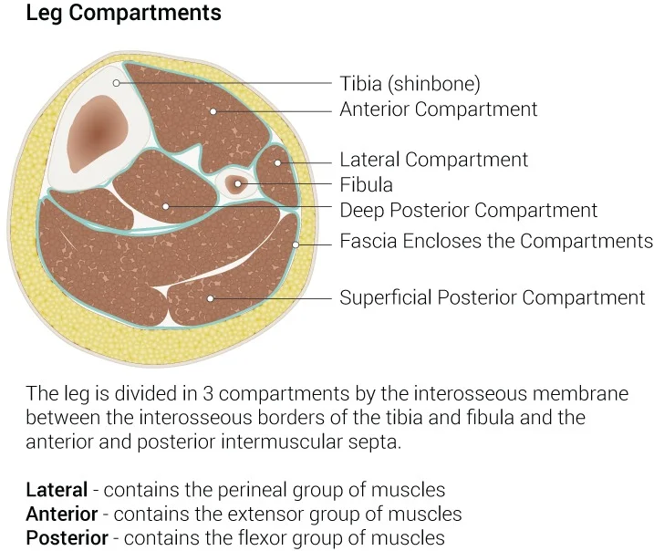
Anatomical layout of the anterior, lateral and posterior leg compartments.
The lower limb is divided into distinct regions by deep fascia, creating compartments that house specific muscle groups and neurovascular structures. In the leg, these compartments are the anterior, lateral, and posterior groups. Understanding these boundaries is critical for diagnosing conditions such as compartment syndrome or understanding the mechanics of gait. [2]
Injuries to the leg often involve multiple compartments. Fractures of the tibia are the most common cause of acute compartment syndrome. The anterior compartment is most frequently affected due to its rigid bony boundaries. Nerve palsies are a significant complication. The deep peroneal nerve supplies the extensor digitorum brevis and provides sensation to the first web space. Injury results in foot drop and sensory loss in this region.
These muscles are held in place by the superior and inferior extensor retinacula, which are thick bands of connective tissue. The anterior tibial artery runs through this compartment, providing blood supply to these muscles. The deep peroneal nerve accompanies the artery, innervating the anterior compartment muscles. Damage to this nerve or its branches can result in foot drop, a condition characterized by the inability to lift the front part of the foot. Foot drop is a classic sign of anterior [1][4]
The posterior compartment is divided into superficial and deep layers by the transverse intermuscular septum. The superficial layer contains the gastrocnemius, soleus, and plantaris, which converge to form the Achilles tendon. This tendon inserts into the calcaneus and is crucial for plantar flexion. The deep posterior compartment contains the tibialis posterior, flexor digitorum longus, and flexor hallucis longus. These muscles are involved in plantar flexion and inversion of the foot. The posterior tibial artery runs through this compartment, supplying the deep muscles. [4]
The lateral compartment contains the fibularis longus and brevis muscles. These muscles evert the foot and assist in plantar flexion. They are supplied by the superficial peroneal nerve. The boundaries of this compartment are the fibula and the deep fascia.
Shin splints, or medial tibial stress syndrome, is a common overuse injury affecting the anterior and medial aspects of the leg. It involves inflammation of the periosteum and muscle attachments, particularly the tibialis anterior. The calf pump mechanism, involving the contraction of the gastrocnemius and soleus, aids venous return from the lower limb. Disruption of this mechanism or compartment integrity can lead to significant clinical issues, including compartment syndrome, which requires urgent fasciotomy to relieve pressure. The anatomical precision of these compartments ensures efficient force transmission and movement, highlighting the importance of detailed anatomical knowledge in clinical practice.
The flexor digitorum longus tendon descends behind the medial malleolus within a dedicated synovial compartment, travels obliquely into the plantar foot superficial to the deltoid ligament, receives a strong slip from the flexor hallucis longus, unites with the quadratus plantae, and divides into four digital tendons inserted into the bases of the distal phalanges of the lateral four toes. Concurrently, the extensor hallucis longus tendon passes through the cruciate crural ligament at the ankle, crosses the anterior tibial vessels, and inserts upon the base of the distal phalanx of the hallux, contributing expansions to the metatarsophalangeal joint and proximal phalanx. Superiorly, the plantaris muscle arises from the lateral linea aspera and oblique popliteal ligament, descending to a narrow tendon inserted near the calcaneal tendon. Deep in the popliteal fossa, the triangular popliteus muscle originates via a robust tendon from the lateral femoral condyle and oblique popliteal ligament, inserting broadly onto the posterior tibia above the soleal line to stabilize the knee joint.
Fluid balance is critical for cellular integrity. The intracellular fluid (ICF) constitutes approximately 60 percent of the total water in the human body. In an average adult male, this accounts for about 25 liters of fluid. This volume is tightly regulated. If intracellular water falls too low, the cytosol becomes too concentrated with solutes. This impairs normal cellular activities. Conversely, if too much water enters, the cell may burst. [5]
The extracellular fluid (ECF) accounts for the remaining one-third of body water. Approximately 20 percent of the ECF is plasma. Plasma transports blood cells, proteins, electrolytes, nutrients, gases, and wastes. Gases and nutrients travel between capillaries and cells through the interstitial fluid (IF). Cells are separated from the IF by a selectively permeable membrane. [5]
Plasma and IF compositions are similar but distinct from the ICF. Plasma has high concentrations of sodium, chloride, bicarbonate, and protein. The IF has high sodium, chloride, and bicarbonate, but lower protein. The ICF has elevated potassium, phosphate, magnesium, and protein. Specifically, the ICF contains high potassium and phosphate (HPO4 2−). Both plasma and ECF contain high sodium and chloride. [5]
Water moves between compartments along an osmotic gradient. This gradient is produced by differences in solute concentration across a semi-permeable membrane. The magnitude is proportional to the solute concentration difference. Water moves by osmosis from high water concentration (low solute) to low water concentration (high solute). In the body, water moves between plasma, IF, and ICF. [5]
Hydrostatic pressure is the force exerted by a fluid against a wall. This force causes fluid movement between compartments. Active transport processes require ATP to move solutes against concentration gradients. An appropriate balance of water and solute concentrations ensures cellular functions. If cytosol becomes too concentrated due to water loss, cell functions deteriorate. If too dilute due to water intake, cell membranes can be damaged. [5]
DefinitionOsmotic Gradient
An osmotic gradient is produced by the difference in concentration of all solutes on either side of a semi-permeable membrane. [5]
DefinitionHydrostatic Pressure
Hydrostatic pressure is the force exerted by a fluid against a wall and causes movement of fluid between compartments.
Figure 1Leg compartments
Long description
Leg Compartments, Tibia (shinbone), Anterior Compartment, Lateral Compartment, Fibula, Deep Posterior Compartment, Fascia Encloses the Compartments, Superficial Posterior Compartment. Contributed Illustration by Beckie P

Figure 1: Leg Compartments, Tibia (shinbone), Anterior Compartment, Lateral Compartment, Fibula, Deep Posterior Compartment, Fascia Encloses the Compartments, Superficial Posterior Compartment (contributed illustration by beckie p).
The leg is anatomically divided into four distinct compartments by deep fascia: the anterior, lateral, superficial posterior, and deep posterior compartments[1]. These compartments house the muscles responsible for moving the foot and toes[1].
Anteriorly, the sharp border of the tibia is palpable beneath the skin. Posteriorly, the gastrocnemius forms the prominent, rounded bulk of the calf, tapering inferiorly into the calcaneal tendon, which inserts into the calcaneal bone[1].
The anterior compartment contains the tibialis anterior, extensor hallucis longus, extensor digitorum longus, and fibularis tertius[1]. These muscles are primarily responsible for dorsiflexion of the foot and extension of the toes. The lateral compartment is situated between the anterior and posterior compartments and contains two muscles: the fibularis longus and the fibularis brevis[1]. These muscles are essential for eversion of the foot.
The posterior aspect of the leg is divided into superficial and deep compartments[1]. The superficial posterior compartment contains the gastrocnemius, the soleus, and the plantaris[1]. The gastrocnemius is the most superficial and visible muscle of the calf[1]. Deep to the gastrocnemius lies the wide, flat soleus muscle[1]. The plantaris muscle runs obliquely between the gastrocnemius and the soleus; notably, no plantaris muscle is observed in about 7 percent of cadaver dissections[1]. All three muscles in this superficial group insert onto the calcaneal tendon, also known as the Achilles tendon, which is a strong tendon that attaches to the calcaneal bone of the ankle[1]. [2]
These muscles are positioned deep to the soleus and are primarily involved in plantarflexion of the foot and flexion of the toes.
DefinitionCompartment syndrome
Increased pressure within a confined osteofascial compartment can compromise circulation and function of the tissues within that space.
| Compartment | Muscles | Primary Action |
|---|---|---|
| Anterior | Tibialis anterior, Extensors | Dorsiflexion |
| Lateral | Fibularis longus/brevis | Eversion |
| Superficial Posterior | Gastrocnemius, Soleus, Plantaris | Plantarflexion |
| Deep Posterior | Popliteus, Flexors, Tibialis posterior | Plantarflexion/Flexion |
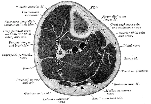
Figure 2Gray440
Long description
Cross-section through middle of leg. (Eycleshymer and Schoemaker.)

Figure 2: Cross-section through middle of leg (Eycleshymer and Schoemaker.).
The ilium, ischium, and pubis form the acetabulum, which receives the head of the femur. The greater trochanter lies laterally, providing an insertion point for the gluteus medius and minimus, while the lesser trochanter lies medially, serving as the insertion for the iliopsoas.
The musculature surrounding these bony landmarks is organised into functional groups. In the gluteal region, the psoas major and iliacus form the iliopsoas. The large and strong gluteus maximus, gluteus medius, and gluteus minimus extend and abduct the femur. Along with the gluteus maximus, the tensor fascia lata muscle forms the iliotibial tract. [1] failed foot revascularisation led to less chance of healing (9.1%) and higher risk of major amputation (36.3%) [2]
DefinitionIliopsoas
The iliopsoas is the primary flexor of the hip joint, formed by the union of the psoas major and iliacus muscles.
The lateral rotators of the femur at the hip are the piriformis, obturator internus, obturator externus, superior gemellus, inferior gemellus, and quadratus femoris. On the medial part of the thigh, the adductor longus, adductor brevis, and adductor magnus adduct the thigh and medially rotate it. The pectineus muscle adducts and flexes the femur at the hip. [1]
| Muscle | Action |
|---|---|
| Gluteus maximus | Extends and abducts the femur |
| Adductor longus | Adducts and medially rotates the thigh |
| Pectineus | Adducts and flexes the femur |
| Piriformis | Lateral rotation of the femur |
These muscles are arranged in distinct compartments defined by deep fascia. The bony landmarks, particularly the trochanters of the femur, provide the necessary mechanical advantage for these muscles to facilitate movement at the hip joint. Understanding these attachments is essential for identifying the boundaries of the thigh compartments and the path of neurovascular structures.
The leg is enclosed by the deep fascia of the leg, known as the crural fascia. This dense connective tissue layer is continuous superiorly with the fascia lata of the thigh and attaches directly to the anterior and medial borders of the tibia. From the deep surface of the crural fascia, two primary intermuscular septa—the anterior and posterior intermuscular septa—pass inward to attach to the fibula, effectively dividing the leg into three distinct anatomical compartments: the anterior, lateral, and posterior compartments. [1]
The anterior compartment lies between the tibia and the anterior intermuscular septum. The lateral compartment is situated between the anterior and posterior intermuscular septa, overlying the fibula. The posterior compartment occupies the space behind the interosseous membrane and the posterior intermuscular septum.
The anterior compartment is bounded by the interosseous membrane posteriorly, the tibia medially, and the anterior intermuscular septum laterally. It contains the dorsiflexors of the ankle and the extensors of the toes. [2]
| Muscle | Origin | Insertion | Action | Nerve |
|---|---|---|---|---|
| Tibialis anterior | Lateral tibia | Medial cuneiform | Dorsiflexion | Deep peroneal |
| Extensor digitorum longus | Fibula | Lateral four toes | Toe extension | Deep peroneal |
| Extensor hallucis longus | Fibula | Distal phalanx (big toe) | Big toe extension | Deep peroneal |
The lateral compartment is the smallest of the three. It is bounded by the anterior intermuscular septum anteriorly and the posterior intermuscular septum posteriorly. It primarily contains the evertors of the foot.
| Muscle | Origin | Insertion | Action | Nerve |
|---|---|---|---|---|
| Peroneus longus | Fibula head | Medial cuneiform | Eversion | Superficial peroneal |
| Peroneus brevis | Fibula shaft | 5th metatarsal base | Eversion | Superficial peroneal |
The posterior compartment is the largest and is subdivided into superficial and deep groups by the transverse intermuscular septum. The superficial group contains the gastrocnemius, soleus, and plantaris, which are primarily plantarflexors. The deep group, located anterior to the transverse septum, contains the flexor hallucis longus, flexor digitorum longus, and tibialis posterior.
DefinitionCompartment syndrome
A condition where increased pressure within a confined osteofascial space compromises circulation and function of the tissues within that space.
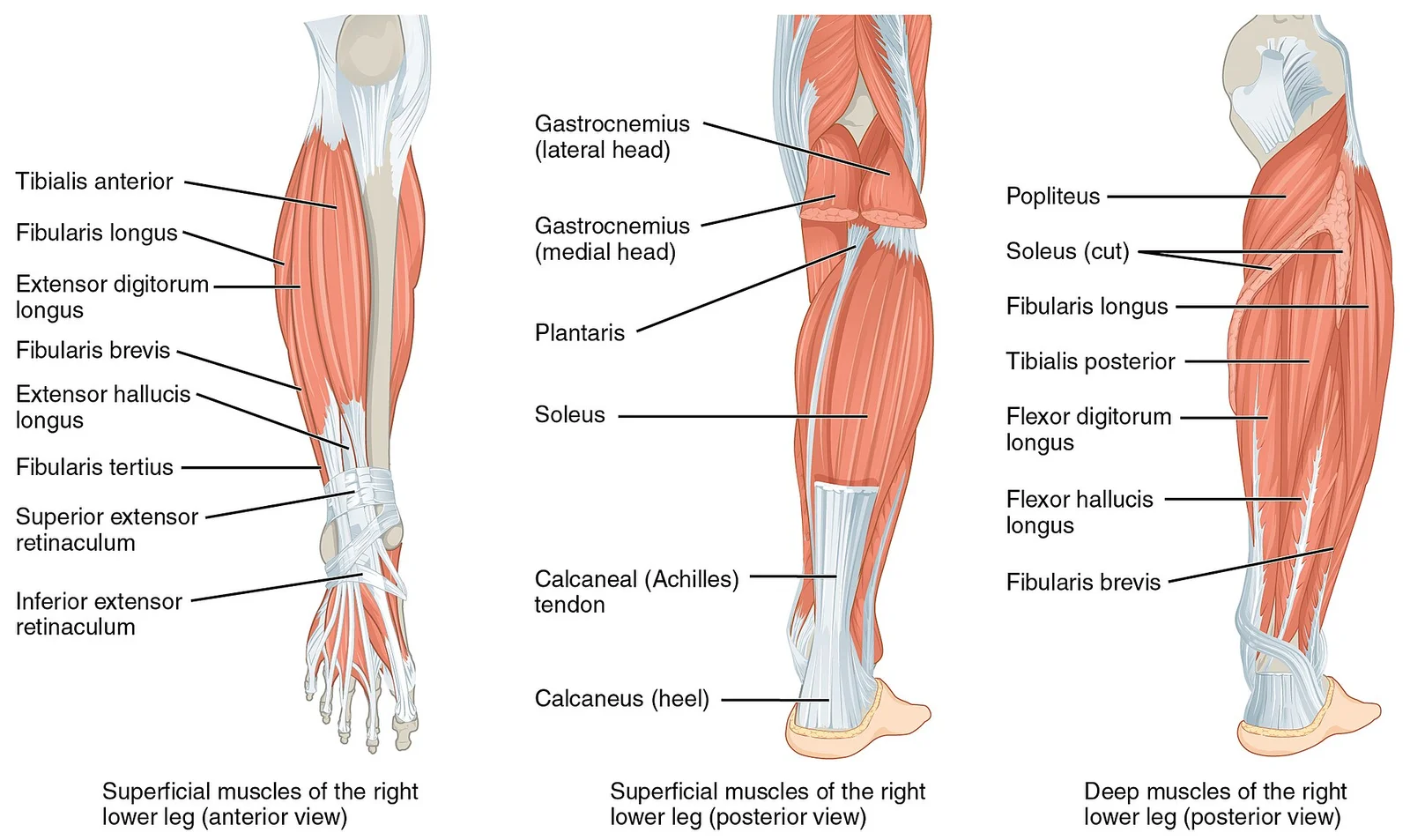
Figure 31123 Muscles of the Leg that Move the Foot and Toes
Long description
The muscles of the anterior compartment of the lower leg are generally responsible for dorsiflexion, and the muscles of the posterior compartment of the lower leg are generally responsible for plantar flexion. The lateral and medial muscles in both compartments invert, evert, and rotate the foot.

Figure 3: The muscles of the anterior compartment of the lower leg are generally responsible for dorsiflexion, and the muscles of the posterior compartment of the lower leg are generally responsible for plantar flexion.
The leg is divided into three fascial compartments: anterior, lateral, and posterior. Each compartment contains specific muscles, nerves, and vessels. Understanding the anatomy requires grouping muscles by their compartmental location, as this dictates their innervation and vascular supply.
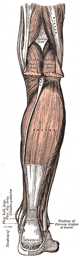
Figure 4Gray438
Long description
Muscles of the back of the leg. Superficial layer.

Figure 4: Muscles of the back of the leg (superficial layer).
Figure 5Gray437
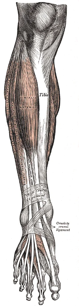
Long description
Muscles of the front of the leg. Labelled illustration from Wikimedia Commons, used under its open licence.

Figure 5: Muscles of the front of the leg.
DefinitionCompartment Syndrome
A surgical emergency where increased pressure within a closed fascial space compromises circulation and tissue function.
Figure 61123 Muscles of the Leg that Move the Foot and Toes b
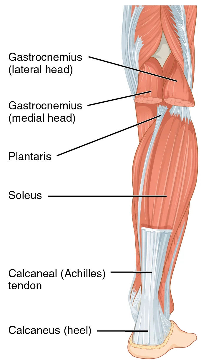
Long description
Caption: The muscles of the anterior compartment of the lower leg are generally responsible for dorsiflexion, and the muscles of the posterior compartment of the lower leg are generally responsible for plantar flexion. T

Figure 6: Caption: The muscles of the anterior compartment of the lower leg are generally responsible for dorsiflexion, and the muscles of the posterior compartment of the lower leg are generally responsible for plantar flexion (t).
The anterior compartment contains muscles that primarily dorsiflex the foot and extend the toes. These are the extensor muscles.
| Muscle | Origin | Insertion | Action | Nerve Supply |
|---|---|---|---|---|
| Tibialis anterior | Lateral condyle and upper lateral tibia | Medial cuneiform and base of first metatarsal | Dorsiflexion and inversion of foot | Deep fibular nerve |
| Extensor digitorum longus | Lateral condyle of tibia and upper fibula | Dorsal digital expansions of toes 2-5 | Extension of toes and dorsiflexion | Deep fibular nerve |
| Extensor hallucis longus | Middle half of anterior fibula | Base of distal phalanx of great toe | Extension of great toe and dorsiflexion | Deep fibular nerve |
| Fibularis tertius | Distal anterior fibula | Dorsal base of fifth metatarsal | Dorsiflexion and eversion | Deep fibular nerve |
The deep fibular nerve supplies all muscles in this compartment. It runs with the anterior tibial artery. Compression here leads to foot drop.
The lateral compartment contains the fibular muscles, which evert the foot.
| Muscle | Origin | Insertion | Action | Nerve Supply |
|---|---|---|---|---|
| Fibularis longus | Head and upper lateral fibula | Medial cuneiform and base of first metatarsal | Eversion and plantarflexion | Superficial fibular nerve |
| Fibularis brevis | Lower lateral fibula | Base of fifth metatarsal | Eversion and plantarflexion | Superficial fibular nerve |
The superficial fibular nerve supplies these muscles. It emerges between the fibularis longus and brevis.
The posterior compartment is divided into superficial and deep layers. The superficial layer contains the gastrocnemius, soleus, and plantaris, which form the triceps surae. These muscles plantarflex the foot. The deep layer contains the tibialis posterior, flexor digitorum longus, and flexor hallucis longus. These muscles invert the foot and flex the toes. The tibial nerve supplies all posterior compartment muscles.
Compartment syndrome can affect any of these compartments. It is caused by increased pressure within the fascia. This pressure reduces capillary perfusion. Ischaemia follows. The deep posterior compartment is particularly susceptible because it is enclosed by the interosseous membrane and the posterior fascia. Symptoms include pain out of proportion to injury, pain on passive stretch, and paresthesia. Early diagnosis is critical to prevent permanent nerve and muscle damage.
The arterial supply to the leg compartments is primarily derived from the popliteal artery, which terminates at the lower border of the popliteus muscle by dividing into the anterior and posterior tibial arteries. These vessels provide the essential perfusion for the anterior, lateral, and posterior compartments of the leg.
The anterior tibial artery is the smaller terminal branch of the popliteal artery. It passes forward between the two heads of the tibialis posterior muscle and through the interosseous membrane to enter the anterior compartment. It descends on the anterior surface of the interosseous membrane, accompanied by the deep peroneal nerve. In the distal leg, it lies on the anterior surface of the tibia before crossing the ankle joint to become the dorsalis pedis artery. The lateral compartment is supplied by branches from the peroneal artery, which pierces the posterior intermuscular septum. [4] [1]
The posterior tibial artery is the larger terminal branch of the popliteal artery. It descends deep to the gastrocnemius and soleus muscles, resting on the tibialis posterior muscle. It provides the peroneal artery, which arises shortly after the popliteal bifurcation and descends along the medial side of the fibula, supplying the deep posterior compartment.
DefinitionSmall Artery Disease
Small artery disease is an arterial disease involving the plantar arch and small arteries, which can be classified into three grades of severity. [2]
| Pattern | Anatomical involvement | Clinical significance |
|---|---|---|
| BTK | Infra-popliteal vessels | Requires recanalisation for wound healing [2] |
| BTA | Below-the-ankle arteries | Associated with foot gangrene and amputation [2] |
| SAD | Plantar arch and small arteries | Extremely severe pattern of PAD |
The venous drainage of the leg compartments follows a hierarchical arrangement, moving from superficial networks to deep systems that accompany the major arteries. Understanding this layout is essential for interpreting imaging and planning surgical interventions, particularly when considering the complex vascular relationships in the lower limb.
Deeply, the anterior tibial veins accompany the anterior tibial artery in the anterior compartment. The posterior tibial veins and peroneal veins are situated within the deep posterior compartment, lying adjacent to their arterial counterparts.
DefinitionSuperficial Veins
The great and small saphenous veins form the superficial venous network, lying within the subcutaneous tissue above the deep fascia.
The deep venous system consists of paired veins that run alongside the major arteries of the leg. These veins are located within the intermuscular septa and compartments, ensuring efficient drainage from the muscle beds. The anterior tibial veins drain the anterior compartment, collecting blood from the tibialis anterior, extensor digitorum longus, and extensor hallucis longus muscles. They join with the posterior tibial veins to form the popliteal vein behind the knee. [1]
NoteClinical Relevance
Deep vein thrombosis often originates in the calf veins, particularly the posterior tibial and peroneal veins, due to stasis within the deep muscular compartments.
The posterior tibial veins drain the deep posterior compartment, which includes the flexor digitorum longus, flexor hallucis longus, and tibialis posterior muscles. These veins are crucial for returning blood from the lateral aspect of the leg. [4]
While this is a salvage procedure for severe ischemia, it highlights the complex interplay between arterial and venous systems in the leg compartments. [2]
These figures underscore the severity of vascular complications and the critical nature of maintaining adequate perfusion to the leg compartments. [3] [2]
| Compartment | Primary Deep Veins | Accompanying Artery |
|---|---|---|
| Anterior | Anterior tibial veins | Anterior tibial artery |
| Lateral | Peroneal (fibular) veins | Peroneal artery |
| Deep Posterior | Posterior tibial veins | Posterior tibial artery |
The management of such complications primarily involves surgical intervention and vascular revision. In some instances, flap revision and debridement were performed to address complications related to microvascular anastomosis, such as venous insufficiency. This highlights the need for meticulous attention to venous drainage during any procedure involving the leg compartments. [3]
The innervation of the leg is derived from the sciatic nerve, which divides into the tibial nerve and the common fibular nerve. These nerves provide motor and sensory supply to the distinct compartments of the leg. In this view, the common fibular nerve winds around the neck of the fibula, where it is vulnerable to injury. It then divides into the superficial fibular nerve, which descends in the lateral compartment, and the deep fibular nerve, which passes into the anterior compartment.
DefinitionCompartment innervation
The anterior compartment is supplied by the deep fibular nerve, the lateral compartment by the superficial fibular nerve, and the posterior compartment by the tibial nerve. [3]
The deep fibular nerve supplies the muscles of the anterior compartment, which are primarily responsible for dorsiflexion of the ankle and extension of the toes. The superficial fibular nerve provides motor innervation to the lateral compartment muscles, which act to evert the foot. The tibial nerve, the larger terminal branch of the sciatic nerve, descends through the posterior compartment to supply the plantar flexors of the ankle and the flexors of the toes. [4]
| Nerve | Compartment | Primary Muscles Supplied | Cutaneous Area |
|---|---|---|---|
| Deep fibular | Anterior | Tibialis anterior, Extensor digitorum longus | First interdigital cleft |
| Superficial fibular | Lateral | Fibularis longus, Fibularis brevis | Dorsum of the foot |
| Tibial | Posterior | Gastrocnemius, Soleus, Tibialis posterior | Sole of the foot |
The cutaneous distribution is highly specific. The deep fibular nerve provides sensation to the skin of the first interdigital cleft. The superficial fibular nerve supplies the majority of the skin on the dorsum of the foot. The tibial nerve continues into the foot, providing sensory innervation to the sole through its medial and lateral plantar branches. Understanding these pathways is essential for clinical assessment, as damage to these nerves results in predictable patterns of motor weakness and sensory loss. [4]
Nerve assessment
- Assess motor functionTest dorsiflexion (deep fibular), eversion (superficial fibular), and plantar flexion (tibial).
- Question: Sensory deficit present?
Yes
- Map dermatome to specific nerve distribution
No
- Monitor for progressive weakness
The lymphatic drainage of the lower limb is a critical pathway for immune surveillance and fluid homeostasis, following a predictable anatomical route that mirrors the venous system but with distinct nodal stations. Understanding this drainage is essential for diagnosing infections, malignancies, and conditions such as lymphoedema. The system is divided into superficial and deep components, which converge at specific inguinal and pelvic nodes.
The medial group lies along the terminal part of the great saphenous vein, while the lateral group accompanies the short saphenous vein.
DefinitionSuperficial Inguinal Nodes
A group of lymph nodes located in the superficial fascia of the upper medial thigh, divided into horizontal and vertical groups.
The superficial lymphatics of the leg follow the course of the superficial veins. The medial superficial lymphatic vessels accompany the great saphenous vein. These vessels drain the skin and subcutaneous tissues of the medial aspect of the foot, the medial side of the leg, and the anterior abdominal wall below the umbilicus. They pass through the saphenous opening to reach the vertical group of superficial inguinal nodes. The lateral superficial lymphatic vessels accompany the short saphenous vein. These vessels drain the lateral aspect of the foot, the lateral side of the leg, and the posterior calf. They typically drain into the popliteal nodes before reaching the deep inguinal nodes, although some may drain directly into the superficial inguinal nodes.
Exam trapPopliteal Nodes
Do not assume all lateral leg drainage goes directly to inguinal nodes; many vessels pass through the popliteal fossa first.
The deep lymphatic vessels accompany the deep veins of the leg, such as the anterior and posterior tibial veins and the peroneal veins. These vessels drain the muscles, bones, and joints of the leg. They ascend through the popliteal fossa and drain into the popliteal nodes. From the popliteal nodes, the lymphatic vessels pass through the adductor canal to reach the deep inguinal nodes. The deep inguinal nodes are located medial to the femoral vein and receive lymph from both the superficial and deep structures of the leg. They also receive efferent vessels from the popliteal nodes.
| Structure | Primary Drainage Site | Secondary Drainage Site |
|---|---|---|
| Medial foot and leg | Superficial inguinal nodes (vertical group) | Deep inguinal nodes |
| Lateral foot and leg | Popliteal nodes | Deep inguinal nodes |
| Deep leg muscles | Popliteal nodes | Deep inguinal nodes |
| Anterior tibial region | Superficial inguinal nodes | Deep inguinal nodes |
The deep inguinal nodes are the final nodal station in the lower limb before lymph enters the external iliac nodes. The external iliac nodes then drain into the common iliac nodes, which converge to form the lumbar (aortic) lymph nodes. This pathway ensures that lymph from the entire lower limb is filtered through multiple nodal stations, providing robust immune protection.
Clinical pearlClinical Relevance
Swelling in the medial thigh suggests involvement of the vertical superficial inguinal nodes, often due to infection in the medial leg or foot.
In summary, the lymphatic drainage of the leg is a complex but orderly system. The superficial lymphatics follow the superficial veins, while the deep lymphatics follow the deep veins. The superficial inguinal nodes receive drainage from the medial leg and foot, while the popliteal nodes receive drainage from the lateral leg and foot. The deep inguinal nodes serve as a convergence point for both superficial and deep drainage before lymph enters the pelvic circulation.
The leg is anatomically divided into three distinct compartments by the interosseous membrane and the crural intermuscular septa. These compartments—anterior, lateral, and posterior—contain specific muscle groups, nerves, and vessels that are essential for lower limb function.
DefinitionFascial compartments
The leg is enclosed by the deep fascia of the leg, which attaches to the anterior and medial borders of the tibia, creating rigid spaces that house the neurovascular and muscular structures.
The anterior compartment lies lateral to the tibia and anterior to the interosseous membrane. It contains the tibialis anterior, extensor digitorum longus, and extensor hallucis longus muscles. The deep peroneal nerve and the anterior tibial artery travel through this space.
The lateral compartment is situated between the anterior and posterior compartments, bounded by the anterior and posterior intermuscular septa. It houses the peroneus longus and brevis muscles, which are supplied by the superficial peroneal nerve.
The posterior compartment is the largest, divided into superficial and deep groups by the transverse intermuscular septum. The superficial group includes the gastrocnemius, soleus, and plantaris. The deep group contains the popliteus, flexor hallucis longus, flexor digitorum longus, and tibialis posterior. The tibial nerve and posterior tibial artery run through the deep posterior compartment.
| Compartment | Muscle | Nerve Supply |
|---|---|---|
| Anterior | Tibialis anterior | Deep peroneal nerve |
| Lateral | Peroneus longus | Superficial peroneal nerve |
| Posterior (deep) | Tibialis posterior | Tibial nerve |
The anterior tibial artery passes through the interosseous membrane to enter the anterior compartment. In the posterior compartment, the posterior tibial artery descends deep to the soleus muscle. At the level of the medial malleolus, the posterior tibial artery (PTA) is identified to document patency and spectral curve patterns. Proceeding distally from the medial margin of the foot, the bifurcation of the PTA into the medial plantar artery (MPA) and lateral plantar artery (LPA) is observed. [4]
Compartment contents
- AnteriorTibialis anterior, EHL, EDL, deep peroneal nerve, anterior tibial artery
- LateralPeroneus longus, peroneus brevis, superficial peroneal nerve
- PosteriorGastrocnemius, soleus, plantaris, popliteus, FHL, FDL, tibialis posterior, tibial nerve, posterior tibial artery
Radiological assessment of the leg compartments relies on understanding the cross-sectional relationships of the fascial boundaries and their contents. The anterior compartment lies between the lateral surface of the tibia and the anterior border of the fibula. On CT and MRI, the muscles appear as distinct bundles separated by low-signal fascial planes. The lateral compartment is situated between the anterior and posterior intermuscular septa of the fibula. The posterior compartment is the largest, divided into superficial and deep groups by the transverse intermuscular septum.
DefinitionCompartment boundaries
The leg is divided into four distinct osteofascial compartments: anterior, lateral, superficial posterior, and deep posterior.
On X-ray, soft tissue swelling can indicate compartment involvement, though plain films lack the resolution to distinguish individual muscle groups. CT provides excellent bony detail and can identify calcification or gas within compartments, while MRI remains the gold standard for soft tissue contrast, allowing clear visualisation of muscle oedema or atrophy. Ultrasound is frequently used to assess vascular patency within these spaces. Doppler ultrasonography is the noninvasive method of choice for assessment of the lower limb arterial system, achieving excellent correlation with arteriography (gold standard), if conducted by experienced professionals. [4]
| Compartment | Muscle | Action | Nerve |
|---|---|---|---|
| Anterior | Tibialis anterior | Dorsiflexion | Deep peroneal |
| Lateral | Peroneus longus | Eversion | Superficial peroneal |
| Superficial Posterior | Gastrocnemius | Plantarflexion | Tibial |
| Deep Posterior | Flexor hallucis longus | Toe flexion | Tibial |
The deep peroneal nerve and anterior tibial artery are located deep within this space. The lateral compartment contains the peroneal muscles, which lie superficial to the fibula. The superficial posterior compartment contains the gastrocnemius and soleus, while the deep posterior compartment contains the tibialis posterior, flexor digitorum longus, and flexor hallucis longus. Doppler ultrasonography is the noninvasive method of choice for assessment of the lower limb arterial system, achieving excellent correlation with arteriography. Although historically limited, mod
Radiological assessment
- Clinical suspicionAssess for pain or neurovascular deficit
- Question: Imaging modality?
Vascular
- Doppler ultrasound
Soft tissue
- MRI
The lower leg is divided into three distinct fascial compartments: anterior, lateral, and posterior. Each compartment contains specific muscles, nerves, and vessels, bounded by the tibia, fibula, and interosseous membrane. Understanding their boundaries is critical for diagnosing compartment syndrome and planning surgical interventions.
The tibia lies anteriorly and medially, forming the anterior border of the anterior compartment. The fibula is lateral, separating the anterior and lateral compartments. The posterior compartment is bounded posteriorly by the skin and subcutaneous tissue, and anteriorly by the interosseous membrane and the posterior surfaces of the tibia and fibula. The anterior tibial artery and deep peroneal nerve run together in the anterior compartment, passing between the tibia and fibula. The posterior tibial artery and tibial nerve are located in the deep posterior compartment, behind the interosseous membrane. The superficial peroneal nerve and peroneal arteries are situated in the lateral compartment. Recognising these relationships allows clinicians to predict the clinical presentation of nerve palsies and vascular compromise following trauma. [2] [4]
DefinitionCompartment Syndrome
A surgical emergency caused by increased pressure within a closed fascial space, leading to reduced perfusion and potential tissue necrosis.
Injuries to the leg often involve multiple compartments. Fractures of the tibia are the most common cause of acute compartment syndrome. The anterior compartment is most frequently affected due to its rigid bony boundaries. Nerve palsies are a significant complication. The deep peroneal nerve supplies the extensor digitorum brevis and provides sensation to the first web space. Injury results in foot drop and sensory loss in this region. The tibial nerve, located in the posterior compartment, supplies the plantar flexors and intrinsic foot muscles. Damage leads to an inability to plantar flex the foot and loss of sensation on the sole. [4]
Surface landmarks are essential for clinical examination. The anterior border of the tibia is palpable subcutaneously along its length. The fibular head is easily palpable laterally and serves as a landmark for the common peroneal nerve. The posterior tibial pulse is palpable behind the medial malleolus. The dorsalis pedis pulse is palpable on the dorsum of the foot, lateral to the extensor hallucis longus tendon. These landmarks guide the assessment of vascular integrity and nerve function. [4]
| Compartment | Key Muscles | Nerve Supply | Arterial Supply | Clinical Relevance |
|---|---|---|---|---|
| Anterior | Tibialis anterior, Extensor digitorum longus | Deep peroneal nerve | Anterior tibial artery | Foot drop, sensory loss in first web space |
| Lateral | Peroneus longus, Peroneus brevis | Superficial peroneal nerve | Peroneal artery | Eversion weakness, sensory loss on dorsum of foot |
| Posterior (Superficial) | Gastrocnemius, Soleus | Tibial nerve | Posterior tibial artery | Plantar flexion weakness |
| Posterior (Deep) | Tibialis posterior, Flexor digitorum longus | Tibial nerve | Posterior tibial artery | Flexion weakness, sensory loss on sole |
Procedures such as fasciotomy are performed to relieve pressure in compartment syndrome. The incision must carefully avoid neurovascular structures. Knowledge of the anatomical course of the nerves and vessels is paramount to prevent iatrogenic injury. The anterior compartment fasciotomy involves an incision along the lateral border of the tibia. The lateral compartment is accessed via an incision over the fibula. The posterior compartment may require two incisions to safely decompress both superficial and deep components.
DefinitionPeripheral Arterial Disease
PAD is defined by atherosclerotic plaques along the peripheral vascular tree, extending from the sub-aortic segment to the foot. [2]
This process can initiate early and often predate the diagnosis of diabetes. [2]
Angiography should not be commonly used as a diagnostic test, but only when a revascularisation procedure by endovascular approach is already planned. [2]
In this specific case, it can be considered the final diagnostic step just before the revascularisation procedure. [2]
Revascularisation should be addressed to recanalise the wound-related artery which supplies the anatomical region of the ulcer. Revascularisation procedures (both surgical bypass and endovascular) should aim to recanalise all the stenotic/occluded arteries that could influence the foot perfusion. [2]
Early revascularisation, preferably within two weeks from the assessment, is essential to achieve the best outcomes. [2]
Early diagnosis of PAD is mandatory for improving outcomes in terms of healing and limb salvage. [2]
When PAD is detected and blood flow is not sufficient, early revascularisation should be performed. [2]
Exam trapAngiography Indication
Do not use angiography for initial diagnosis; reserve it for cases where endovascular revascularisation is already planned.
Clinical pearlTiming of Intervention
For ischaemic diabetic foot ulcers, aim for revascularisation within two weeks of assessment to optimise healing. [2]
Students often struggle with the compartmentalisation of the lower limb, leading to several recurring errors in clinical assessment and anatomical reasoning.
- Misidentifying the boundaries: A common mistake is failing to recognise that the deep fascia of the leg is inelastic. Students often assume that swelling can be accommodated by the skin, forgetting that the four distinct compartments (anterior, lateral, superficial posterior, and deep posterior) are bound by rigid osseofascial structures.
- Assuming pain is always local: Beginners often look for signs of trauma only at the site of pain. In reality, pain in the anterior compartment can be referred or masked by nerve involvement, necessitating a systematic examination of all four compartments.
- Over-reliance on distal pulses: Students frequently believe that the presence of a dorsalis pedis pulse rules out compartment syndrome. This is a dangerous trap; pulses are often preserved until the very late stages of the condition because the intracompartmental pressure rarely exceeds systolic blood pressure.
- Neglecting the deep posterior compartment: This is the most frequently missed compartment during clinical examination. Students often focus on the anterior compartment, forgetting that the deep posterior compartment contains the tibialis posterior, which is critical for foot function.
- Ignoring passive stretch: The most sensitive clinical sign is pain on passive stretch of the muscles within the affected compartment. Students often rely on active movement, which may be preserved even when the compartment is under significant pressure.
- Misunderstanding the '5 Ps': Relying on pallor, pulselessness, paraesthesia, paralysis, and poikilothermia is a mistake. These are late signs of irreversible ischaemia. Early diagnosis relies on the clinical suspicion of pain out of proportion to the injury.
Exam trapThe Pulse Fallacy
The presence of a palpable pedal pulse does not exclude compartment syndrome, as intracompartmental pressure is usually lower than systemic arterial pressure.
Anatomical knowledge of leg compartments is essential for safe practice in the operating theatre, at the bedside, and in diagnostic imaging.
- Bedside assessment: Clinicians use the anatomical boundaries to perform targeted palpation. In patients with suspected ischaemia or trauma, the clinician assesses the tension of the muscle groups. If the patient has BTA arterial disease, the risk of tissue necrosis is high, and clinicians must monitor for signs of "desert foot," where blood flow is absent below the ankle. [2]
- Operating theatre: Surgeons use these boundaries to perform fasciotomies. Knowledge of the intermuscular septa is vital to ensure all four compartments are released. In cases of microsurgical free flap transfer, surgeons must be aware of the risk of steal syndrome, where blood is diverted from native tissue to the flap, potentially causing limb ischaemia.
- Imaging: Radiologists use Doppler ultrasonography to assess the patency of pedal arteries. A patent plantar arch is a key prognostic indicator for the healing of trophic ulcers and the success of vascular reconstructions. [4]
- Examinations: In clinical exams, students are expected to map the muscles to their respective compartments.
| Compartment | Muscle | Action | Nerve |
|---|---|---|---|
| Anterior | Tibialis anterior | Dorsiflexion | Deep peroneal |
| Lateral | Peroneus longus | Eversion | Superficial peroneal |
| Superficial Posterior | Gastrocnemius | Plantarflexion | Tibial |
| Deep Posterior | Tibialis posterior | Inversion | Tibial |
Clinical Assessment of Leg Ischaemia
- Assess pedal pulsesCheck dorsalis pedis and posterior tibial
- Question: Pulses absent?
Yes
- Doppler ultrasoundAssess plantar arch patency
No
- MonitorObserve for signs of SAD or BTA disease
- The leg is divided into four rigid compartments: anterior, lateral, superficial posterior, and deep posterior.
- The deep fascia is inelastic, meaning pressure increases can lead to compartment syndrome.
- Pain on passive stretch is the most sensitive clinical sign of compartment syndrome.
- Pedal pulses are often preserved in early compartment syndrome.
- The plantar arch is a critical predictor of healing in patients with peripheral arterial disease.
- Steal syndrome is a rare but significant complication in free flap reconstruction where native tissue becomes ischaemic.
- Doppler ultrasonography provides high-resolution anatomical detail of pedal arteries.
Answer each one in your head first, then open the answer.
Which compartment contains the tibialis anterior muscle?
Show answerHide answer
Anterior compartmentWhat is the most sensitive clinical sign of compartment syndrome?
Show answerHide answer
Pain on passive stretchDoes a normal pedal pulse exclude compartment syndrome?
Show answerHide answer
NoWhat is the term for the absence of blood flow below the ankle?
Show answerHide answer
Desert footWhich imaging modality is used to assess pedal artery patency?
Show answerHide answer
Doppler ultrasonography
- Betts JG, Young KA, Wise JA, et al. Appendicular Muscles of the Pelvic Girdle and Lower Limbs. OpenStax, Anatomy and Physiology 2e (CC BY-NC-SA 4.0). Accessed .
- Meloni M, Vas PRJ. Peripheral Arterial Disease in Diabetic Foot: One Disease with Multiple Patterns. Journal of clinical medicine 2025. Accessed .
- Karamitros G, Iliadis I, Pensy RA, Lamaris GA. Steal Syndrome in Free Flap Microvascular Reconstruction of the Lower Extremity: Systematic Review of Incidence, Risk Factors, and Surgical Management. Bioengineering (Basel, Switzerland) 2025. Accessed .
- Takahashi LA, França GJ, Valle CED, Ferreira LRC. Assessment of the pedal arteries with Duplex Scanning. Jornal vascular brasileiro 2020. Accessed .
- Betts JG, Young KA, Wise JA, et al. Body Fluids and Fluid Compartments. OpenStax, Anatomy and Physiology 2e (CC BY-NC-SA 4.0). Accessed .
Figure credits
- Figure 1: Beckie Palmer, via Wikimedia Commons, CC BY 4.0, CC BY 4.0.
- Figure 2: Henry Vandyke Carter, via Wikimedia Commons, Public domain, Public domain.
- Figure 3: OpenStax, via Wikimedia Commons, CC BY 4.0, CC BY 4.0. resized.
- Figure 4: Henry Vandyke Carter, via Wikimedia Commons, Public domain, Public domain.
- Figure 5: Henry Vandyke Carter, via Wikimedia Commons, Public domain, Public domain.
- Figure 6: J. Gordon Betts, Kelly A. Young, James A. Wise, Eddie Johnson, Brandon Poe, Dean H. Kruse, Oksana Korol, Jody E. Johnson, Mark Womble, Peter DeSaix, via Wikimedia Commons, CC BY 4.0, CC BY 4.0.
Educational content for medical students and doctors. It is not a substitute for clinical judgement or local guidelines.