Popliteal fossa
Anatomical space behind the knee containing neurovascular structures.
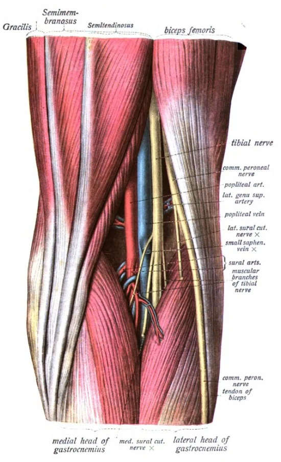
The popliteal fossa is a diamond-shaped depression located posterior to the knee joint. It serves as a critical conduit for neurovascular structures passing between the thigh and the leg. The boundaries are defined by the surrounding musculature. The superolateral boundary is formed by the biceps femoris tendon. The superomedial boundary consists of the semitendinosus and semimembranosus tendons. The inferolateral boundary is created by the lateral head of the gastrocnemius muscle. The inferomedial boundary is formed by the medial head of the gastrocnemius muscle. The floor of the fossa is formed by the popliteal surface of the femur and the capsule of the knee joint. The roof is formed by the popliteal fascia and the skin.
The popliteal artery is the direct continuation of the femoral artery. It passes through the adductor hiatus and enters the popliteal fossa. It is accompanied by the tibial nerve and the common fibular nerve. The artery terminates at the lower border of the popliteus muscle. Here, it divides into the anterior and posterior tibial arteries. The anterior tibial artery passes between the tibia and fibula. The posterior tibial artery provides blood to the muscles and integument on the posterior surface of the tibial region. [1]
The posterior tibial vein drains the posterior surface of the tibia and joins the popliteal vein. The fibular vein drains the muscles and integument in proximity to the fibula and also joins the popliteal vein.
Clinical conditions often involve the structures within this fossa. A popliteal aneurysm is a dilation of the popliteal artery. This condition can lead to thrombosis or embolism. A Baker cyst, also known as a popliteal cyst, is a fluid-filled swelling. It arises from the knee joint capsule, typically between the medial head of the gastrocnemius and the semimembranosus tendon. This cyst can cause pain and swelling in the popliteal fossa. It may rupture, causing symptoms similar to deep vein thrombosis.
DefinitionPopliteal Fossa
A diamond-shaped space posterior to the knee containing the popliteal artery, vein, and tibial nerve.
| Boundary | Structure |
|---|---|
| Superolateral | Biceps femoris tendon |
| Superomedial | Semitendinosus and semimembranosus |
| Inferolateral | Lateral head of gastrocnemius |
| Inferomedial | Medial head of gastrocnemius |
| Floor | Popliteal surface of femur |
Popliteal Vessels
- Femoral ArteryPasses through adductor hiatus
- Question: Enters Popliteal Fossa?
Yes
- Popliteal ArteryBranches into anterior/posterior tibial
No
- Continue distally
Figure 1Sobo 1909 718
Long description
An anatomical illustration from Sobotta's Human Anatomy 1908

Figure 1: An anatomical illustration from Sobotta's Human Anatomy 1908.
The popliteal fossa is a diamond-shaped depression located at the posterior aspect of the knee joint. It serves as a critical conduit for neurovascular structures passing between the thigh and the leg. Understanding its surface anatomy is essential for clinical procedures such as venous access and nerve blocks.
The floor is formed by the popliteal surface of the femur and the capsule of the knee joint. The contents, including the popliteal artery and vein, lie deep to the fascia, protected by the surrounding musculotendinous boundaries.
Palpable landmarks define the borders of this space. The biceps femoris tendon is easily palpable on the lateral side, particularly when the knee is flexed against resistance. Medially, the tendons of the semitendinosus and semimembranosus can be felt, although they are often obscured by the medial head of the gastrocnemius. The medial and lateral heads of the gastrocnemius form the prominent bulges on either side of the lower part of the fossa. These muscles originate from the femur and insert into the calcaneus via the calcaneal tendon. Their bulk creates the characteristic shape of the popliteal region.
Surface markings are used to locate deeper structures. The midpoint of the popliteal fossa corresponds to the level of the knee joint line. The popliteal pulse is typically palpated in the centre of the fossa, deep to the skin and fascia. It is best felt with the knee slightly flexed to relax the surrounding muscles. The saphenous nerve, a branch of the femoral nerve, emerges near the medial border and supplies sensation to the medial aspect of the leg. The tibial nerve and common fibular nerve are also present but lie deeper and are not directly palpable.
DefinitionPopliteal Fossa
A diamond-shaped depression behind the knee, bounded by hamstring and calf muscles, containing major neurovascular structures.
The great saphenous vein is a prominent surface vessel located on the medial surface of the leg and thigh that collects blood from the superficial portions of these areas. While primarily associated with the medial leg, its termination near the saphenous opening is relevant to venous drainage patterns in the lower limb. This is distinct from the lower limb venous system and should not be confused with popliteal drainage. [1]
| Boundary | Structure |
|---|---|
| Superolateral | Biceps femoris tendon |
| Superomedial | Semimembranosus tendon |
| Inferolateral | Lateral head of gastrocnemius |
| Inferomedial | Medial head of gastrocnemius |
| Floor | Popliteal surface of femur, knee joint capsule |
| Roof | Skin, superficial fascia, popliteal fascia |
The popliteal fossa is a diamond-shaped depression located at the posterior aspect of the knee joint. It serves as a critical conduit for neurovascular structures passing between the thigh and the leg. Understanding its boundaries requires identifying the specific bony landmarks that define its corners.
The superior corners of the fossa are defined by the condyles of the femur. The fibular head is particularly prominent and serves as a key palpable landmark. The tibial condyles articulate with the femoral condyles to form the knee joint. These bony structures provide the rigid framework against which the soft tissue boundaries of the fossa are tensioned.
The floor of the popliteal fossa is formed by the popliteal surface of the femur and the capsule of the knee joint. The popliteal surface of the femur is a triangular area located just above the condyles. It provides attachment for the popliteus muscle and the posterior capsule of the knee. The knee joint capsule reinforces the floor, separating the fossa from the joint space. This bony floor is relatively flat and smooth, allowing the neurovascular structures to glide over it during knee flexion and extension.
The boundaries of the popliteal fossa are formed by muscles rather than bones, but these muscles attach to the bony landmarks described above. The superolateral boundary is formed by the biceps femoris tendon, which attaches to the head of the fibula.
The shape of the fossa is maintained by the tension of these tendons and muscles. The bony landmarks provide the anchor points for these soft tissue structures. The head of the fibula is especially important as it lies close to the common fibular nerve, which winds around it. This proximity makes the fibular head a critical landmark for clinical examination and potential injury. The condyles of the femur and tibia provide the stable base for the knee joint, ensuring that the forces transmitted through the fossa are properly distributed.
DefinitionPopliteal Fossa
A diamond-shaped depression at the back of the knee, bounded by muscles and containing neurovascular structures.
The popliteal fossa is bounded superiorly by the biceps femoris laterally and the semitendinosus and semimembranosus medially. Inferiorly, it is bounded by the medial and lateral heads of the gastrocnemius. The floor is formed by the popliteal surface of the femur and the posterior capsule of the knee joint. The roof is formed by the popliteal fascia, which is reinforced by the deep fascia of the leg.
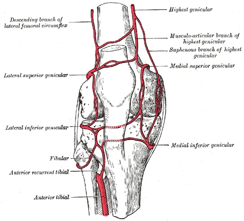
Figure 2Gray552
Long description
Circumpatellar anastomosis. Labelled illustration from Wikimedia Commons, used under its open licence.

Figure 2: Circumpatellar anastomosis.
| Landmark | Location | Clinical Significance |
|---|---|---|
| Head of Fibula | Lateral side, proximal leg | Common fibular nerve winding point |
| Lateral Condyle of Femur | Superior, lateral | Origin of lateral gastrocnemius |
| Medial Condyle of Femur | Superior, medial | Origin of medial gastrocnemius |
| Popliteal Surface of Femur | Posterior distal femur | Floor of the fossa |
These bony landmarks are essential for understanding the anatomy of the popliteal fossa. They provide the structural framework for the muscles and tendons that form the boundaries. They also serve as reference points for clinical procedures such as nerve blocks and vascular access. The head of the fibula is particularly vulnerable to injury due to its superficial location. Fractures of the fibular head can damage the common fibular nerve, leading to foot drop. Understanding the relationship between these bones and the surrounding structures is crucial for accurate diagnosis and treatment.
The popliteal fossa is a diamond-shaped depression located at the posterior aspect of the knee joint. It serves as a critical conduit for neurovascular structures passing between the thigh and the leg. The boundaries of this fossa are defined by specific muscular attachments that create a protective compartment for the contents within.
The fascia provides a rigid boundary that can lead to increased pressure within the compartment if swelling occurs, a condition known as compartment syndrome.
DefinitionPopliteal Fossa
A diamond-shaped depression behind the knee, bounded by muscles and containing major neurovascular structures.
The floor is formed by the popliteal surface of the femur, the capsule of the knee joint, and the popliteus muscle. These muscular boundaries create a tunnel-like structure that protects the delicate neurovascular contents from external trauma.
| Boundary | Structure |
|---|---|
| Superolateral | Biceps femoris |
| Superomedial | Semimembranosus and semitendinosus |
| Inferolateral | Lateral head of gastrocnemius |
| Inferomedial | Medial head of gastrocnemius |
| Floor | Popliteal surface of femur, knee joint capsule, popliteus |
| Roof | Popliteal fascia, skin |
The contents of the popliteal fossa include the tibial nerve, common fibular (peroneal) nerve, popliteal artery, and popliteal vein. The popliteal artery is a continuation of the femoral artery and gives off several branches, including the genicular arteries, which supply the knee joint.
Exam trapNerve Order
Remember the mnemonic 'NTVA' for the structures from superficial to deep: Nerve, Vein, Artery.
Clinical significance arises from the proximity of these structures to the knee joint. Popliteal artery entrapment syndrome is a rare condition where the artery is compressed by adjacent muscles, leading to ischemia. Baker's cysts, or popliteal cysts, are fluid-filled swellings that can develop in the bursa between the medial head of the gastrocnemius and the semimembranosus tendon. These cysts can cause pain and swelling in the popliteal fossa and may rupture, leading to symptoms mimicking deep vein thrombosis. Understanding the anatomy of the popliteal fossa is essential for diagnosing and managing these conditions.
The popliteal fossa is a diamond-shaped depression located at the posterior aspect of the knee joint. It serves as a critical conduit for neurovascular structures passing between the thigh and the leg. The boundaries of this fossa are defined by specific muscle groups that create its lateral and medial walls. Understanding the arrangement of these muscles is essential for interpreting clinical signs and surgical approaches in the lower limb.
As it approaches the apex of the popliteal fossa, the sciatic nerve typically divides into its two major terminal branches: the tibial nerve and the common fibular nerve. This anatomical arrangement explains why injuries to the lateral aspect of the knee often affect the common fibular nerve.
The muscles forming the boundaries of the popliteal fossa are grouped into medial and lateral compartments. The medial boundary is formed by the semimembranosus and semitendinosus muscles. The lateral boundary is formed by the biceps femoris muscle. The roof is formed by the popliteal fascia.
| Muscle | Origin | Insertion | Action | Nerve Supply |
|---|---|---|---|---|
| Semimembranosus | Ischial tuberosity | Medial condyle of tibia | Knee flexion, hip extension | Tibial nerve |
| Semitendinosus | Ischial tuberosity | Medial surface of tibia | Knee flexion, hip extension | Tibial nerve |
| Biceps Femoris (Long Head) | Ischial tuberosity | Head of fibula | Knee flexion, hip extension | Tibial nerve (long head) |
| Biceps Femoris (Short Head) | Linea aspera | Head of fibula | Knee flexion | Common fibular nerve |
| Popliteus | Lateral condyle of femur | Posterior surface of tibia | Unlocks knee joint | Tibial nerve |
They are innervated by the tibial division of the sciatic nerve. The sciatic nerve itself arises from the sacral plexus and descends through the thigh, providing motor innervation to the posterior compartment. In the popliteal fossa, the sciatic nerve bifurcates. The common fibular nerve winds around the neck of the fibula, where it is susceptible to injury. This detailed anatomical knowledge is fundamental for diagnosing nerve palsies and understanding the biomechanics of the knee joint.
The arterial supply to the popliteal fossa is derived from the continuation of the femoral artery as it passes through the adductor hiatus. The femoral artery becomes the popliteal artery, which serves as the primary vascular conduit for the posterior knee region. This vessel is deep within the fossa, lying anterior to the knee joint capsule and the popliteus muscle. The popliteal artery is a significant source of collateral circulation around the knee. [1]
The popliteal artery is centrally located, descending from the adductor canal. Lateral to the artery lies the lateral head of the gastrocnemius muscle. Medial to the artery is the medial head of the gastrocnemius and the semimembranosus tendon. The small saphenous vein runs along the lateral aspect of the fossa.
The popliteal artery gives off several important branches within the fossa. These include the superior medial genicular artery, the superior lateral genicular artery, the middle genicular artery, the inferior medial genicular artery, and the inferior lateral genicular artery. These genicular arteries form an anastomotic network around the knee joint, ensuring blood supply even if the main trunk is compromised.
DefinitionPopliteal Artery
The continuation of the femoral artery through the adductor hiatus, supplying the posterior knee and giving rise to genicular branches and the terminal tibial arteries.
The course of the popliteal artery is straight and vertical, lying on the floor of the fossa. It is covered by skin, superficial fascia, and the popliteal fascia. Deep to the artery are the popliteus muscle and the capsule of the knee joint. The artery is vulnerable to injury in posterior knee dislocations due to its fixed position. The genicular branches anastomose with branches of the femoral artery, such as the descending genicular artery, and branches of the anterior tibial artery, creating a rich collateral network. This network is crucial for maintaining perfusion to the lower leg in cases of proximal arterial occlusion.
| Branch | Origin | Course | Termination/Termination |
|---|---|---|---|
| Superior Medial Genicular | Popliteal | Winds around medial condyle | Anastomoses with other geniculars |
| Superior Lateral Genicular | Popliteal | Winds around lateral condyle | Anastomoses with other geniculars |
| Middle Genicular | Popliteal | Pierces posterior capsule | Supplies knee joint structures |
| Anterior Tibial | Popliteal | Passes through interosseous membrane | Anterior compartment of leg |
| Posterior Tibial | Popliteal | Descends in posterior leg | Medial malleolus region |
| Fibular (Peroneal) | Popliteal | Descends along fibula | Lateral malleolus region |
This bifurcation is a critical landmark in vascular surgery. The anterior tibial artery supplies the anterior compartment of the leg, while the posterior tibial artery supplies the posterior compartment. Understanding these relationships is vital for diagnosing and managing vascular injuries in the knee region.
The venous system operates on a complementary principle to the arterial supply, returning oxygen-depleted blood to the right atrium. While arteries deliver oxygenated blood, veins carry blood that has passed through systemic capillaries, resulting in lower oxygen concentration. This return journey is not as rigidly parallel as arterial distribution; there is significant variability in the venous circulation compared to the arterial system. Understanding this variability is crucial for clinical practice, even though standard anatomical teaching focuses on the most common patterns. [1]
The central structure is the popliteal vein. Superiorly, the popliteal vein continues as the femoral vein as it passes through the adductor hiatus. Inferiorly, it is formed by the union of the anterior and posterior tibial veins. Lateral to the popliteal vein lies the small saphenous vein, which often drains into it. Medial structures include the tibial nerve and the medial sural cutaneous vein.
The primary vessel is the popliteal vein. It is a deep vein that accompanies the popliteal artery. Its origin is at the lower border of the popliteus muscle, where it is formed by the union of the anterior and posterior tibial veins. The course of the vein is upward through the popliteal fossa. Its termination is at the adductor hiatus, where it becomes the femoral vein. This transition marks the entry into the thigh's venous system.
Superficial veins also contribute to drainage. The small saphenous vein ascends posterior to the lateral malleolus and runs up the back of the leg. It typically pierces the deep fascia in the popliteal fossa to drain into the popliteal vein. This connection is a key site for venous reflux in clinical conditions. The great saphenous vein drains into the femoral vein in the thigh, not directly into the popliteal system, although it is part of the broader lower limb drainage.
DefinitionPopliteal Vein
The deep vein of the posterior knee, formed by the union of the tibial veins and terminating as the femoral vein.
The systemic venous return ultimately flows into the right atrium. Most blood enters via the superior or inferior vena cava. Blood from the lower limbs, including the popliteal region, flows into the inferior vena cava. This pathway ensures that deoxygenated blood from the legs is returned to the heart for pulmonary gas exchange. However, for the lower limb, the route is strictly through the inferior vena cava. [1]
| Structure | Type | Relation | Termination |
|---|---|---|---|
| Popliteal Vein | Deep | Anterior to artery | Femoral Vein |
| Small Saphenous Vein | Superficial | Lateral aspect | Popliteal Vein |
| Anterior Tibial Vein | Deep | Anterior compartment | Popliteal Vein |
| Posterior Tibial Vein | Deep | Posterior compartment | Popliteal Vein |
Understanding these relationships allows for accurate identification of structures in both anatomical dissection and clinical imaging. The popliteal vein is the central conduit, receiving blood from the deep leg muscles and superficial tributaries. Its position relative to the artery and nerves is consistent, making it a reliable landmark. Variations in the drainage pattern, such as the small saphenous vein draining into the great saphenous vein, are important to note but do not change the primary pathway to the inferior vena cava.
The large nerve lying in the midline of the fossa is the tibial nerve. It descends vertically through the centre of the popliteal fossa, lying superficial to the popliteal vein and then the popliteal artery. It gives off muscular branches to the gastrocnemius, plantaris, and soleus muscles. It continues inferiorly to become the posterior tibial nerve. The other major nerve is the common fibular (peroneal) nerve. It lies along the lateral border of the fossa, running along the medial border of the biceps femoris tendon. It winds around the neck of the fibula, where it is subcutaneous and vulnerable to injury. It divides into the superficial and deep fibular nerves.
The tibial nerve provides the motor supply to the posterior compartment of the leg. This includes the gastrocnemius, soleus, plantaris, popliteus, tibialis posterior, flexor digitorum longus, and flexor hallucis longus. These muscles are responsible for plantar flexion and inversion of the foot. The tibial nerve also provides cutaneous supply via the medial sural cutaneous nerve. This nerve joins with the peroneal communicating branch to form the sural nerve. The sural nerve supplies the skin of the posterolateral leg and the lateral side of the foot.
The deep fibular nerve innervates the tibialis anterior, extensor digitorum longus, extensor hallucis longus, and fibularis tertius. These muscles dorsiflex and extend the toes. The superficial fibular nerve innervates the fibularis longus and fibularis brevis. These muscles evert the foot.
| Nerve | Motor Supply | Cutaneous Supply |
|---|---|---|
| Tibial | Gastrocnemius, soleus, plantaris, popliteus, tibialis posterior, flexor digitorum longus, flexor hallucis longus | Medial sural cutaneous nerve (via sural nerve) |
| Common Fibular | Tibialis anterior, extensor digitorum longus, extensor hallucis longus, fibularis tertius, fibularis longus, fibularis brevis | Lateral sural cutaneous nerve, superficial fibular nerve |
Lesions of the tibial nerve result in loss of plantar flexion and inversion. The foot assumes a dorsiflexed and everted position. Sensory loss occurs on the sole of the foot. Lesions of the common fibular nerve result in foot drop. The patient cannot dorsiflex or evert the foot. Sensory loss occurs on the anterolateral leg and dorsum of the foot.
Clinical pearlClinical Pearl
The common fibular nerve is superficial at the neck of the fibula, making it susceptible to compression from casts or prolonged leg crossing.
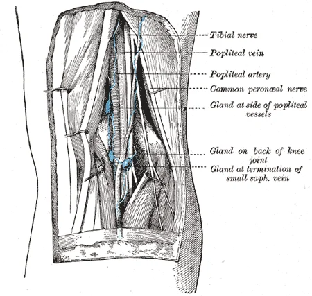
Figure 3Gray609
Long description
Lymph glands of popliteal fossa. (Poirier and Charpy.)

Figure 3: Lymph glands of popliteal fossa (Poirier and Charpy.).
The lymphatic drainage of the lower limb converges at the popliteal fossa, a critical anatomical junction for immune surveillance and fluid return. Understanding this pathway is essential for identifying the spread of infection or malignancy from the distal lower extremity. The primary collectors are the popliteal lymph nodes, which are situated within the fatty tissue of the fossa. These nodes are not merely passive filters but active participants in the immune response, receiving afferent vessels from the superficial and deep systems of the leg and foot.
The central structure is the popliteal vein, which serves as the primary landmark. Inferiorly, the small saphenous vein drains the lateral aspect, its tributaries converging towards the popliteal region. The gastrocnemius muscles form the lateral and medial boundaries of the fossa, providing a muscular backdrop to the neurovascular bundle.
The popliteal nodes are divided into superficial and deep groups based on their position relative to the fascia. They receive lymph from the lateral side of the foot and the posterior aspect of the leg. The deep nodes are located more medially, surrounding the popliteal vessels. They collect lymph from the deep structures of the leg, including the bones, joints, and muscles. Afferent vessels from the superficial nodes often pass through the deep nodes before draining into the iliac nodes. This hierarchical arrangement ensures that lymph is filtered multiple times, reducing the risk of systemic spread of pathogens or cancer cells.
Clinical significance arises when these nodes become enlarged due to infection or metastasis. Palpation of the popliteal nodes can be challenging due to their deep location, but tenderness or swelling may indicate pathology in the lower limb. In cases of melanoma or squamous cell carcinoma of the foot, the popliteal nodes may be the first site of metastatic spread. Therefore, accurate knowledge of this drainage pattern is vital for staging and treatment planning. The lymphatic vessels themselves are thin-walled and valveless in some segments, relying on muscle contraction and arterial pulsation for flow. This dependence on surrounding structures highlights the importance of maintaining normal anatomical relationships for effective lymphatic return.
The boundaries are formed superiorly by the biceps femoris laterally and the semimembranosus and semitendinosus medially. Inferiorly, the boundaries are the lateral and medial heads of the gastrocnemius.
It runs vertically down the middle of the fossa. The common fibular (peroneal) nerve runs along the lateral border, wrapping around the neck of the fibula.
The neurovascular bundle is central. It lies deep to the popliteal vein. This arrangement is critical for surgical access and understanding vascular supply to the knee and leg.
Clinical relevance is high due to the proximity of these structures to the knee joint. Injury to the popliteal artery can lead to severe ischaemia of the lower leg. Understanding the spatial relationships is vital for interpreting imaging and performing procedures such as knee arthroscopy or vascular surgery.
The fossa also contains the popliteal lymph nodes, which drain the lower limb. These nodes are located within the fat of the fossa and are important in the spread of infection or malignancy from the leg.
In summary, the popliteal fossa is a complex anatomical region containing vital nerves and vessels. Its boundaries are defined by specific muscles, and its contents are arranged in a predictable pattern. Mastery of this anatomy is fundamental for medical students and clinicians dealing with lower limb pathology.
Understanding the popliteal fossa on imaging requires correlating surface landmarks with deep structural relationships. The fossa is a diamond-shaped depression located at the posterior aspect of the knee joint. Its boundaries are defined by specific muscles, which remain consistent across imaging modalities. The superolateral boundary is formed by the biceps femoris tendon. The superomedial boundary is created by the semimembranosus and semitendinosus tendons. The inferolateral boundary is defined by the lateral head of the gastrocnemius muscle. The inferomedial boundary is formed by the medial head of the gastrocnemius muscle.
Posterior to the femur lies the popliteal fossa. The tibial nerve is located posterolateral to the vessels. The gastrocnemius heads form the posterior muscular bulk.
X-ray imaging primarily evaluates the bony architecture of the knee. It is useful for detecting fractures of the distal femur or proximal tibia. It can also reveal joint effusions or loose bodies within the joint space. However, X-rays do not visualise the soft tissue contents of the popliteal fossa directly. Soft tissue swelling may be inferred from the displacement of fat planes.
Computed tomography (CT) provides detailed cross-sectional images of the bony and soft tissue structures. It is particularly useful for assessing complex fractures and foreign bodies. CT angiography can visualise the popliteal artery and its branches. It is highly sensitive for detecting arterial dissections or aneurysms. The high contrast resolution allows for clear differentiation between the artery, vein, and nerve.
Magnetic resonance imaging (MRI) is the gold standard for evaluating soft tissue pathology in the popliteal fossa. It provides excellent contrast resolution for muscles, tendons, ligaments, and neurovascular structures. MRI is superior for detecting popliteal artery entrapment syndrome. It can identify Baker’s cysts and their communication with the joint space. MRI is also essential for assessing meniscal tears and ligamentous injuries that may extend into the fossa.
Ultrasound is a dynamic, real-time imaging modality. It is ideal for evaluating superficial structures and guiding interventions. Ultrasound can distinguish between solid and cystic masses in the popliteal fossa. It is highly accurate for diagnosing Baker’s cysts. Doppler ultrasound can assess blood flow in the popliteal artery and vein. It is useful for detecting deep vein thrombosis. Ultrasound can also visualise nerve compression in real-time.
Clinical pearlImaging Choice
MRI is the preferred modality for soft tissue pathology, while ultrasound is best for dynamic assessment and cyst diagnosis.
| Modality | Primary Utility | Key Structures Visualised |
|---|---|---|
| X-ray | Bony anatomy | Femur, tibia, joint space |
| CT | Fractures, vascular calcification | Bone detail, arterial lumen |
| MRI | Soft tissue pathology | Nerves, vessels, cysts, muscles |
| Ultrasound | Dynamic assessment, cysts | Superficial vessels, nerve compression |
The popliteal fossa is a diamond-shaped depression located at the posterior aspect of the knee joint. It serves as a critical conduit for neurovascular structures passing between the thigh and the leg. Understanding its boundaries and contents is essential for interpreting clinical signs and performing safe interventions.
The superior boundaries are formed by the semimembranosus muscle medially and the biceps femoris muscle laterally. The inferior boundaries are defined by the medial and lateral heads of the gastrocnemius muscle. The floor of the fossa is formed by the popliteal surface of the femur and the capsule of the knee joint. The roof is formed by the popliteal fascia and the skin. The common fibular nerve winds around the neck of the fibula, lateral to the biceps femoris tendon. Recognising these relationships allows one to predict the pattern of injury in trauma.
Injuries to the popliteal fossa often result from hyperextension trauma or direct blunt force. The popliteal artery is tethered at the adductor hiatus and the soleal arch, making it susceptible to tearing during knee dislocation. Such injuries require immediate vascular assessment. Nerve palsies are also common; the common fibular nerve is particularly vulnerable as it courses around the fibular neck, leading to foot drop if damaged. The tibial nerve, being more protected, is less frequently injured but can suffer from compression syndromes.
Surface landmarks are vital for clinical procedures. The popliteal pulse is palpable in the midline of the fossa, deep to the fascia. This landmark guides the placement of needles for popliteal nerve blocks used in anaesthesia for lower limb surgery. Ultrasound imaging is increasingly used to visualise these structures in real-time, reducing the risk of vascular puncture. This knowledge is crucial for interpreting Doppler ultrasound studies or angiograms.
The clinical significance of the popliteal fossa extends to the diagnosis of Baker’s cysts, which are fluid-filled swellings arising from the gastrocnemio-semimembranosus bursa. These cysts can compress adjacent structures, causing pain and swelling. Differentiating a Baker’s cyst from a deep vein thrombosis is a common diagnostic challenge. Physical examination involves assessing the tension of the cyst and checking for signs of inflammation. Imaging confirms the diagnosis and rules out other pathologies such as tumours or abscesses.
| Structure | Origin | Course | Relations | Termination |
|---|---|---|---|---|
| Popliteal Artery | Continuation of femoral artery | Passes through adductor hiatus | Deep to vein and nerve | Bifurcates into anterior/posterior tibial arteries |
| Tibial Nerve | Sciatic nerve | Descends midline of fossa | Superficial to artery | Divides into medial/lateral plantar nerves |
| Common Fibular Nerve | Sciatic nerve | Winds around fibular neck | Lateral to biceps femoris tendon | Divides into superficial/deep fibular nerves |
Clinical pearlClinical Pearl
Always assess distal pulses and neurological function in any knee trauma to rule out popliteal vessel or nerve injury.
The popliteal fossa is a diamond-shaped space located at the posterior aspect of the knee joint. It serves as a critical conduit for neurovascular structures passing between the thigh and the leg. Understanding its boundaries and contents is essential for clinical examinations and surgical procedures involving the knee.
The boundaries of the popliteal fossa are defined by specific muscle groups. The superolateral boundary is formed by the biceps femoris tendon. The superomedial boundary consists of the semitendinosus and semimembranosus tendons. The inferolateral boundary is created by the lateral head of the gastrocnemius muscle. The inferomedial boundary is formed by the medial head of the gastrocnemius muscle. The floor of the fossa is formed by the popliteal surface of the femur and the capsule of the knee joint.
The contents of the popliteal fossa are arranged in a specific order from superficial to deep. This arrangement is crucial for safe needle insertion during procedures such as popliteal nerve blocks.
Clinical pearlNerve Block
When performing a popliteal nerve block, the tibial nerve is the primary target. It is located superficially, making it easier to access than the common fibular nerve.
Popliteal artery aneurysms are a significant clinical concern. These aneurysms can compress adjacent nerves, leading to neurological deficits. They also pose a risk of thromboembolism, which can result in acute limb ischaemia. Compression of the tibial nerve may cause weakness in plantar flexion and sensory loss in the sole of the foot.
Exam trapCommon Error
Do not confuse the popliteal artery with the femoral artery. The popliteal artery is the continuation of the femoral artery after it passes through the adductor hiatus.
Anatomical variations in the popliteal fossa are common. This variation can predispose the nerve to injury during surgical procedures. Awareness of these variations is vital for avoiding iatrogenic damage.
DefinitionAdductor Hiatus
The adductor hiatus is an opening in the adductor magnus tendon that allows the femoral artery to pass into the popliteal fossa.
The popliteal fossa contains vital neurovascular structures. Its boundaries are formed by hamstring and calf muscles. The tibial nerve, popliteal vein, and popliteal artery are arranged from superficial to deep. Clinical conditions such as aneurysms and nerve compression require careful anatomical knowledge for diagnosis and treatment.
Students frequently confuse the anatomical boundaries of the popliteal fossa, leading to errors in identifying contained structures. The superior boundaries are formed by the semimembranosus and semitendinosus muscles medially, and the biceps femoris muscle laterally. A common error is omitting the gastrocnemius heads from the inferior boundaries. The correct inferior boundaries are the medial and lateral heads of the gastrocnemius and the plantaris muscle. Without this precise definition, the space is incorrectly perceived as larger or shallower than it is.
Another frequent mistake involves the neurovascular contents. Students often list the popliteal artery and vein as separate entities running parallel without noting their relationship. In reality, the popliteal vein lies posterior to the artery, and the tibial nerve lies posterior to the vein. This order from anterior to posterior is artery, vein, nerve (AVN). Misremembering this sequence leads to incorrect interpretations of clinical signs such as pulse deficits or nerve palsies.
A third pitfall is the failure to distinguish between the common peroneal nerve and the tibial nerve courses. The common peroneal nerve winds around the neck of the fibula, a site vulnerable to injury. The tibial nerve runs more medially and deeply. Confusing these paths results in incorrect localisation of motor and sensory deficits in the lower leg and foot.
The fourth error relates to the genicular arteries. Students often overlook the anastomosis around the knee joint. The genicular arteries branch from the popliteal artery and contribute to the rich collateral circulation around the knee. Ignoring this network leads to an incomplete understanding of blood supply in cases of arterial occlusion.
The fifth pitfall is the misidentification of the popliteal lymph nodes. These nodes are located in the fat of the popliteal fossa, draining the lower limb. They are often missed in dissection or imaging reviews. Their enlargement can indicate infection or malignancy in the leg or foot, making their identification clinically relevant.
The sixth error involves the saphenous nerve. This nerve is a branch of the femoral nerve and runs subcutaneously along the medial aspect of the knee and leg. It is not a primary content of the deep popliteal fossa but is often confused with the tibial nerve. Its sensory distribution is distinct and important for anaesthetic techniques.
The seventh pitfall is the assumption of uniformity in venous drainage. Individual variations in circulation patterns are not uncommon. Students must recognise that venous anatomy can vary, affecting surgical approaches and imaging interpretations. [1]
The eighth error is neglecting the role of the deep veins in thermoregulation. In both the neck and limb regions, there are often both superficial and deeper levels of veins. The deeper veins generally correspond to the complementary arteries. Understanding this helps explain physiological responses to temperature changes. [1]
Knowledge of the popliteal fossa is critical at the bedside for assessing vascular and neurological status. Clinicians palpate the popliteal pulse to evaluate peripheral arterial disease. The pulse is located deep in the fossa, between the tendons of the semimembranosus and biceps femoris. Absence or weakness of this pulse suggests arterial obstruction proximal to the knee.
In the operating theatre, the popliteal fossa is accessed for vascular surgeries such as bypass grafting. Surgeons must carefully dissect through the fascia to avoid injuring the tibial nerve and popliteal vessels. The AVN relationship is crucial to prevent iatrogenic damage. Additionally, the fossa is approached for excision of Baker’s cysts, which are fluid-filled sacs that can cause pain and swelling.
On imaging, the popliteal fossa is evaluated using ultrasound, CT, and MRI. Ultrasound is the first-line modality for assessing deep vein thrombosis (DVT) in the popliteal vein. The vein is compressible; non-compressibility indicates thrombosis. CT and MRI are used to visualise soft tissue masses, such as lipomas or sarcomas, which may arise in the fossa. The clear anatomical boundaries help in localising these lesions.
In examinations, students are tested on the boundaries and contents of the popliteal fossa. Questions often require identification of structures on diagrams or in dissection specimens. Understanding the origin, course, relations, branches, and termination of the popliteal artery is essential. The artery is a continuation of the femoral artery and terminates by dividing into the anterior and posterior tibial arteries.
DefinitionPopliteal Fossa Boundaries
The popliteal fossa is a diamond-shaped space behind the knee, bounded superiorly by the semimembranosus and semitendinosus medially and the biceps femoris laterally, and inferiorly by the medial and lateral heads of the gastrocnemius and the plantaris.
| Muscle | Origin | Insertion | Action | Nerve Supply |
|---|---|---|---|---|
| Semimembranosus | Ischial tuberosity | Medial condyle of tibia | Knee flexion, hip extension | Tibial nerve |
| Semitendinosus | Ischial tuberosity | Medial surface of tibia | Knee flexion, hip extension | Tibial nerve |
| Biceps Femoris | Ischial tuberosity (long head); Linea aspera (short head) | Head of fibula | Knee flexion, hip extension | Tibial nerve (long head); Common peroneal nerve (short head) |
| Gastrocnemius (Medial) | Lateral condyle of femur | Calcaneus via Achilles tendon | Plantar flexion, knee flexion | Tibial nerve |
| Gastrocnemius (Lateral) | Lateral condyle of femur | Calcaneus via Achilles tendon | Plantar flexion, knee flexion | Tibial nerve |
| Plantaris | Lateral condyle of femur | Calcaneus via Achilles tendon | Weak plantar flexion, knee flexion | Tibial nerve |
Popliteal Fossa Assessment
- StartPatient supine, knee slightly flexed
- Question: Palpate Pulse?
Present
- Assess character
Absent
- Consider arterial occlusion
Exam trapNerve Vulnerability
The common peroneal nerve winds around the neck of the fibula, making it highly susceptible to injury from fractures or compression.
The superficial veins do not normally have direct arterial counterparts, but in addition to returning blood, they also make contributions to the maintenance of body temperature. When the ambient temperature is warm, more blood is diverted to the superficial veins where heat can be more easily dissipated to the environment. In colder weather, there is more constriction of the superficial veins and blood is diverted deeper where the body can retain more of the heat. This physiological principle is relevant in understanding local blood flow changes during surgery or trauma. [1]
The hepatic portal system carries blood to the liver for processing before it enters circulation. While not directly in the popliteal fossa, understanding systemic circulation is vital for interpreting haemodynamic changes in lower limb conditions. [1]
NoteVenous Variations
Individual variations in circulation patterns are not uncommon. Always consider anatomical variants during surgical planning.
The popliteal fossa is bounded by hamstring muscles superiorly and gastrocnemius muscles inferiorly. The neurovascular bundle follows the order: artery, vein, nerve (AVN) from anterior to posterior. The common peroneal nerve is vulnerable at the neck of the fibula. The popliteal artery terminates by dividing into anterior and posterior tibial arteries. Deep veins correspond to complementary arteries and play a role in thermoregulation. Superficial veins contribute to body temperature maintenance through vasodilation and vasoconstriction. Ultrasound is the primary imaging modality for assessing popliteal vein thrombosis. Anatomical variations in venous patterns are common and should be anticipated.
Answer each one in your head first, then open the answer.
What is the order of structures from anterior to posterior in the popliteal fossa?
Show answerHide answer
Artery, Vein, NerveWhich muscle forms the medial superior boundary of the popliteal fossa?
Show answerHide answer
SemimembranosusWhere does the common peroneal nerve wind around?
Show answerHide answer
Neck of the fibulaWhat is the primary imaging modality for popliteal vein thrombosis?
Show answerHide answer
UltrasoundWhich veins contribute to body temperature maintenance?
Show answerHide answer
Superficial veins
- Betts JG, Young KA, Wise JA, et al. Circulatory Pathways. OpenStax, Anatomy and Physiology 2e (CC BY-NC-SA 4.0). Accessed .
Figure credits
Educational content for medical students and doctors. It is not a substitute for clinical judgement or local guidelines.