Knee joint
The knee is the body's largest synovial joint, comprising three articulations within a single capsule.
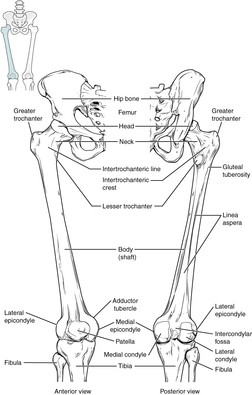
Figure 1810 Femur and Patella
Long description
The femur is the single bone of the thigh region. It articulates superiorly with the hip bone at the hip joint, and inferiorly with the tibia at the knee joint. The patella only articulates with the distal end of the femur.

Figure 1: The femur is the single bone of the thigh region.
Figure 2Gray345
Long description
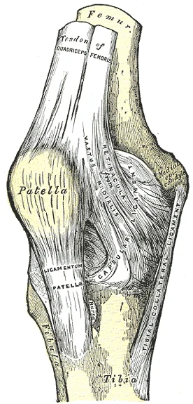
Right knee-joint. Anterior view. Labelled illustration from Wikimedia Commons, used under its open licence.

Figure 2: Right knee-joint (anterior view).
Figure 3Gray350
Long description
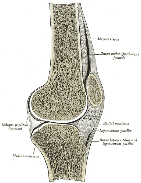
Sagittal section of right knee-joint. Labelled illustration from Wikimedia Commons, used under its open licence.

Figure 3: Sagittal section of right knee-joint.
The knee joint is the largest joint of the body. It is a synovial joint, which is the most common type of joint in the body. A key structural characteristic for a synovial joint is the presence of a joint cavity. Unlike fibrous or cartilaginous joints, the articulating bone surfaces are not directly connected by fibrous connective tissue or cartilage. This gives the bones the ability to move smoothly against each other, allowing for increased joint mobility. The ability of the bones to move smoothly means that each synovial joint is functionally classified as a diarthrosis. [1] [2]
The knee actually consists of three articulations enclosed within a single articular capsule. The femoropatellar joint is found between the patella and the distal femur. The medial tibiofemoral joint and lateral tibiofemoral joint are located between the medial and lateral condyles of the femur and the medial and lateral condyles of the tibia. The patella is a sesamoid bone incorporated into the tendon of the quadriceps femoris muscle of the anterior thigh. It serves to protect this tendon from [1] [3]
The knee functions primarily as a hinge joint, allowing flexion and extension of the leg. This action is generated by both rolling and gliding motions of the femur on the tibia. The medial and lateral tibiofemoral joints are modified hinge joints that allow for knee extension and flexion. During these movements, the condyles of the femur both roll and glide over the surface of the tibia. In addition, some rotation of the leg is available when the knee is flexed, but not when extended. [1]
Because of the shape of the medial femoral condyle, the knee must “screw home” to fully extend. The tibia externally rotates to achieve proper alignment. As the knee comes into full extension, a slight medial rotation of the femur serves to “lock” the knee into its most stable, weight-bearing position. The reverse motion, a small lateral rotation of the femur, is required to initiate knee flexion. [1]
DefinitionScrew-home mechanism
The tibia externally rotates to achieve proper alignment as the knee extends, locking it into a stable position.
Clinical pearlKnee Stability
The locking mechanism provides stability during standing and weight bearing.
| Articulation | Location |
|---|---|
| Femoropatellar | Between patella and distal femur |
| Medial tibiofemoral | Between medial condyles of femur and tibia |
| Lateral tibiofemoral | Between lateral condyles of femur and tibia |
Knee Movement Logic
- StartAssess knee position
- Question: Is knee extended?
Yes
- Rotation is restrictedTibia is locked externally
No
- Rotation is availableLeg can rotate when flexed
Figure 4Gray346
Long description
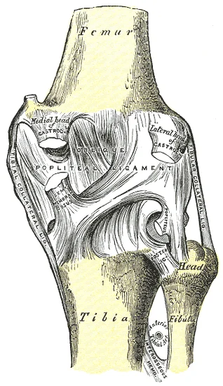
Right knee-joint. Posterior view. Labelled illustration from Wikimedia Commons, used under its open licence.

Figure 4: Right knee-joint (posterior view).
Between these bony surfaces lie the medial and lateral menisci, which are C-shaped fibrocartilaginous discs. These menisci serve to provide padding and improve the fit between the bones. The patellar articular surface is located on the anterior aspect of the distal femur, allowing the patella to glide during knee extension. The femoral condyles are rounded and project downwards, while the tibial plateaus are relatively flat but concave, accommodating the femoral condyles. The menisci sit on the tibial plateaus, deepening the socket for the femoral condyles. This arrangement allows for the knee's significant range of motion while maintaining stability. The articular surfaces are covered by hyaline cartilage, which reduces friction and absorbs shock during movement. The patella acts as a sesamoid bone, increasing the leverage of the quadriceps tendon. The alignment of these surfaces is crucial for normal knee function. Misalignment or damage to these surfaces can lead to joint instability and pain. Understanding this anatomy is essential for diagnosing injuries such as meniscal tears or ligamentous disruptions.
DefinitionArticular Surfaces
The bony surfaces that come into contact within a synovial joint, covered by articular cartilage to facilitate smooth movement and reduce friction.
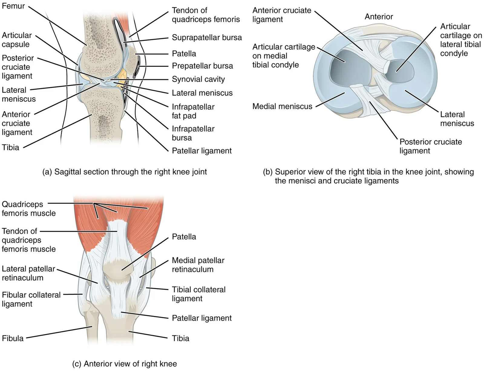
Synovial joints are characterised by the presence of a joint cavity, inside of which the bones articulate with each other. The articulating surfaces of the bones at a synovial joint are not directly connected to each other by connective tissue or cartilage. Friction between the bones is reduced by a thin layer of articular cartilage covering the surfaces of the bones. The knee joint has multiple ligaments that provide support, particularly in the extended position. Outside of the articular capsule, located at the sides of the knee, are two extrinsic ligaments. As it crosses the knee, the tibial collateral ligament is firmly attached on its deep side to the articular capsule and to the medial meniscus. [2]
| Ligament | Location | Attachment | Function |
|---|---|---|---|
| Fibular collateral | Lateral | Lateral femoral epicondyle to fibular head | Resists hyperextension and rotation |
| Tibial collateral | Medial | Medial femoral epicondyle to medial tibia | Resists hyperextension and rotation |
| Anterior cruciate | Intracapsular | Anterior tibia to femur | Resists hyperextension |
| Posterior cruciate | Intracapsular | Posterior tibia to femur | Prevents anterior sliding of femur |
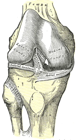
Inside the knee are two intracapsular ligaments, the anterior cruciate ligament and posterior cruciate ligament. These ligaments are anchored inferiorly to the tibia at the intercondylar eminence. The cruciate ligaments are named for whether they are attached anteriorly or posteriorly to this tibial region. Each ligament runs diagonally upward to attach to the inner aspect of a femoral condyle. The posterior cruciate ligament is the stronger ligament. It serves to support the knee when it is flexed and weight bearing. The anterior cruciate ligament resists hyperextension of the knee. The posterior cruciate ligament prevents anterior sliding of the femur. The medial and lateral menisci, located between the femoral and tibial condyles, are articular discs that provide padding and improve the fit between the bones. [1] [3]
Figure 5917 Knee Joint
Long description
(a) The knee joint is the largest joint of the body. (b)–(c) It is supported by the tibial and fibular collateral ligaments located on the sides of the knee outside of the articular capsule, and the anterior and posterior cruciate ligaments found inside the capsule. The medial and lateral menisci pr

Figure 5: (a) The knee joint is the largest joint of the body.
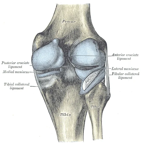
Figure 6Gray352
Long description
Capsule of right knee-joint (distended). Posterior aspect.

Figure 6: Capsule of right knee-joint (distended) (posterior aspect).
The knee joint is a synovial joint, characterised by the presence of a joint cavity. The walls of this space are formed by the articular capsule, a fibrous connective tissue structure that is attached to each bone just outside the area of the bone’s articulating surface. Friction between the bones at a synovial joint is prevented by the presence of the articular cartilage, a thin layer of hyaline cartilage that covers the entire articulating surface of each bone. However, unlike at a cartilaginous joint, the articular cartilages of each bone are not continuous with each other. Instead, the articular cartilage acts like a Teflon® coating over the bone surface, allowing the articulating bones to move smoothly against each other without damaging the underlying bone tissue. [2]
Lining the inner surface of the articular capsule is a thin synovial membrane. The cells of this membrane secrete synovial fluid (synovia = “a thick fluid”), a thick, slimy fluid that provides lubrication to further reduce friction between the bones of the joint. This fluid also provides nourishment to the articular cartilage, which does not contain blood vessels. [2]
DefinitionArticular Capsule
A fibrous connective tissue structure attached to each bone just outside the area of the bone’s articulating surface, forming the walls of the joint cavity.
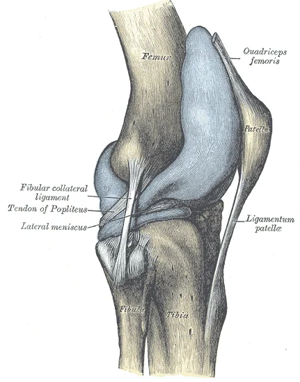
Figure 7Gray351
Long description
Capsule of right knee-joint (distended). Lateral aspect.

Figure 7: Capsule of right knee-joint (distended) (lateral aspect).
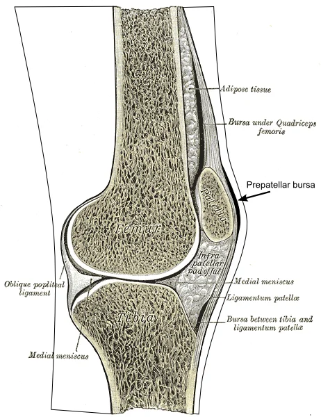
The articular capsule surrounds the joint, attaching to the femur and tibia. The synovial membrane lines the inner surface of the capsule, excluding the articular cartilage. The synovial cavity is the space between the articulating bones, filled with synovial fluid. A subcutaneous bursa is located between the skin and an underlying bone, allowing skin to move smoothly over the bone. Examples include the prepatellar bursa located over the kneecap and the olecranon bursa at the tip of the elbow. A submuscular bursa is found between a muscle and an underlying bone, or between adjacent muscles. [2]
Additional structures located outside of a synovial joint serve to prevent friction between the bones of the joint and the overlying muscle tendons or skin. A bursa (plural = bursae) is a thin connective tissue sac filled with lubricating liquid. They are located in regions where skin, ligaments, muscles, or muscle tendons can rub against each other, usually near a body joint. Bursae reduce friction by separating the adjacent structures, preventing them from rubbing directly against each other. [2]
At many synovial joints, additional support is provided by the muscles and their tendons that act across the joint. A tendon is the dense connective tissue structure that attaches a muscle to bone. As forces acting on a joint increase, the body will automatically increase the overall strength of contraction of the muscles crossing that joint, thus allowing the muscle and its tendon to serve as a “dynamic ligament” to resist forces and support the joint. This type of indirect support by muscles is very important at the shoulder joint, for example, where the ligaments are relatively weak. [2]
The femur is the single bone of the thigh. The patella is the kneecap and articulates with the distal femur. The tibia is the larger, weight-bearing bone located on the medial side of the leg, and the fibula is the thin bone of the lateral leg. [3]
| Structure | Description |
|---|---|
| Articular Capsule | Fibrous connective tissue attached to bone outside articulating surface |
| Synovial Membrane | Thin membrane lining inner capsule, secretes synovial fluid |
| Articular Cartilage | Thin hyaline cartilage covering articulating surfaces |
| Bursa | Thin connective tissue sac filled with lubricating liquid |
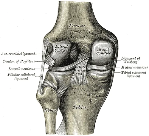
Figure 8Gray348
Long description
Left knee-joint from behind, showing interior ligaments.

Figure 8: Left knee-joint from behind, showing interior ligaments.
Figure 9Gray347
Long description
Right knee-joint, from the front, showing interior ligaments.

Figure 9: Right knee-joint, from the front, showing interior ligaments.
Figure 10Gray348-2
Long description
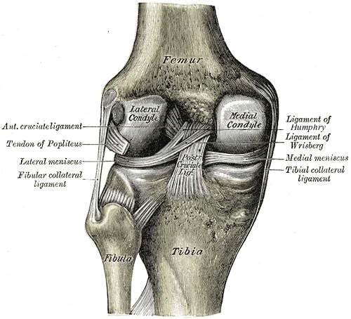
Left knee-joint from behind, showing interior ligaments including the meniscofemoral ligaments Humphry and Wrisberg.

Figure 10: Left knee-joint from behind, showing interior ligaments including the meniscofemoral ligaments Humphry and Wrisberg.
Understanding their spatial relationships is essential for interpreting clinical tests and imaging.
The knee joint is reinforced by several strong ligaments that strengthen the posterior capsule walls. These structures work in concert to provide static stability, complementing the dynamic support provided by surrounding muscles. The primary ligaments are the collateral ligaments, the cruciate ligaments, and the patellar ligament. [1]
The collateral ligaments are located on the sides of the knee. In the fully extended knee position, both collateral ligaments are taut (tight), thus serving to stabilize and support the extended knee and preventing side-to-side or rotational motions between the femur and tibia. This tension locks the knee in extension, allowing for efficient weight-bearing with minimal muscular effort. The medial collateral ligament (MCL) is broad and flat, attaching to the medial epicondyle of the femur and the medial condyle of the tibia. The lateral collateral ligament (LCL) is cord-like, attaching to the lateral epicondyle of the femur and the head of the fibula. [1]
Figure 11Gray349
Long description
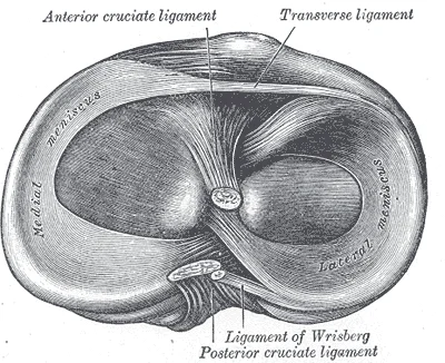
Head of right tibia seen from above, showing menisci and attachments of ligaments.

Figure 11: Head of right tibia seen from above, showing menisci and attachments of ligaments.
The cruciate ligaments are named for their attachment to the intercondylar area of the tibia. They cross each other within the knee joint, hence the name 'cruciate'. The ACL becomes tight when the knee is extended, and thus resists hyperextension. It also prevents anterior translation of the tibia relative to the femur. In this position, the posterior cruciate ligament prevents the femur from sliding anteriorly off the top of the tibia. This mechanism is crucial during activities such as walking downhill or descending stairs. [1]
Although not ligaments, the menisci are fibrocartilaginous structures that deepen the articular surface of the tibia. They act as shock absorbers and improve congruence between the femoral condyles and the tibial plateau. The medial meniscus is larger and C-shaped, while the lateral meniscus is smaller and O-shaped. They are attached to the tibial plateau and the joint capsule. [1]
A sesamoid bone is a bone that is incorporated into the tendon of a muscle where that tendon crosses a joint. The patella is found in the tendon of the quadriceps femoris muscle, the large muscle of the anterior thigh that passes across the anterior knee to attach to the tibia. The patella articulates with the patellar surface of the femur and thus prevents rubbing of the muscle tendon against the distal femur. The patella also lifts the tendon away from the knee joint, which increases the leverage power of the quadriceps femoris muscle as it acts across the knee. Continuing from the patella to the anterior tibia just below the knee is the patellar ligament. Acting via the patella and patellar ligament, the quadriceps femoris is a powerful muscle that acts to extend the leg at the knee. It also serves as a “dynamic ligament” to provide very important support and stabilization for the knee joint. [3] [1]
| Ligament | Attachments | Function |
|---|---|---|
| Medial Collateral | Medial femoral epicondyle to medial tibial condyle | Prevents valgus stress; stabilizes extended knee |
| Lateral Collateral | Lateral femoral epicondyle to fibular head | Prevents varus stress; stabilizes extended knee |
| Anterior Cruciate | Anterior tibia to lateral femoral condyle | Prevents anterior tibial translation; resists hyperextension |
| Posterior Cruciate | Posterior tibia to medial femoral condyle | Prevents posterior tibial translation |
| Patellar | Patella to tibial tuberosity | Transmits quadriceps force; extends knee |
Figure 12Prepatellar bursa
Long description
Prepatellar bursa. Labelled illustration from Wikimedia Commons, used under its open licence.

Figure 12: Prepatellar bursa.
Bursae are connective tissue sacs containing lubricating fluid that serve to reduce friction between structures. They are classified by their anatomical position relative to surrounding tissues. Subcutaneous bursae prevent friction between the skin and an underlying bone. Submuscular bursae protect muscles from rubbing against a bone or another muscle. A subtendinous bursa prevents friction between bone and a muscle tendon. [2]
Tendon sheaths are similar in structure to a bursa, but smaller. They are connective tissue sacs that surround a muscle tendon at places where the tendon crosses a joint. They contain a lubricating fluid that allows for smooth motions of the tendon during muscle contraction and joint movements. [2]
The knee joint itself is a complex hinge mechanism. During initial flexion from a fully extended position, the knee “unlocks” by the tibia internally rotating. The knee is well constructed for weight bearing in its extended position. However, it is vulnerable to injuries associated with hyperextension, twisting, or blows to the medial or lateral side of the joint. These injuries are particularly likely while weight bearing. [1]
The resulting small medial rotation of the femur serves to “lock” the knee into its fully extended and most stable position. Flexion of the knee is initiated by a slight lateral rotation of the femur on the tibia. This lateral rotation motion “unlocks” the knee. This motion is produced by the popliteus muscle of the posterior leg. [1]
The proximal end of the tibia is greatly expanded. The two sides of this expansion form the medial condyle of the tibia and the lateral condyle of the tibia. The top surface of each condyle is smooth and flattened. These areas articulate with the medial and lateral condyles of the femur to form the knee joint. Between the articulating surfaces of the tibial condyles is the intercondylar eminence. This is an irregular, elevated area that serves as the inferior attachment point for two supporting ligaments of the knee. [3]
Clinical pearlLocking Mechanism
The knee locks in extension via medial femoral rotation for stability, and unlocks via lateral rotation initiated by the popliteus muscle.
| Feature | Description |
|---|---|
| Joint Type | Hinge (functionally uniaxial) |
| Primary Movement | Bending and straightening |
| Locking Mechanism | Medial rotation of femur |
| Unlocking Muscle | Popliteus |
| Vulnerability | Hyperextension, twisting, blows |
The knee joint is a synovial joint, and like other joints in the lower limb, it is robust to provide the stability needed to support body weight during activities such as running, jumping, or kicking[1]. While the upper limb joints prioritise mobility, the knee balances strength with a specific range of motion. The primary movements occurring at this hinge joint are flexion and extension, which take place in the sagittal plane.
The muscles producing these movements are grouped by their action. Flexion is primarily performed by the hamstrings, which originate from the ischial tuberosity and insert on the tibia and fibula. Extension is driven by the quadriceps femoris group, which originates from the ilium and femur and inserts via the patellar ligament onto the tibial tuberosity.
| Muscle Group | Primary Action | Nerve Supply |
|---|---|---|
| Quadriceps Femoris | Extension | Femoral nerve |
| Hamstrings | Flexion | Tibial nerve |
| Gastrocnemius | Flexion (assists) | Tibial nerve |
The locking mechanism is crucial for weight-bearing. When the knee is fully extended, the tibia rotates externally. This rotation tightens the ligaments and creates a stable, locked position. To unlock the knee, the popliteus muscle contracts to internally rotate the tibia. This allows flexion to begin. The suprapatellar bursa, which separates the tendon of the large anterior thigh muscle from the distal femur just above the knee, reduces friction during these movements. The joint capsule and li [1]
The knee joint is the largest joint of the body. It actually consists of three articulations. [4]
The patella, a sesamoid bone incorporated into the tendon of the quadriceps femoris muscle, serves to protect this tendon from rubbing against the distal femur during knee movements. The medial and lateral tibiofemoral joints are modified hinge joints that allow for knee extension and flexion. During these movements, the condyles of the femur both roll and glide over the surface of the tibia. [4]
A key factor in knee stability is the "screw home" mechanism that occurs during full extension. Due to the shape of the medial femoral condyle, the knee must "screw home" to fully extend, and the tibia externally rotates to achieve proper alignment. As the knee comes into full extension, a slight medial rotation of the femur serves to "lock" the knee into its most stable, weight-bearing position. The reverse motion, a small lateral rotation of the femur, is required to initiate knee flexion. [4]
The knee functions primarily as a hinge joint, allowing flexion and extension of the leg. This action is generated by both rolling and gliding motions of the femur on the tibia. In addition, some rotation of the leg is available when the knee is flexed, but not when extended. When the knee is flexed, some rotation of the leg is available. [4]
Synovial joints, like the knee, have a joint cavity - a fluid-filled space where the articulating surfaces of the bones contact each other. Unlike fibrous or cartilaginous joints, the articulating bone surfaces at a synovial joint are not directly connected to each other with fibrous connective tissue or cartilage, allowing for increased joint mobility. All synovial joints are functionally classified as diarthrosis joints, which are freely moveable joints. [5]
The vascular and neural integrity of the knee joint is governed by Hilton’s law, which states that the nerves supplying a joint also innervate the muscles acting on that joint and the skin overlying it. This principle ensures coordinated proprioception and motor control during movement. The stability of the knee relies on a complex interplay between bony congruence, ligamentous support, and muscular tension, all of which are modulated by this shared innervation. [1]
The primary arterial supply to the knee joint is derived from the popliteal artery, which gives rise to several genicular branches. These include the superior medial genicular artery, the superior lateral genicular artery, the inferior medial genicular artery, and the inferior lateral genicular artery. These vessels anastomose extensively around the knee, forming a rich collateral network that ensures blood flow even during extreme flexion or extension. The descending genicular artery, a branch of the femoral artery, also contributes to the medial supply. Venous drainage follows the arterial pathways, ultimately draining into the popliteal vein.
Neural innervation is provided by multiple sources, including the femoral nerve, the tibial nerve, the common fibular nerve, and the obturator nerve. The femoral nerve supplies the anterior capsule via its articular branches. The tibial nerve provides innervation to the posterior capsule. The common fibular nerve contributes to the lateral aspect of the joint. The obturator nerve supplies the medial aspect. This diverse innervation pattern supports the complex sensory and motor functions required for knee stability and movement.
DefinitionHilton's Law
The nerves supplying a joint also innervate the muscles acting on that joint and the skin overlying it.
The stability of the knee is maintained by the ligaments and muscles, which are themselves innervated by these same nerves. This integrated system allows for rapid reflexive adjustments to maintain joint integrity during dynamic activities. Disruption of this blood and nerve supply can lead to significant functional impairment, highlighting the clinical importance of understanding this anatomy. [1]
The knee joint is a complex synovial articulation that can be visualised using various imaging modalities. Plain radiography remains the initial investigation of choice for assessing bony alignment and gross pathology. Magnetic resonance imaging (MRI) provides superior soft tissue contrast, allowing detailed evaluation of ligaments, menisci, and cartilage. Computed tomography (CT) is particularly useful for characterising intra-articular fractures and bony abnormalities.
The femur is positioned superiorly, articulating with the tibia inferiorly. The medial collateral ligament (MCL) is a broad, flat band extending from the medial epicondyle of the femur to the medial condyle of the tibia. The lateral collateral ligament (LCL) is a cord-like structure extending from the lateral epicondyle of the femur to the head of the fibula. Understanding these relationships is crucial for interpreting knee imaging and diagnosing pathology. [1]
DefinitionImaging Anatomy
The study of anatomical structures as visualised by medical imaging techniques, essential for diagnosing joint pathology.
Plain radiographs typically include anteroposterior, lateral, and skyline views. The anteroposterior view assesses joint space narrowing, osteophyte formation, and alignment. The lateral view evaluates the patellofemoral joint and posterior structures. The skyline view, also known as the sunrise view, assesses the patellofemoral articulation. MRI is the gold standard for soft tissue evaluation. It can detect ligament tears, meniscal injuries, and cartilage defects with high sensitivity and specificity. CT arthrography may be used when MRI is contraindicated or inconclusive. Ultrasound is useful for evaluating superficial structures such as the patellar tendon and bursae.
| Modality | Primary Use | Key Structures Visualised |
|---|---|---|
| Plain Radiography | Initial assessment, bony pathology | Joint space, osteophytes, fractures, alignment |
| MRI | Soft tissue evaluation | Ligaments, menisci, cartilage, bone marrow |
| CT | Bony detail, complex fractures | Intra-articular fractures, bony anatomy |
| Ultrasound | Superficial structures | Tendons, bursae, effusions |
Interpretation of knee imaging requires knowledge of normal anatomical variants and common pathologies. Degenerative changes such as osteoarthritis are characterised by joint space narrowing, subchondral sclerosis, and osteophyte formation. Ligamentous injuries are graded based on the extent of fibre disruption. Meniscal tears are classified by morphology and location. Understanding these features allows accurate diagnosis and appropriate management planning.
The knee joint is a complex synovial hinge that permits flexion, extension, and slight rotation. Clinical assessment begins with inspection for swelling or deformity, followed by palpation of the joint line, patella, and collateral ligaments.
DefinitionJoint Stability
Stability is maintained by the articular capsule, intrinsic ligaments, and dynamic muscular control, resisting side-to-side movements and hyperextension.
Physical examination aims to isolate specific ligamentous and meniscal injuries. The anterior drawer test assesses the integrity of the anterior cruciate ligament (ACL). With the knee flexed at 90 degrees and the foot fixed, anterior translation of the tibia relative to the femur is observed. Excessive anterior movement indicates an ACL tear. The posterior drawer test evaluates the posterior cruciate ligament (PCL). Posterior translation of the tibia suggests PCL injury, often caused by a direct blow to the proximal tibia with a flexed knee.
Exam trapDrawer Sign
Do not confuse the anterior drawer test with the Lachman test; the latter is more sensitive for acute ACL injuries as it minimizes hamstring co-contraction.
Collateral ligament integrity is assessed using stress tests. The valgus stress test evaluates the medial collateral ligament (MCL). With the knee extended and then flexed to 30 degrees, lateral pressure is applied to the ankle to open the medial joint space. Gapping indicates MCL injury. Conversely, the varus stress test assesses the lateral collateral ligament (LCL). Medial pressure at the ankle opens the lateral joint space. The McMurray test detects meniscal tears. The examiner flexes the knee, then extends it while rotating the tibia internally or externally. A palpable or audible click a
Ligamentous injuries often occur in combination. The 'unhappy triad' involves simultaneous injury to the ACL, MCL, and medial meniscus, typically resulting from a lateral blow to the extended knee. Dislocation of the knee is a rare but severe injury, often associated with neurovascular damage to the popliteal artery and tibial nerve. Fractures of the tibial plateau or patella can also disrupt joint congruity.
| Test | Structure Assessed | Positive Finding |
|---|---|---|
| Anterior Drawer | Anterior Cruciate Ligament | Excessive anterior tibial translation |
| Posterior Drawer | Posterior Cruciate Ligament | Excessive posterior tibial translation |
| Valgus Stress | Medial Collateral Ligament | Medial joint space opening |
| Varus Stress | Lateral Collateral Ligament | Lateral joint space opening |
| McMurray | Menisci | Click or pain on rotation |
Joint aspiration is indicated for diagnostic analysis of effusions or therapeutic relief of pressure. The fluid is analysed for cell count, crystals, and culture. In the context of applied anatomy, understanding the relations of the popliteal fossa is crucial. The popliteal artery lies deep to the popliteal fascia and is at risk during posterior knee dislocations or surgical approaches. The tibial nerve and common fibular nerve also traverse this region, with the common fibular nerve being particularly vulnerable as it winds around the neck of the fibula. [1]
NoteClinical Pearl
Always assess distal pulses and neurological function in knee dislocations due to the high risk of popliteal artery injury.
The patella acts as a sesamoid bone within the quadriceps tendon, increasing the leverage of the quadriceps muscle. Fractures here can result from direct trauma or violent quadriceps contraction. The articular cartilage of the femoral condyles and tibial plateau is critical for smooth movement. Degeneration leads to osteoarthritis, characterised by pain, stiffness, and reduced range of motion. Understanding these anatomical relationships allows for accurate diagnosis and appropriate management of knee pathologies. [1]
This unique geometry permits the 'screw-home mechanism', a final external rotation of the tibia on the femur during full extension, which locks the joint for stability in standing. [1]
The joint capsule is reinforced by several ligaments. The anterior cruciate ligament (ACL) prevents anterior translation of the tibia relative to the femur and is tested using the anterior drawer test. The posterior cruciate ligament (PCL) prevents posterior translation and is tested using the posterior drawer test. [1]
Exam trapACL vs PCL
Do not confuse the tests. The anterior drawer tests the ACL; the posterior drawer tests the PCL. Remember 'Anterior' ACL, 'Anterior' drawer.
The medial collateral ligament (MCL) is attached to the medial meniscus, making it more susceptible to injury during valgus stress. [1]
The medial meniscus is larger and less mobile than the lateral meniscus. [1]
DefinitionMeniscal Function
Menisci distribute load, absorb shock, and provide proprioceptive feedback. They are avascular in the inner two-thirds, limiting healing potential.
The popliteus muscle unlocks the knee by internally rotating the tibia on the femur (or externally rotating the femur on the tibia). [1]
NotePopliteus Injury
Isolated popliteus injuries are rare but can occur with hyperextension or direct trauma. Pain is typically felt in the posterolateral corner.
The blood supply to the knee joint is rich, derived from the genicular arteries, which are branches of the popliteal artery. This anastomosis ensures adequate perfusion even if one vessel is compromised. [1]
Clinical pearlGenicular Anastomosis
The genicular anastomosis involves branches from the femoral, popliteal, and anterior tibial arteries, ensuring collateral circulation around the knee.
0>
The anterior capsule is innervated by the femoral nerve, while the posterior capsule receives innervation from the tibial nerve. [1]
Exam trapInnervation Trap
Do not assume the common fibular nerve innervates the anterior capsule. It primarily supplies the lateral side and posterior capsule via its branches.
Bursae reduce friction around the knee. The prepatellar bursa is superficial to the patella, while the infrapatellar bursa is deep to the patellar ligament. Inflammation of the prepatellar bursa is known as 'housemaid's knee'. [1]
DefinitionBursae
Bursae are fluid-filled sacs that reduce friction between moving parts. They are not part of the joint cavity but communicate with it in some cases.
0>
The quadriceps tendon inserts into the patella, which then continues as the patellar ligament to the anterior tibia just below the knee. [1]
Clinical pearlPatellar Leverage
The patella increases the moment arm of the quadriceps tendon, enhancing the efficiency of knee extension by up to 50%.
The hamstrings act as antagonists to the quadriceps, providing deceleration and stability. [1]
NoteHamstring Insertion
The biceps femoris inserts laterally on the fibular head, while the semitendinosus and semimembranosus insert medially on the tibia. This distinction is crucial for surgical approaches. [1]
The popliteal fossa contains the popliteal artery, vein, and tibial nerve. [1]
0>
The knee joint capsule is reinforced by the patellar ligament anteriorly and the oblique popliteal ligament posteriorly. [1]
DefinitionOblique Popliteal Ligament
This ligament reinforces the posterior capsule and prevents hyperextension. It is an important landmark in posterior knee surgery.
0>
This bursa can accumulate fluid in cases of effusion, leading to a bulge above the patella. [1]
Clinical pearlSuprapatellar Effusion
Fluid in the suprapatellar bursa is a sign of knee joint effusion. It can be detected by the 'bulge sign' or 'ballottement' test.
0>
NoteCoronary Ligaments
These ligaments attach the menisci to the tibial margin. They are not true ligaments but rather synovial reflections.
0>
Exam trapCruciate Ligament Position
The cruciate ligaments are inside the joint capsule but outside the synovial membrane. This distinction is important for arthroscopic surgery.
0>
DefinitionRange of Motion
Normal knee flexion is 135 degrees, and extension is 0 degrees. Hyperextension is rare and usually indicates ligamentous laxity.
0>
Clinical pearlPatellofemoral Joint
This joint is prone to maltracking and chondromalacia. Pain is typically anterior and worsened by activities like squatting or climbing stairs.
0>
NoteMeniscal Mobility
This makes it less prone to tearing than the medial meniscus.
0>
Exam trapMedial Meniscus Injury
The medial meniscus is injured more frequently because it is attached to the MCL. This limits its ability to move away from compressive forces.
0>
0>
Clinical pearlMuscular Stabilization
The quadriceps and hamstrings provide dynamic stability. Weakness in either group can lead to joint instability and increased risk of injury.
0>
NoteJoint Innervation
The knee joint receives innervation from multiple nerves. This redundancy ensures sensory feedback and motor control during complex movements.
0>
Exam trapComplex Anatomy
The knee's complexity often leads to misdiagnosis. A thorough understanding of its anatomy is essential for accurate clinical assessment.
0>
Students frequently confuse the structural roles of the lower limb bones, particularly regarding weight-bearing responsibilities. A critical error is assuming the fibula contributes significantly to axial load transmission. The fibula is the slender bone located on the lateral side of the leg[1]. It does not bear weight. It serves primarily for muscle attachments and thus is largely surrounded by muscles[1]. Only the proximal and distal ends of the fibula can be palpated[3]
Another frequent misconception involves the mechanics of the foot arches. Students often overlook the dynamic nature of the longitudinal arches. These ligaments have elasticity, which allows them to stretch somewhat during weight bearing, thus allowing the longitudinal arches to spread[1]. The stretching of these ligaments stores energy within the foot, rather than passing these forces into the leg[1]. Contraction of the foot muscles also plays an important role in this energy absorption[1]. When the weight is removed, the elastic ligaments recoil and pull the ends of the arches closer together[1]. This recovery of the arches releases the stored energy and improves the energy efficiency of walking[1]. Failure to appreciate this elastic recoil leads to incorrect assumptions about static support alone. [3]
Pathological changes in these arches are often misunderstood as immediate structural failures. Stretching of the ligaments that support the longitudinal arches can lead to pain[1]. This can occur in overweight individuals, with people who have jobs that involve standing for long periods of time (such as a waitress), or walking or running long distances[1]. If stretching of the ligaments is prolonged, excessive, or repeated, it can result in a gradual lengthening of the supporting ligaments, with subsequent depression or collapse of the longitudinal arches, particularly on the medial side of the foot[1]. This condition is called pes planus (“flat foot” or “fallen arches”)[1]. Students must distinguish between transient fatigue and permanent ligamentous laxity. [3]
Confusion also arises regarding the composition of the digits. Students may overestimate the number of bones in the toes. The toes contain 14 small bones, each of which is a phalanx bone of the foot[1]. This count is fixed and distinct from the hand, which has more phalanges. [3]
Finally, students often misinterpret the functional limitations of spinal joints when extrapolating to the knee or foot. In the neck, the articular processes of cervical vertebrae are flattened and generally face upward or downward. This orientation provides the cervical vertebral column with extensive ranges of motion for flexion, extension, lateral flexion, and rotation. In the thoracic region, the downward projecting and overlapping spinous processes, along with the attached thoracic cage, greatly limit flexion, extension, and lateral flexion. The lumbar region allows for considerable extension, flexion, and lateral flexion, but the orientation of the articular processes largely prohibits rotation. These regional variations highlight that joint mobility is dictated by articular process orientation, a principle that applies to the zygapophysial joints. In addition to being held together by the intervertebral discs, adjacent vertebrae also articulate with each other at synovial joints formed between the superior and inferior articular processes called zygapophysial joints (facet joints). These are plane joints that provide for only limited motions between the vertebrae. The orientation of the articular processes at these joints varies in different regions of the vertebral column and serves to determine the types of motions available in each vertebral region. The cervical and lumbar regions have the greatest ranges of motions. [1]
Exam trapWeight Bearing
The fibula does not bear weight. It serves primarily for muscle attachments and thus is largely surrounded by muscles[1].
DefinitionPes Planus
This condition is called pes planus (“flat foot” or “fallen arches”)[1].
Knowledge of lower limb anatomy is essential at the bedside for assessing gait and stability. The foot has a transverse arch, a medial longitudinal arch, and a lateral longitudinal arch[1]. The transverse arch forms the medial-lateral curvature of the mid-foot[1]. It is formed by the wedge shapes of the cuneiform bones and bases (proximal ends) of the first to fourth metatarsal bones[1]. This arch helps to distribute body weight from side to side within the foot, thus allowing the foot to accommodate uneven terrain[1]. Clinicians use this understanding to evaluate patients with medial ankle pain or arch collapse. [3]
In the operating theatre, precise identification of bony landmarks is crucial for surgical access and fixation. The expanded distal end of the fibula is the lateral malleolus[1]. This structure is frequently involved in ankle fractures. Surgeons must recognise that the head of the fibula forms the proximal end and articulates with the underside of the lateral condyle of the tibia[1]. The distal fibula articulates with the fibular notch of the tibia[1]. Understanding these articulations prevents iatrogenic injury to the tibiofibular syndesmosis. [3]
On imaging, radiologists rely on the distinction between weight-bearing and non-weight-bearing structures. The leg contains the large tibia on the medial side and the slender fibula on the lateral side[1]. The tibia bears the weight of the body, whereas the fibula does not bear weight[1]. The interosseous border of each bone is the attachment site for the interosseous membrane of the leg, the connective tissue sheet that unites the tibia and fibula[1]. Pathologies such as stress fractures are more common in the tibia due to its load-bearing role. The fibula is the slender bone located on the lateral side of the leg[1]. It does not bear weight[1]. It serves primarily for muscle attachments and thus is largely surrounded by muscles[1]. Only the proximal and distal ends of the fibula can be palpated[1]. This palpability aids in physical examination and landmark identification for injections or nerve blocks. [3]
In examinations, students must accurately describe the functional anatomy of the foot arches. These ligaments have elasticity, which allows them to stretch somewhat during weight bearing, thus allowing the longitudinal arches to spread[1]. The stretching of these ligaments stores energy within the foot, rather than passing these forces into the leg[1]. Contraction of the foot muscles also plays an important role in this energy absorption[1]. When the weight is removed, the elastic ligaments recoil and pull the ends of the arches closer together[1]. This recovery of the arches releases the stored energy and improves the energy efficiency of walking[1]. Questions often test the mechanism of energy storage and release.
NoteArch Function
This arch helps to distribute body weight from side to side within the foot, thus allowing the foot to accommodate uneven terrain[1].
Arch Assessment
- Assess Foot ArchPatient standing
- Question: Arch collapsed?
Yes
- Check for Pes PlanusMedial side depression
No
- Normal ArchElastic recoil present
Clinical pearlPalpation
Only the proximal and distal ends of the fibula can be palpated[1].
The fibula does not bear weight. It serves primarily for muscle attachments and thus is largely surrounded by muscles[1]. The leg contains the large tibia on the medial side and the slender fibula on the lateral side[1]. The tibia bears the weight of the body, whereas the fibula does not bear weight[1]. The toes contain 14 small bones, each of which is a phalanx bone of the foot[1]. The foot has a transverse arch, a medial longitudinal arch, and a lateral longitudinal arch[1]. The transverse arch forms the medial-lateral curvature of the mid-foot[1]. It is formed by the wedge shapes of the cuneiform bones and bases (proximal ends) of the first to fourth metatarsal bones[1]. This arch helps to distribute body weight from side to side within the foot, thus allowing the foot to accommodate uneven terrain[1].
Answer each one in your head first, then open the answer.
Which bone in the leg does not bear weight?
Show answerHide answer
The fibulaWhat is the expanded distal end of the fibula called?
Show answerHide answer
The lateral malleolusHow many phalanx bones are in the toes?
Show answerHide answer
14What condition is called pes planus?
Show answerHide answer
Flat foot or fallen archesWhich arch helps distribute body weight from side to side?
Show answerHide answer
The transverse arch
- Betts JG, Young KA, Wise JA, et al. Anatomy of Selected Synovial Joints. OpenStax, Anatomy and Physiology 2e (CC BY-NC-SA 4.0). Accessed .
- Betts JG, Young KA, Wise JA, et al. Synovial Joints. OpenStax, Anatomy and Physiology 2e (CC BY-NC-SA 4.0). Accessed .
- Betts JG, Young KA, Wise JA, et al. Bones of the Lower Limb. OpenStax, Anatomy and Physiology 2e (CC BY-NC-SA 4.0). Accessed .
- Betts JG, Young KA, Wise JA, et al. Anatomy of Selected Synovial Joints. OpenStax, Anatomy and Physiology 2e (CC BY-NC-SA 4.0). Accessed .
- Betts JG, Young KA, Wise JA, et al. Synovial Joints. OpenStax, Anatomy and Physiology 2e (CC BY-NC-SA 4.0). Accessed .
Figure credits
- Figure 1: OpenStax College, via Wikimedia Commons, CC BY 3.0, CC BY 3.0.
- Figure 2: Henry Vandyke Carter, via Wikimedia Commons, Public domain, Public domain.
- Figure 3: Henry Vandyke Carter, via Wikimedia Commons, Public domain, Public domain.
- Figure 4: Henry Vandyke Carter, via Wikimedia Commons, Public domain, Public domain.
- Figure 5: OpenStax College, via Wikimedia Commons, CC BY 3.0, CC BY 3.0. resized.
- Figure 6: Henry Vandyke Carter, via Wikimedia Commons, Public domain, Public domain.
- Figure 7: Henry Vandyke Carter, via Wikimedia Commons, Public domain, Public domain.
- Figure 8: Henry Vandyke Carter, via Wikimedia Commons, Public domain, Public domain.
- Figure 9: Henry Vandyke Carter, via Wikimedia Commons, Public domain, Public domain.
- Figure 10: Henry Vandyke Carter, via Wikimedia Commons, Public domain, Public domain.
- Figure 11: Henry Vandyke Carter, via Wikimedia Commons, Public domain, Public domain.
- Figure 12: Mikael Häggström. When using this image in external works, it may be cited as: Häggström, Mikael (2014). "Medical gallery of Mikael Häggström 2014". WikiJournal of Medicine 1 (2). DOI:10.15347/wjm/2014.008. ISSN 2002-4436. Public Domain. or By Mikael Häggström, used with permission., via Wikimedia Commons, Public domain, Public domain.
Educational content for medical students and doctors. It is not a substitute for clinical judgement or local guidelines.