Adductor canal
The adductor canal is a fibromuscular tunnel in the middle third of the thigh containing neurovascular structures.
The adductor canal, also known as the subsartorial canal, is an aponeurotic tunnel located in the middle third of the thigh. It serves as a critical passageway for neurovascular structures transitioning from the anterior thigh to the posterior compartment. The roof of the canal is composed of the sartorius muscle and a thick layer of fascia, known as the subsartorial aponeurosis, which connects the sartorius to the adductor longus. This arrangement creates a protective sheath for the contents within. [1]
The canal begins at the apex of the femoral triangle, where the sartorius muscle crosses over the adductor longus. The adductor hiatus is a large opening in the tendon of the adductor magnus muscle, situated near the junction of the middle and distal thirds of the thigh. This hiatus allows the passage of the femoral artery and vein into the popliteal fossa, where they become the popliteal artery and vein.
The contents of the adductor canal include the femoral artery, the femoral vein, and the saphenous nerve. These structures are embedded in loose connective tissue, allowing for some movement during knee flexion and extension. The canal provides a protected route for these vessels, shielding them from direct trauma and ensuring consistent blood flow to the lower leg.
DefinitionAdductor Hiatus
A large opening in the adductor magnus tendon that transmits the femoral vessels to the popliteal fossa.
The boundaries are clinically significant for regional anaesthesia. The adductor canal block targets the saphenous nerve and the nerve to the vastus medialis within this tunnel. The precision of the block relies on understanding the anatomical relationships between the sartorius, vastus medialis, and adductor muscles. This technique is preferred for post-operative pain management in knee surgery due to its preservation of quadriceps strength, unlike femoral nerve blocks which may cause motor weakness.
| Muscle | Role in Canal |
|---|---|
| Vastus medialis | Lateral wall |
| Adductor longus | Medial wall (proximal) |
| Adductor magnus | Medial wall (distal) |
| Sartorius | Roof |
The adductor canal is a vital anatomical landmark for vascular access and surgical approaches. Knowledge of its boundaries ensures safe navigation during procedures involving the femoral vessels. The transition from the femoral triangle to the popliteal fossa occurs through this confined space, making it a key area for understanding lower limb circulation.
The adductor canal spans the interval between the femoral triangle and the popliteal fossa, linking the hip and knee joints through its neurovascular contents. The hip joint itself is the articulation where the pelvic girdle and the lower limb come together, with the heads of the femurs secured in the acetabula of the pelvis[3]. This ball-and-socket arrangement fixes the body’s centre of gravity in the pelvic region, providing the stability required for weight-bearing and locomotion[3]. The muscles that form the walls and contents of the adductor canal cross the hip joint and produce its principal movements. The adductor longus, adductor brevis and adductor magnus adduct the thigh and can medially or laterally rotate it depending on foot placement; the adductor longus also flexes the thigh while the adductor magnus extends it[3]. The pectineus, which lies in the femoral triangle at the canal’s proximal end, adducts and flexes the femur at the hip[3]. The gracilis, a strap-like muscle of the medial compartment, adducts the thigh and additionally flexes the leg at the knee joint[3].
Distally, the adductor canal terminates at the adductor hiatus, an opening in the adductor magnus that transmits the femoral vessels into the popliteal fossa behind the knee. This anatomical relationship explains why pathology or intervention at the canal level can influence knee joint sensation and motor function. The saphenous nerve, a branch of the femoral nerve that traverses the canal, provides cutaneous innervation to the medial aspect of the knee and lower leg. In the context of total knee arthroplasty, the adductor canal block targets this nerve and the nerve to vastus medialis to provide analgesia while preserving quadriceps strength. Ultrasound guidance for the block places the transducer at the mid-thigh, approximately midway between the anterior superior iliac spine and the patella, aligning the needle trajectory with the femoral artery deep to sartorius. For single-shot blockade, 15 mL of 0.25–0.5% bupivacaine or 15–30 mL of 0.2–0.5% ropivacaine is infiltrated around the saphenous nerve. Continuous techniques use a 17- or 18-gauge Tuohy cannula inserted lateral to the artery, with catheter advancement after saline or local anaesthetic expansion of the canal. [4]
DefinitionAdductor hiatus
The distal opening of the adductor canal in the adductor magnus, transmitting the femoral vessels into the popliteal fossa.
Clinical pearlHip-knee linkage
Muscles forming the adductor canal walls (adductors, vastus medialis) act on both hip and knee joints; blockade at the canal level spares proximal motor fibres to quadriceps, preserving knee extension.
| Muscle | Hip joint action | Knee joint action |
|---|---|---|
| Adductor longus | Adduction, flexion, medial/lateral rotation* | — |
| Adductor brevis | Adduction, medial/lateral rotation* | — |
| Adductor magnus | Adduction, extension, medial/lateral rotation* | — |
| Pectineus | Adduction, flexion | — |
| Gracilis | Adduction | Flexion |
*Rotation direction depends on foot placement[3].
Adductor canal block landmark sequence
- Identify landmarksMid-thigh, midway between anterior superior iliac spine and patella
- Transducer placementHigh-frequency linear probe transverse to limb axis
- Locate femoral arteryDeep to sartorius, vein in short axis
- Identify saphenous nerveHyperechoic structure lateral to artery within canal
- Question: Block type?
Single-shot
- Inject 15 mL 0.25–0.5% bupivacaine or 15–30 mL 0.2–0.5% ropivacaine around SN
Continuous
- Insert 17–18G Tuohy lateral to arteryExpand canal with saline/LA, thread catheter, inject per protocol
The adductor canal, also known as Hunter’s canal, is an aponeurotic tunnel in the middle third of the thigh. It serves as a critical pathway for neurovascular structures passing from the femoral triangle to the adductor hiatus. To locate it clinically, one must first identify the prominent palpable landmarks of the anterior thigh. The sartorius muscle forms the roof and anterior boundary of the canal. Its tendon is easily visible and palpable as it crosses the thigh obliquely from the anterior superior iliac spine to the medial aspect of the knee.
DefinitionAdductor Canal
A fibrous tunnel in the middle third of the thigh, bounded by the sartorius anteriorly and the adductor longus and magnus posteriorly, containing the femoral artery and saphenous nerve.
These muscles are deep to the sartorius and can be palpated as firm structures along the medial aspect of the thigh. The lateral wall is formed by the vastus medialis muscle, part of the quadriceps group. This muscle lies lateral to the femoral vessels and is palpable on the anteromedial thigh.
Figure 1Adductor canal
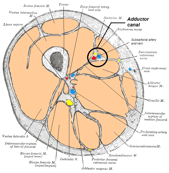
Long description
Adductor canal , horizontal section. Labelled illustration from Wikimedia Commons, used under its open licence.

Figure 1: Adductor canal , horizontal section.
The sartorius muscle is outlined in red, running from the hip to the knee. The femoral artery is marked in red, running centrally within the canal. The adductor hiatus is indicated at the distal end, where the artery passes through the adductor magnus to become the popliteal artery.
The saphenous nerve, a branch of the femoral nerve, emerges from under the medial border of the sartorius in the distal third of the thigh. It becomes subcutaneous and can be palpated just anterior to the medial condyle of the femur. This landmark is crucial for performing saphenous nerve blocks.
| Structure | Role in Canal |
|---|---|
| Sartorius | Roof and anterior boundary |
| Vastus Medialis | Lateral wall |
| Adductor Longus | Floor (proximal) |
| Adductor Magnus | Floor (distal) |
Understanding these surface relationships allows for accurate clinical examination and intervention. The canal is not visible as a distinct structure on the surface but is defined by the muscles that bound it. Palpation of the sartorius and the adductor muscles provides the necessary reference points. The neurovascular bundle within the canal is protected by the overlying fascia and muscle, but it remains accessible for diagnostic and therapeutic procedures.
The adductor canal, also known as Hunter’s canal, is an aponeurotic tunnel in the middle third of the thigh. It serves as a critical passageway for neurovascular structures transitioning from the femoral triangle to the popliteal fossa. Understanding its bony and muscular boundaries is essential for regional anaesthesia and surgical approaches to the lower limb.
The femur is the central bony landmark, specifically the medial aspect of the distal shaft. The adductor magnus forms the posterior boundary. The sartorius muscle overlies the canal, forming its roof.
The adductor canal is bounded by specific muscles that create its walls. The lateral wall is formed by the vastus medialis muscle, part of the quadriceps group. The roof is completed by the sartorius muscle, which covers the canal anteriorly. These muscular boundaries create a protective tunnel for the femoral vessels.
The contents of the adductor canal include the femoral artery, femoral vein, and saphenous nerve. The femoral artery is the largest vessel, running centrally within the canal. The saphenous nerve, a branch of the femoral nerve, runs with the vessels. The nerve to vastus medialis also passes through the canal. These structures are protected by the muscular boundaries. The canal ends at the adductor hiatus, where the femoral artery and vein pass through the adductor magnus tendon to enter the popliteal fossa. The saphenous nerve continues distally to supply sensation to the medial leg and foot.
DefinitionAdductor Canal
A fibrous tunnel in the middle third of the thigh that transmits the femoral artery, vein, and saphenous nerve from the femoral triangle to the popliteal fossa.
The clinical significance of the adductor canal lies in its use for regional anaesthesia. Femoral nerve blocks can be performed at the level of the canal to provide anaesthesia to the knee and medial leg. The proximity of the saphenous nerve to the femoral artery makes it a reliable landmark for needle placement. Understanding the anatomical relationships ensures safe and effective anaesthetic techniques. The canal’s muscular boundaries provide a stable platform for surgical access to the femoral vessels.
| Structure | Role in Canal |
|---|---|
| Sartorius | Forms the anterior roof |
| Vastus Medialis | Forms the lateral wall |
| Adductor Longus | Forms part of the posterior wall |
| Adductor Magnus | Forms the posterior wall and adductor hiatus |
The adductor canal, also known as Hunter’s canal, is an aponeurotic tunnel in the middle third of the thigh. It serves as a critical passageway for neurovascular structures moving from the femoral triangle to the popliteal fossa. Understanding its boundaries requires a clear grasp of the thigh’s compartmental organisation. The thigh muscles that move the femur, tibia, and fibula are divided into medial, anterior, and posterior compartments. [1]
The roof of the adductor canal is formed by the sartorius muscle and the subsartorial aponeurosis. The sartorius is a long, ribbon-like muscle that crosses the thigh obliquely. It forms the lateral boundary of the femoral triangle and continues to cover the adductor canal. Deep to the sartorius, the subsartorial aponeurosis provides a firm, fibrous cover. This aponeurosis extends from the sartorius to the adductor longus and magnus muscles.
The adductor longus originates from the pubis and inserts into the linea aspera of the femur. The adductor magnus is a large, triangular muscle that also attaches to the linea aspera and the adductor tubercle. These muscles form the medial and posterior boundaries of the canal. The lateral boundary is formed by the vastus medialis muscle. The vastus medialis is one of the four muscles of the quadriceps femoris.
The femoral artery lies centrally within the canal. The femoral vein lies medial to the artery. The nerve of the vastus medialis also runs in the canal. The adductor hiatus is the distal opening where the femoral artery passes into the popliteal fossa. The adductor magnus muscle forms the posterior wall of this hiatus. [1]
Similar to the thigh muscles, the muscles of the leg are divided by deep fascia into compartments. Although the leg has three compartments, the thigh has three main groups: anterior, medial, and posterior. The adductor canal is a key feature of the anterior compartment’s deep anatomy. It protects the femoral vessels from compression during knee flexion. The canal ends at the adductor hiatus, where the femoral artery becomes the popliteal artery. This transition is clinically significant for vascular access and surgical approaches. [1]
DefinitionAdductor Canal
An aponeurotic tunnel in the middle third of the thigh, formed by the sartorius and adductor muscles, containing the femoral artery, vein, and saphenous nerve.
| Boundary | Structure |
|---|---|
| Roof | Sartorius muscle and subsartorial aponeurosis |
| Floor | Adductor longus and adductor magnus |
| Lateral | Vastus medialis |
| Medial | Adductor longus and magnus |
The adductor canal, also known as Hunter’s canal, is an aponeurotic tunnel in the middle third of the thigh. It serves as a critical passageway for neurovascular structures moving from the femoral triangle to the popliteal fossa. Understanding the muscular boundaries is essential for locating the saphenous nerve and the femoral artery within this confined space.
The muscles surrounding the adductor canal define its anatomical limits. These muscles are grouped by their compartmental origin and function, primarily involving thigh flexion, extension, and adduction.
The vastus medialis forms the anterior wall of the adductor canal. It originates from the intertrochanteric line and the medial lip of the linea aspera of the femur. Its fibres converge to insert into the patella and the tibial tuberosity via the patellar ligament. The primary action is extension of the leg at the knee. It is supplied by the femoral nerve.
The adductor longus contributes to the lateral and anterior boundaries. It originates from the body of the pubis, just below the pubic crest. The muscle inserts into the middle third of the linea aspera of the femur. Its actions include adduction, flexion, and lateral rotation of the thigh at the hip. It is innervated by the obturator nerve. In the context of the adductor canal, it lies lateral to the femoral vessels. [1]
The adductor magnus forms the posterior and medial walls. It has multiple origins: the inferior pubic ramus, the ischial ramus, and the ischial tuberosity. It inserts into the adductor tubercle of the femur and the linea aspera. The muscle adducts and flexes the thigh. It receives dual innervation from the obturator nerve and the tibial division of the sciatic nerve. The adductor hiatus, an opening in the adductor magnus, allows the femoral artery to pass into the popliteal fossa. [1]
The saphenous nerve is the longest cutaneous branch of the femoral nerve. It does not have a muscular origin but travels through the adductor canal. It accompanies the femoral artery and vein within the canal before piercing the deep fascia to become subcutaneous. It provides sensory innervation to the medial aspect of the leg and foot. [1]
The femoral artery is central. The saphenous nerve is anterior to the artery.
| Muscle | Origin | Insertion | Action | Nerve Supply |
|---|---|---|---|---|
| Vastus Medialis | Intertrochanteric line, linea aspera | Tibial tuberosity | Knee extension | Femoral nerve |
| Adductor Longus | Pubis | Linea aspera | Thigh adduction | Obturator nerve |
| Adductor Magnus | Ischial tuberosity, pubic ramus | Adductor tubercle | Thigh adduction | Obturator, Sciatic |
The adductor canal, also known as Hunter’s canal, is an aponeurotic tunnel in the middle third of the thigh. It serves as a critical conduit for neurovascular structures passing from the femoral triangle to the popliteal fossa. Understanding its arterial supply requires tracing the femoral artery as it transitions into the popliteal artery. Before this transition, the artery gives off several important branches within the canal. The most significant branch is the descending genicular artery, which arises just before the artery exits the canal. This vessel supplies the knee joint and surrounding soft tissues. Other branches include muscular branches that supply the quadriceps femoris muscle, particularly the vastus medialis. The artery itself is accompanied by the femoral vein and the saphenous nerve. It provides sensory innervation to the medial aspect of the leg and foot. The roof is formed by the sartorius muscle and the subsartorial fascia. The lateral wall is formed by the vastus medialis muscle. It begins at the apex of the femoral triangle and ends at the adductor hiatus. The artery is pulsatile and can be palpated in the femoral triangle. In the adductor canal, it lies deep to the sartorius muscle. The artery is accompanied by the femoral vein, which lies medial to the artery. The canal is a common site for vascular access procedures. The femoral artery is often cannulated in the femoral triangle before it enters the canal. This block provides analgesia to the knee and medial leg. The descending genicular artery is a key landmark for this block. It arises from the femoral artery just before the adductor hiatus. The artery supplies the knee joint via its articular branches. The saphenous nerve is the target for sensory block. It provides sensation to the medial leg and foot. The canal is a vital pathway for blood supply to the lower limb. The adductor canal is a key anatomical structure in the thigh. It is essential for understanding lower limb anatomy and regional anaesthesia. The femoral artery is the main arterial supply. It passes through the adductor hiatus to become the popliteal artery. The descending genicular artery is a major branch. It arises just before the hiatus. The saphenous nerve is the main neural component. It provides sensory innervation to the medial leg. The canal is bounded by the sartorius muscle. The roof is formed by the sartorius and subsartorial fascia. The lateral wall is formed by the vastus medialis. The posterior wall is formed by the adductor magnus. The canal is approximately 15 cm long. It extends from the femoral triangle to the adductor hiatus. The femoral vein lies medial to the artery. The canal is a common site for vascular access. The femoral artery is often cannulated in the femoral triangle. The descending genicular artery is a key landmark. It arises from the femoral artery just before the adductor hiatus. The artery supplies the knee joint via its articular branches. The saphenous nerve is the target for sensory block. It provides sensation to the medial leg and foot. The canal is a vital pathway for blood supply to the lower limb. The adductor canal is a key anatomical structure in the thigh. It is essential for understanding lower limb anatomy and regional anaesthesia. [2]
Figure 2Common femoral and subsartorial artery and vein
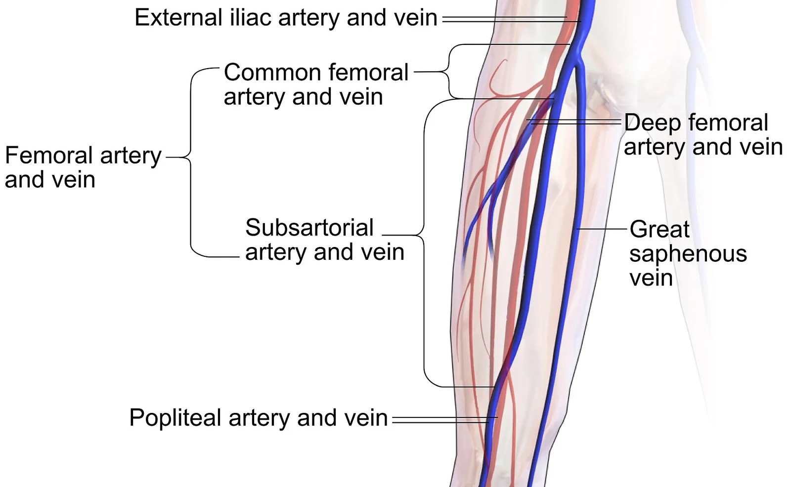
Long description
edit Segments of the femoral artery and femoral vein: The common femoral artery and vein (arteria & vena femoralis communis) proximally to the branching points of the deep femoral artery and vein, respectively. The subsa

Figure 2: Edit Segments of the femoral artery and femoral vein: The common femoral artery and vein (arteria & vena femoralis communis) proximally to the branching points of the deep femoral artery and vein, respectively (the subsa).
The femoral artery is the central structure, running vertically through the canal. It is accompanied by the femoral vein, which lies medial to it. The sartorius muscle forms the roof of the canal, covering these structures. The vastus medialis muscle forms the lateral wall. The adductor longus and adductor magnus muscles form the medial and posterior walls. The descending genicular artery is a branch of the femoral artery, arising just before the adductor hiatus. It supplies the knee joint. The adductor hiatus is the opening in the adductor magnus muscle through which the femoral artery passes to become the popliteal artery. The femoral vessels are the main arterial supply to the lower limb. The femoral vein lies medial to the artery. The canal is a common site for vascular access. The femoral artery is often cannulated in the femoral triangle. The descending genicular artery is a key landmark. It arises from the femoral artery just before the adductor hiatus. The artery supplies the knee joint via its articular branches. The saphenous nerve is the target for sensory block. It provides sensation to the medial leg and foot. The canal is a vital pathway for blood supply to the lower limb. The adductor canal is a key anatomical structure in the thigh. It is essential for understanding lower limb anatomy and regional anaesthesia.
The venous drainage of the adductor canal is a critical component of lower limb physiology, ensuring the efficient return of deoxygenated blood from the deep structures of the thigh to the central circulation. Understanding this pathway requires tracing the flow from the deep veins accompanying the neurovascular bundle within the canal, through the femoral vein, and finally into the pelvic venous system.
The surrounding musculature, including the vastus medialis and the adductor longus, frames the canal, providing context for the spatial relationships.
As the femoral vein enters the adductor canal, it lies in close proximity to the femoral artery, typically positioned medial to it.
Deep veins of the thigh, such as the profunda femoris vein (deep vein of the thigh), drain into the femoral vein. The profunda femoris vein typically joins the femoral vein at the level of the distal third of the thigh, just before or within the proximal part of the adductor canal. This junction is a common site for thrombus formation, particularly in patients with prolonged immobilisation.
Upon exiting the adductor canal through the adductor hiatus, the femoral vein continues its ascent into the pelvis, where it becomes the external iliac vein. This transition marks the end of the adductor canal segment of the venous system. The efficiency of this drainage is vital for preventing oedema and maintaining tissue health in the lower limb. Any obstruction or compression of the femoral vein within the adductor canal can lead to significant clinical consequences, including deep vein thrombosis and subsequent pulmonary embolism.
The venous anatomy of the adductor canal is therefore not merely a passive conduit but a dynamic system integrated with the muscular and neural structures of the thigh. Its understanding is essential for both clinical practice and surgical safety.
The adductor canal, also known as Hunter’s canal, is an aponeurotic tunnel in the middle third of the thigh. It serves as a critical passageway for neurovascular structures moving from the femoral triangle to the popliteal fossa. Understanding the nerves within this space requires a clear view of their origin, course, and distribution. The primary nerve is the saphenous nerve, a branch of the femoral nerve. It is the only sensory branch of the femoral nerve that extends beyond the knee.
The nerve continues distally, piercing the deep fascia below the knee to become subcutaneous. It then travels down the medial aspect of the leg, accompanying the great saphenous vein.
This motor branch does not travel within the canal itself but exits proximally. Therefore, the contents of the canal are primarily sensory and vascular.
This distribution is crucial for clinical examination of sensory deficits. Lesions of the saphenous nerve can result in numbness in this specific area.
| Nerve | Type | Muscles Innervated | Skin Area Supplied |
|---|---|---|---|
| Saphenous | Sensory | None | Medial leg, medial foot, medial malleolus |
| Nerve to Vastus Medialis | Motor | Vastus Medialis | None |
The anatomical relationships are key for surgical approaches and nerve blocks. The saphenous nerve lies medial to the femoral artery within the canal. This position makes it accessible for ultrasound-guided blocks. The nerve is protected by the sartorius muscle above and the adductor magnus muscle below. Knowledge of these relations prevents iatrogenic injury during procedures.
The terminal branch of the saphenous nerve emerges below the adductor hiatus. It runs superficially along the medial leg. This subcutaneous course makes it vulnerable to trauma. Clinicians must consider this pathway when assessing medial leg injuries. The nerve does not provide motor function to any muscles in the leg. Its role is purely sensory.
Understanding the adductor canal anatomy is essential for regional anaesthesia. The saphenous nerve block targets this specific nerve. It provides analgesia for medial leg surgeries. The block is often combined with other blocks for knee surgery. The simplicity of the anatomy allows for reliable technique.
It retains the sensory characteristics of the femoral nerve. This continuity explains the pattern of sensory loss in femoral nerve injuries.
The adductor canal acts as a conduit. It protects the neurovascular bundle. The structures are compact and well-defined. This organization facilitates efficient transmission of signals and blood flow. The saphenous nerve is the key sensory component. Its pathway is predictable and consistent.
The lymphatic drainage of the adductor canal region is a critical pathway for immune surveillance and fluid balance in the lower limb. Understanding this drainage is essential for diagnosing infections, malignancies, and lymphatic disorders affecting the thigh.
DefinitionLymphatic Drainage
The movement of lymph fluid from tissues into lymphatic vessels, eventually returning to the venous circulation via the thoracic duct or right lymphatic duct.
| Structure | Drainage Destination | Clinical Significance |
|---|---|---|
| Adductor Canal Lymphatics | Deep Inguinal Nodes | Infection spread from lower limb |
| Deep Inguinal Nodes | External Iliac Nodes | Metastasis from lower limb cancers |
| External Iliac Nodes | Common Iliac Nodes | Systemic lymphatic spread |
The deep inguinal nodes are located just below the inguinal ligament, medial to the femoral vein.
Clinical pearlClinical Pearl
Infections in the lower limb often present with swollen inguinal lymph nodes, indicating involvement of the deep inguinal nodes.
Disruption or obstruction of lymphatic drainage in the adductor canal can lead to lymphoedema, characterized by swelling of the lower limb. This condition can result from surgical trauma, radiation therapy, or malignancy.
Exam trapCommon Trap
Do not confuse the deep inguinal lymph nodes with the superficial inguinal lymph nodes, which drain the skin of the lower limb but not the deep structures of the thigh.
Diagnosis of lymphatic disorders in the adductor canal region typically involves clinical examination, ultrasound, or lymphoscintigraphy. Management depends on the underlying cause and may include compression therapy, surgical intervention, or treatment of the primary pathology.
NoteNote
Regular monitoring of lymphatic drainage is important in patients with a history of lower limb surgery or radiation to detect early signs of lymphoedema.
The adductor canal, also known as Hunter’s canal, is an aponeurotic tunnel in the middle third of the thigh. It serves as a critical conduit for neurovascular structures passing from the femoral triangle to the popliteal fossa. Understanding its boundaries and contents is essential for regional anaesthesia and surgical approaches to the knee.
The reader should identify the sartorius muscle forming the anterior roof. Deep to this lies the vastus medialis laterally and the adductor longus and magnus medially. Within this space, the femoral artery transitions into the popliteal artery. The saphenous nerve, a sensory branch, runs alongside the artery.
The primary contents are the femoral artery, the femoral vein, and the saphenous nerve. It provides sensation to the medial aspect of the leg and foot.
| Structure | Position | Role |
|---|---|---|
| Vastus medialis | Anterior/Lateral | Forms the anterior and lateral walls |
| Adductor longus | Medial | Forms the medial wall |
| Adductor magnus | Posterior | Forms the floor |
| Sartorius | Anterior | Forms the roof |
Compression or injury within the adductor canal can lead to significant deficits. Damage to the femoral artery may compromise blood flow to the lower limb. Injury to the saphenous nerve results in sensory loss over the medial leg. This is commonly seen in knee surgeries or prolonged compression. Understanding the precise anatomical relationships allows for accurate nerve blocks and safe surgical dissection. The canal’s compact nature means that swelling or haematoma can rapidly increase pressure, potentially leading to compartment syndrome. Therefore, careful monitoring of limb perfusion is vital after any trauma or procedure in this region.
Understanding the adductor canal requires correlating cross-sectional anatomy with standard imaging planes. The canal is an aponeurotic tunnel in the middle third of the thigh, visible on ultrasound, CT, and MRI. It serves as a critical landmark for regional anaesthesia and vascular assessment.
Ultrasound is the primary modality for visualising the adductor canal in real-time. It offers high resolution of superficial structures and dynamic assessment of needle placement.
Deep to the sartorius lies the subsartorial aponeurosis, which is often difficult to distinguish as a separate layer but defines the anterior boundary.
Within the canal, the femoral artery is the most consistent landmark. It appears as a pulsatile, anechoic circular structure. Posterior to the vessels lies the adductor longus muscle. In a correct scan, the nerve appears as a hyperechoic, honeycomb-like structure.
Computed tomography (CT) and magnetic resonance imaging (MRI) provide detailed anatomical context, particularly for surgical planning or investigating pathology.
MRI offers superior soft tissue contrast. T1-weighted images show the fat planes clearly. The subsartorial bursa, if present, appears as a fluid signal intensity structure between the sartorius and the underlying vessels. The nerve bundles are isointense to muscle on T1 and hyperintense on T2-weighted images. This helps distinguish the saphenous nerve from surrounding adipose tissue.
| Structure | Ultrasound Appearance | CT/MRI Location |
|---|---|---|
| Sartorius Muscle | Hyperechoic, striated | Anterior boundary |
| Femoral Artery | Anechoic, pulsatile | Central, lateral to vein |
| Femoral Vein | Compressible, anechoic | Medial to artery |
| Saphenous Nerve | Hyperechoic honeycomb | Anterior to artery |
| Adductor Longus | Hyperechoic, striated | Posterior boundary |
Recognising these structures is vital for performing adductor canal blocks. The goal is to deposit local anaesthetic around the saphenous nerve and the nerve to vastus medialis. This technique provides effective analgesia for knee surgery while preserving quadriceps strength.
DefinitionAdductor Canal Block
A regional anaesthesia technique targeting the saphenous nerve within the adductor canal to provide knee analgesia.
The adductor canal, also known as Hunter’s canal, is an aponeurotic tunnel in the middle third of the thigh. It serves as a critical conduit for neurovascular structures passing from the femoral triangle to the popliteal fossa. Understanding its boundaries and contents is essential for interpreting thigh trauma and planning regional anaesthesia.
DefinitionAdductor Canal
An aponeurotic tunnel in the middle third of the thigh containing the femoral artery, femoral vein, and saphenous nerve.
The boundaries define the safe zone for surgical access. The anterior wall is formed by the sartorius muscle, which can be retracted laterally to expose the canal. The roof is the strong subsartorial fascia, which is continuous with the deep fascia of the thigh.
| Structure | Role in Canal |
|---|---|
| Sartorius | Anterior roof |
| Vastus medialis | Lateral wall |
| Adductor longus | Posterior wall (proximal) |
| Adductor magnus | Posterior wall (distal) |
Injuries to the adductor canal often result from penetrating trauma or iatrogenic damage during surgery.
Procedures such as femoral nerve blocks or vascular access may involve this region. Surface landmarks for locating the canal include the apex of the femoral triangle and the adductor tubercle. The femoral pulse is palpable in the femoral triangle but becomes deeper within the canal. Ultrasound imaging is frequently used to visualise the canal and guide interventions, identifying the artery as a pulsatile, anechoic structure surrounded by hyperechoic fascia.
The adductor hiatus, the distal opening of the canal, is formed by the adductor magnus tendon. The femoral artery passes through this hiatus to become the popliteal artery. Obstruction or aneurysm at this site can compromise distal blood flow, leading to ischaemia of the lower leg. Surgical exposure of the femoral artery in the thigh requires careful dissection through the sartorius to avoid damaging the adjacent vein and nerve.
The adductor canal, also known as Hunter’s canal, is a critical anatomical space in the middle third of the thigh. It serves as a passageway for neurovascular structures moving from the femoral triangle to the popliteal fossa. Understanding its boundaries and contents is essential for regional anaesthesia and surgical approaches. [2]
The sartorius acts as a roof, creating a fibrous layer that can be palpated in the distal thigh. This arrangement creates a triangular space that tapers distally.
The canal contains the femoral artery, the femoral vein, and the saphenous nerve. The femoral artery gives off the descending genicular artery within this space. This branch supplies the knee joint and surrounding structures.
This is a rare but serious complication of trauma or tight dressings. Nerve blocks in this region are used for post-operative pain relief after knee arthroplasty.
Students often confuse the femoral nerve with the saphenous nerve. The femoral nerve exits the canal proximally. Another common error is misidentifying the posterior boundary. It is not the adductor brevis but the adductor longus and magnus. The sartorius muscle is often overlooked as the anterior boundary. It is crucial for locating the canal during physical examination.
The length of the adductor canal varies between individuals. This affects the approach for nerve blocks. Some individuals may have an accessory nerve supply. This can alter the sensory distribution in the medial leg. Surgeons must be aware of these variations to avoid complications.
The adductor canal is a vital corridor for thigh neurovascular structures. Its boundaries are defined by specific muscles. The contents include the femoral vessels and the saphenous nerve. Clinical procedures often target this area for anaesthesia. Accurate knowledge prevents injury to these structures.
Students frequently confuse the bony boundaries of the nasal cavity with the deeper structures of the skull base. A primary error involves misidentifying the contribution of the ethmoid bone. The ethmoid bone is a single, midline bone that forms the roof and lateral walls of the upper nasal cavity, the upper portion of the nasal septum, and contributes to the medial wall of the orbit. It is crucial to distinguish this from the inferior nasal conchae, which are separate, paired bones. The right and left inferior nasal conchae form a curved bony plate that projects into the nasal cavity space from the lower lateral wall. The inferior concha is the largest of the nasal conchae and can easily be seen when looking into the anterior opening of the nasal cavity. Confusing these leads to errors in understanding drainage pathways. [2]
Another frequent mistake relates to the nasal septum's composition. The nasal septum consists of both bone and cartilage components. The upper portion of the septum is formed by the perpendicular plate of the ethmoid bone. The lower and posterior parts of the septum are formed by the triangular-shaped vomer bone. Students often overlook the anterior nasal septum, which is formed by the septal cartilage, a flexible plate that fills in the gap between the perpendicular plate of the ethmoid and vomer bones. This cartilage also extends outward into the nose where it separates the right and left nostrils. The septal cartilage is not found in the dry skull, a fact often tested in practical exams. [2]
Confusion also arises regarding the mandible. The mandible forms the lower jaw and is the only moveable bone of the skull. At the time of birth, the mandible consists of paired right and left bones, but these fuse together during the first year to form the single U-shaped mandible of the adult skull. Each side of the mandible consists of a horizontal body and posteriorly, a vertically oriented ramus of the mandible. The outside margin of the mandible, where the body and ramus come together is called the angle of the mandible. Misidentifying the ramus as part of the zygomatic arch is a common error. The short temporal process of the zygomatic bone projects posteriorly, where it forms the anterior portion of the zygomatic arch. [2]
Orbital boundaries are another source of error. The walls of each orbit include contributions from seven skull bones. The frontal bone forms the roof and the zygomatic bone forms the lateral wall and lateral floor. The medial floor is primarily formed by the maxilla, with a small contribution from the palatine bone. The ethmoid bone and lacrimal bone make up much of the medial wall and the sphenoid bone forms the posterior orbit. Students often forget the palatine bone's minor role in the medial floor. [2]
Finally, cranial fossae boundaries are frequently mixed up. The anterior cranial fossa is the most anterior and the shallowest of the three cranial fossae. It overlies the orbits and contains the frontal lobes of the brain. Anteriorly, the anterior fossa is bounded by the frontal bone, which also forms the majority of the floor for this space. The lesser wings of the sphenoid bone form the prominent ledge that marks the boundary between the anterior and middle cranial fossae. The posterior cranial fossa is the most posterior and deepest portion of the cranial cavity. It contains the cerebellum of the brain. The posterior fossa is bounded anteriorly by the petrous ridges, while the occipital bone forms the floor and posterior wall. It is divided at the midline by the large foramen magnum (“great aperture”), the opening that provides for passage of the spinal cord. [2]
This knowledge is used at the bedside for assessing facial trauma and nasal obstruction. Understanding the nasal septum's composition is vital for epistaxis management. The anterior nasal septum is formed by the septal cartilage, a flexible plate that fills in the gap between the perpendicular plate of the ethmoid and vomer bones. Knowledge of the ethmoid bone's position helps in evaluating sinusitis. The ethmoid bone is a single, midline bone that forms the roof and lateral walls of the upper nasal cavity, the upper portion of the nasal septum, and contributes to the medial wall of the orbit.
In the operating theatre, precise identification of orbital walls is critical for decompression surgeries. The walls of each orbit include contributions from seven skull bones. The frontal bone forms the roof and the zygomatic bone forms the lateral wall and lateral floor. The medial floor is primarily formed by the maxilla, with a small contribution from the palatine bone. The ethmoid bone and lacrimal bone make up much of the medial wall and the sphenoid bone forms the posterior orbit. Surgeons must avoid the thin lamina papyracea of the ethmoid to prevent orbital complications.
On imaging, radiologists use these landmarks to classify fractures. The zygomatic bone is also known as the cheekbone. Each of the paired zygomatic bones forms much of the lateral wall of the orbit and the lateral-inferior margins of the anterior orbital opening. The short temporal process of the zygomatic bone projects posteriorly, where it forms the anterior portion of the zygomatic arch. CT scans clearly show the vomer's position. The unpaired vomer bone, often referred to simply as the vomer, is triangular-shaped and forms the posterior-inferior part of the nasal septum. The vomer is best seen when looking from behind into the posterior openings of the nasal cavity. In this view, the vomer is seen to form the entire height of the nasal septum. [2]
In examinations, students must describe the mandible's development and structure. The mandible forms the lower jaw and is the only moveable bone of the skull. At the time of birth, the mandible consists of paired right and left bones, but these fuse together during the first year to form the single U-shaped mandible of the adult skull. Each side of the mandible consists of a horizontal body and posteriorly, a vertically oriented ramus of the mandible. The outside margin of the mandible, where the body and ramus come together is called the angle of the mandible.
Understanding cranial fossae is essential for neurosurgical planning. The anterior cranial fossa is the most anterior and the shallowest of the three cranial fossae. It overlies the orbits and contains the frontal lobes of the brain. Anteriorly, the anterior fossa is bounded by the frontal bone, which also forms the majority of the floor for this space. The lesser wings of the sphenoid bone form the prominent ledge that marks the boundary between the anterior and middle cranial fossae. Located in the floor of the anterior cranial fossa at the midline is a portion of the ethmoid bone, consisting of the upward projecting crista galli and to either side of this, the cribriform plates. The posterior cranial fossa is the most posterior and deepest portion of the cranial cavity. It contains the cerebellum of the brain. The posterior fossa is bounded anteriorly by the petrous ridges, while the occipital bone forms the floor and posterior wall. It is divided at the midline by the large foramen magnum (“great aperture”), the opening that provides for passage of the spinal cord. [2]
The ethmoid bone forms the roof and lateral walls of the upper nasal cavity, the upper portion of the nasal septum, and contributes to the medial wall of the orbit. The inferior concha is the largest of the nasal conchae and can easily be seen when looking into the anterior opening of the nasal cavity. The nasal septum consists of both bone and cartilage components, with the upper portion formed by the perpendicular plate of the ethmoid bone. The anterior nasal septum is formed by the septal cartilage, a flexible plate that fills in the gap between the perpendicular plate of the ethmoid and vomer bones. The mandible forms the lower jaw and is the only moveable bone of the skull. The walls of each orbit include contributions from seven skull bones. The anterior cranial fossa is the most anterior and the shallowest of the three cranial fossae. The posterior cranial fossa is the most posterior and deepest portion of the cranial cavity.
Answer each one in your head first, then open the answer.
Which bone forms the upper portion of the nasal septum?
Show answerHide answer
Perpendicular plate of the ethmoid boneWhat is the only moveable bone of the adult skull?
Show answerHide answer
MandibleWhich bone forms the lateral wall and lateral floor of the orbit?
Show answerHide answer
Zygomatic boneWhat structure forms the anterior portion of the zygomatic arch?
Show answerHide answer
Short temporal process of the zygomatic boneWhich cranial fossa contains the cerebellum?
Show answerHide answer
Posterior cranial fossa
- Betts JG, Young KA, Wise JA, et al. Appendicular Muscles of the Pelvic Girdle and Lower Limbs. OpenStax, Anatomy and Physiology 2e (CC BY-NC-SA 4.0). Accessed .
- Betts JG, Young KA, Wise JA, et al. The Skull. OpenStax, Anatomy and Physiology 2e (CC BY-NC-SA 4.0). Accessed .
- Betts JG, Young KA, Wise JA, et al. Appendicular Muscles of the Pelvic Girdle and Lower Limbs. OpenStax, Anatomy and Physiology 2e (CC BY-NC-SA 4.0). Accessed .
- Koh IJ, Choi YJ, Kim MS, et al. Femoral Nerve Block versus Adductor Canal Block for Analgesia after Total Knee Arthroplasty. Knee surgery & related research 2017;29(2):87-95. doi:10.5792/ksrr.16.039. Accessed .
Figure credits
- Figure 1: Mikael Häggström. When using this image in external works, it may be cited as: Häggström, Mikael (2014). "Medical gallery of Mikael Häggström 2014". WikiJournal of Medicine 1 (2). DOI:10.15347/wjm/2014.008. ISSN 2002-4436. Public Domain. or By Mikael Häggström, used with permission., via Wikimedia Commons, Public domain, Public domain.
- Figure 2: Source image by BruceBlaus. Derivative by Mikael Häggström, M.D., via Wikimedia Commons, CC BY-SA 3.0, CC BY-SA 3.0. resized.
Educational content for medical students and doctors. It is not a substitute for clinical judgement or local guidelines.