Femoral triangle
A triangular subinguinal region containing femoral vessels, nerve and lymphatics.
The femoral triangle is a clinically important subfascial space in the upper anteromedial thigh, shaped like an inverted triangle. It is formed at the junction between the hip and the leg and contains the femoral nerve, femoral artery, femoral vein, and deep inguinal lymph nodes. Its boundaries are: superiorly, the inguinal ligament; laterally, the medial border of the sartorius muscle; and medially, the medial border of the adductor longus muscle. The floor is formed by the iliopsoas, pectineus [1][2]
The sartorius is a band-like muscle that extends from the anterior superior iliac spine to the medial side of the proximal tibia. It forms the lateral boundary of the triangle. The iliopsoas (psoas major plus iliacus) lies on the floor, passing deep to the inguinal ligament to insert on the lesser trochanter of the femur. The pectineus, located in the femoral triangle, adducts and flexes the femur at the hip. The adductor longus forms the medial boundary and also lies on the floor; it flexes the thigh, while the adductor magnus extends it. [1]
Figure 1Gray430
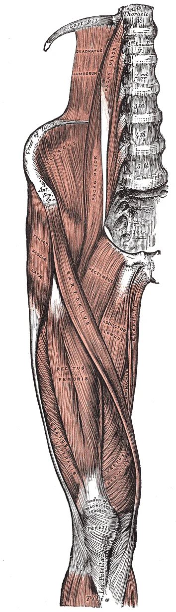
Long description
Muscles of the iliac and anterior femoral regions.

Figure 1: Muscles of the iliac and anterior femoral regions.
The apex of the triangle is where the sartorius crosses the adductor longus, some distance below the inguinal ligament. The triangle is continuous distally with the adductor (subsartorial) canal, which transmits the femoral vessels to the popliteal fossa. The saphenofemoral junction is the point where the great saphenous vein pierces the cribriform fascia to drain into the femoral vein, just below and lateral to the pubic tubercle. This junction is a common site for venous reflux and varic [2]
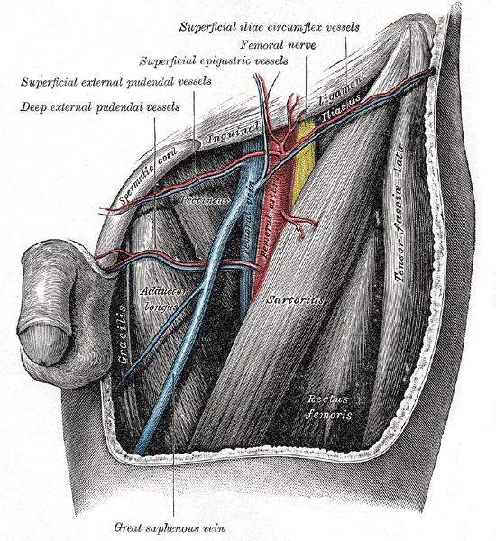
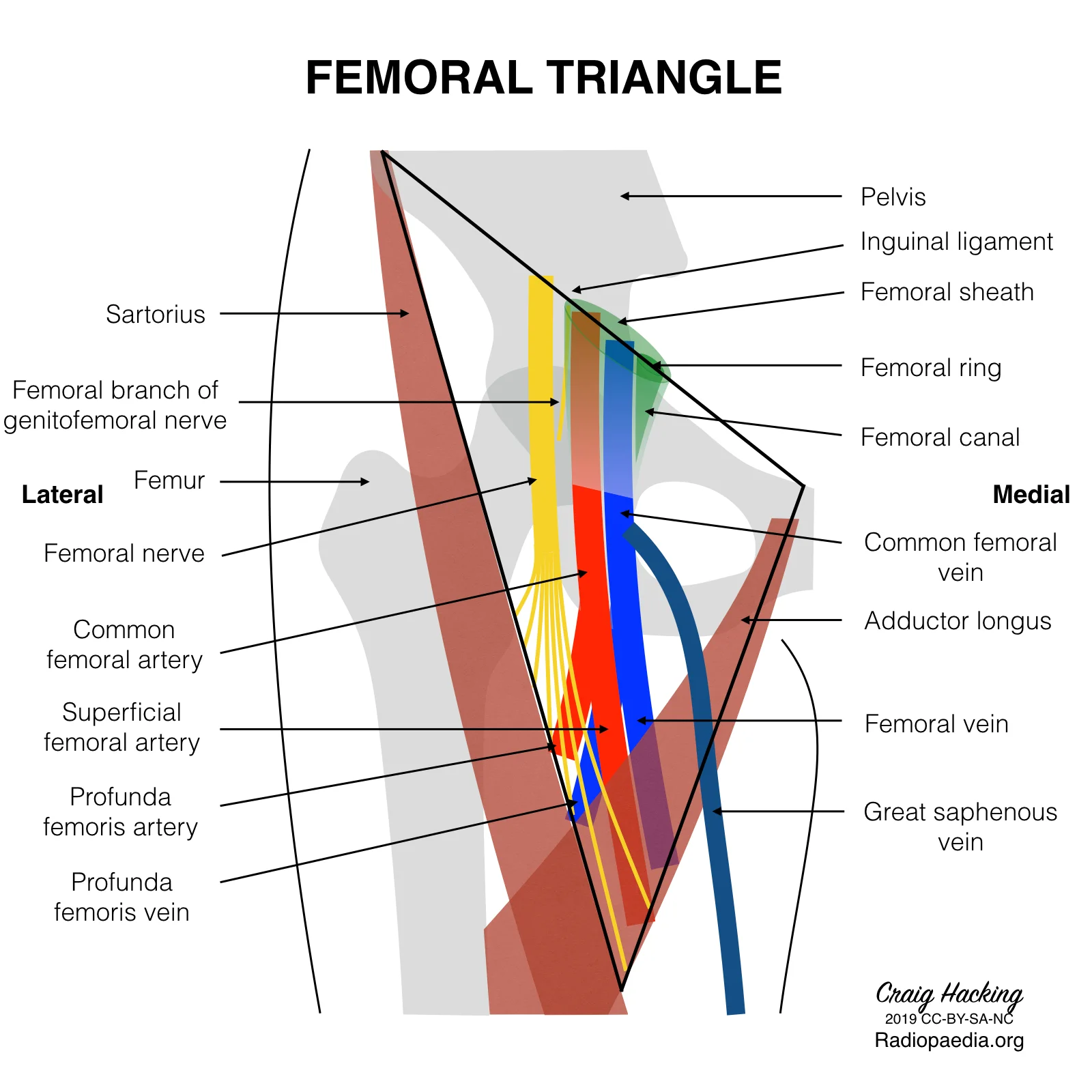
The contents of the femoral triangle, from lateral to medial, are the femoral nerve, femoral artery, femoral vein, and the deep inguinal lymph nodes. This order is remembered by the mnemonic "NAVEL" (Nerve, Artery, Vein, Empty space, Lymphatics). The femoral nerve lies lateral to the artery, and divides into anterior and posterior divisions that supply the anterior thigh muscles, including the quadriceps femoris and sartorius. [1] [2]
Figure 2Gray546
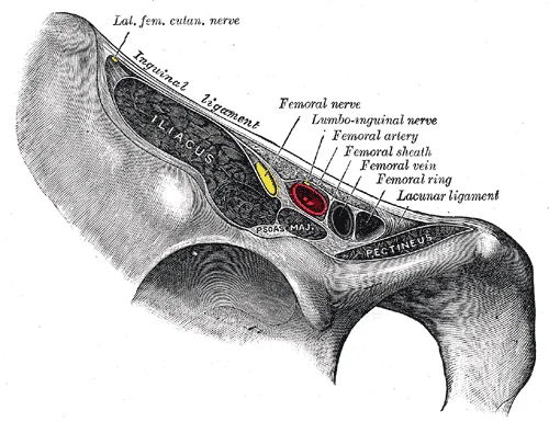
Long description
Structures passing behind the inguinal ligament.

Figure 2: Structures passing behind the inguinal ligament.
It then passes posterior to the knee near the popliteal fossa, where it is renamed the popliteal artery. [2]
| Muscle | Origin | Insertion | Action | Nerve supply |
|---|---|---|---|---|
| Iliopsoas (psoas major + iliacus) | Lumbar vertebrae; iliac fossa | Lesser trochanter of femur | Flexes thigh at hip | Femoral nerve (L1–L3) |
| Sartorius | Anterior superior iliac spine | Medial side of proximal tibia | Flexes, abducts, laterally rotates thigh; flexes leg | Femoral nerve (L2–L3) |
| Pectineus | Superior pubic ramus | Pectineal line of femur | Adducts and flexes thigh | Femoral nerve (L2–L3) |
| Adductor longus | Body of pubis | Middle third of linea aspera | Adducts, flexes, medially rotates thigh | Obturator nerve (L2–L4) |
Clinical relevance: the femoral triangle is the site for femoral artery catheterisation, where the artery is palpated at the mid-inguinal point. Femoral hernia emerges medial to the femoral vessels, through the femoral ring. The saphenofemoral junction is a key site for varicose vein ligation. The femoral nerve block anaesthetises the anterior thigh. The adductor longus tendon is a surgical landmark for the medial boundary)Skip to content
The femoral triangle is a subfascial space in the superior third of the anterior thigh. Its surface anatomy is defined by palpable bony and muscular landmarks that allow for the clinical localization of the neurovascular bundle.
The superior boundary of the triangle is marked by the inguinal ligament, which runs from the anterior superior iliac spine to the pubic tubercle. The lateral boundary is formed by the medial border of the sartorius muscle, while the medial boundary is the medial border of the adductor longus muscle.
Figure 1 shows the surface anatomy of the femoral triangle in an anterior view of the right thigh. The figure illustrates the relationship between the superficial landmarks and the underlying vessels.
The inguinal ligament forms the superior edge of the region. Just inferior to this ligament, the femoral artery is the central structure of the neurovascular bundle. Medial to the femoral artery lies the femoral vein. Further medial to the vein is the femoral canal, which contains lymphatic vessels. Lateral to the femoral artery is the femoral nerve.
In patients without excessive adipose tissue, certain vessels in the region may be palpable. [2]
Clinical pearlClinical Correlation
The mid-inguinal point is the standard surface landmark used to locate the femoral artery for pulse palpation or arterial cannulation.
| Muscle | Origin | Insertion | Action | Nerve Supply |
|---|---|---|---|---|
| Sartorius | Anterior superior iliac spine | Pes anseris (proximal medial tibia) | Flexes, abducts, and laterally rotates hip; flexes knee | Femoral nerve |
| Adductor longus | Pubic crest and tubercle | Linea aspera of femur | Adducts thigh | Obturator nerve |
The femoral triangle is a clinically vital region in the upper medial thigh, and its boundaries are best understood by first identifying its bony framework. The triangle is not a joint, but a topographic space whose floor is formed by muscles and whose roof is fascia; however, its superior border is defined by the inguinal ligament, which runs from the anterior superior iliac spine (ASIS) to the pubic tubercle. Both of these bony landmarks are palpable in the living patient and are essential for locating the femoral artery for pulse assessment or catheterisation.
The medial side of the femur, specifically the adductor longus muscle arising from the pubis, forms the medial boundary, but the bony reference is the pubic symphysis and the pubic tubercle. The floor of the triangle is formed by the iliopsoas muscle laterally and the pectineus muscle medially, both of which attach to the femur and the pelvis.
The pecten pubis is a faint ridge on the superior pubic ramus, just medial to the pubic tubercle.
The inguinal ligament itself is not a bone but a thickened lower edge of the external oblique aponeurosis, and it attaches to the ASIS and pubic tubercle. The femoral head, which articulates with the acetabulum of the hip bone, lies deep to the triangle's floor, and the femoral neck is a common fracture site in the elderly; the bony landmarks of the triangle guide the surgeon's approach to this region. [3]
The femoral triangle is situated in the superior aspect of the thigh. The deep fascia of the thigh, known as the fascia lata, invests the muscles and separates the thigh into three distinct compartments: anterior, medial, and posterior. [1]
Figure 3Gray545
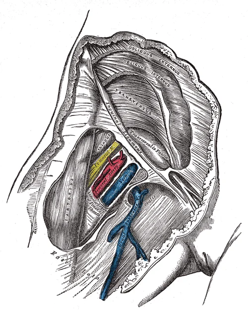
Long description
Femoral sheath laid open to show its three compartments.

Figure 3: Femoral sheath laid open to show its three compartments.
DefinitionFascia Lata
The deep fascia of the thigh that divides the region into anterior, medial, and posterior compartments.
The muscles within these compartments are grouped by their primary actions and nerve supplies. The anterior compartment is primarily responsible for knee extension and hip flexion, while the medial compartment focuses on adduction of the femur. The posterior compartment contains the muscles responsible for knee flexion.
| Muscle | Origin/Location | Insertion/Action | Nerve Supply |
|---|---|---|---|
| Sartorius | Anterior compartment | Flexes hip/knee | Not specified |
| Quadriceps femoris | Anterior compartment | Extend the knee | Not specified |
| Pectineus | Medial compartment | Adducts and flexes femur | Not specified |
| Adductor longus | Medial compartment | Adducts and medially rotates thigh | Not specified |
| Adductor brevis | Medial compartment | Adducts and medially rotates thigh | Not specified |
| Adductor magnus | Medial compartment | Adducts and medially rotates thigh | Not specified |
| Gracilis | Medial compartment | Adducts thigh, flexes leg at knee | Not specified [1] |
On the lateral aspect of the superior thigh, the tensor fascia latae is a thick, squarish muscle. It works as a synergist with the iliopsoas and gluteus medius to flex and abduct the thigh. It also pulls on the iliotibial tract (band) to make it taut, which stabilises the lateral aspect of the knee. [1]
Thigh Compartment Division
- Deep Fascia (Fascia Lata)Invests the thigh muscles
- Question: Compartment?
Anterior
- Quadriceps and Sartorius
Medial
- Adductors, Pectineus, Gracilis
Posterior
- Hamstrings
The femoral triangle is a subfascial space in the upper anteromedial thigh. Its boundaries are the inguinal ligament superiorly, the sartorius laterally, and the adductor longus medially. Its contents, from lateral to medial, are the femoral nerve, femoral artery, femoral vein, and the femoral canal (mnemonic: NAVEL). The femoral sheath encloses the femoral artery, femoral vein, and femoral canal, but not the femoral nerve, which lies lateral to the sheath.
Figure 4Gray547
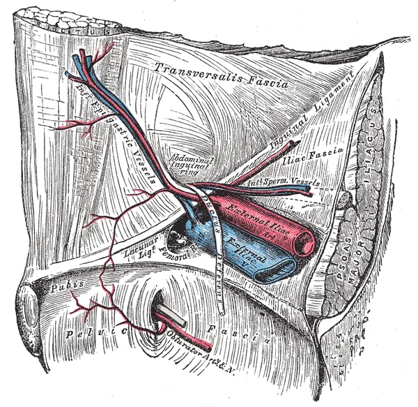
Long description
The relations of the femoral and abdominal inguinal rings, seen from within the abdomen. Right side.

Figure 4: The relations of the femoral and abdominal inguinal rings, seen from within the abdomen (right side).
| Muscle | Origin | Insertion | Action | Nerve supply |
|---|---|---|---|---|
| Iliopsoas (psoas major + iliacus) | Psoas: T12–L5 vertebrae; Iliacus: iliac fossa | Lesser trochanter of femur | Flexes hip; stabilises hip joint | Femoral nerve (L1–L3) and anterior rami of L1–L3 |
| Sartorius | Anterior superior iliac spine (ASIS) | Upper medial tibia (pes anserinus) | Flexes, abducts, laterally rotates thigh; flexes knee | Femoral nerve (L2–L3) |
| Pectineus | Superior pubic ramus | Pectineal line of femur | Adducts and flexes thigh | Femoral nerve (L2–L3); occasionally obturator nerve |
| Adductor longus | Body of pubis | Middle third of linea aspera | Adducts and flexes thigh | Obturator nerve (L2–L4) |
It lies lateral to the femoral vein and medial to the femoral nerve. It gives off the profunda femoris (deep artery of thigh), which supplies the adductor and hamstring muscles. The femoral artery continues through the adductor canal to become the popliteal artery.
The sartorius forms the lateral border, running obliquely from the ASIS to the medial tibia. The adductor longus forms the medial border, descending from the pubis. The iliopsoas and pectineus form the floor.
Clinical pearlemergency care point
The femoral artery is cannulated for angiography and cardiac catheterisation; the femoral vein is used for central venous access. The femoral nerve block anaesthetises the anterior thigh and knee.
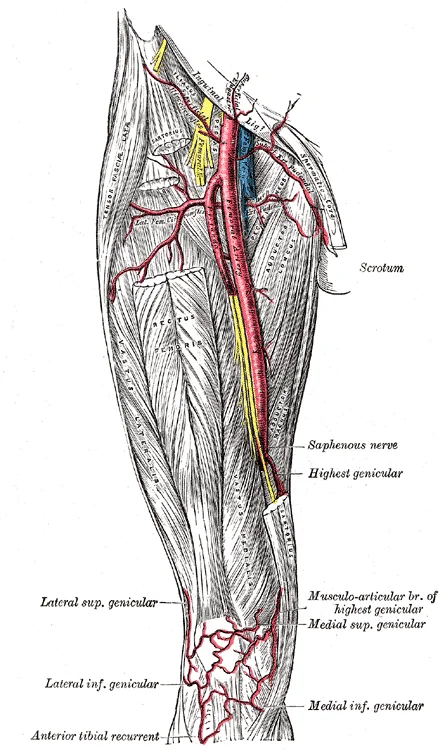
The arterial supply of the femoral triangle is dominated by the femoral artery, the main continuation of the external iliac artery. The external iliac artery passes beneath the inguinal ligament, midway between the anterior superior iliac spine and the pubic symphysis, and becomes the femoral artery at the mid-inguinal point. This is the key landmark: the femoral artery enters the triangle at its base, the inguinal ligament, and lies lateral to the femoral vein and medial to the femoral nerve. [2]
Figure 5Gray550
Long description
The femoral artery. Labelled illustration from Wikimedia Commons, used under its open licence.

Figure 5: The femoral artery.
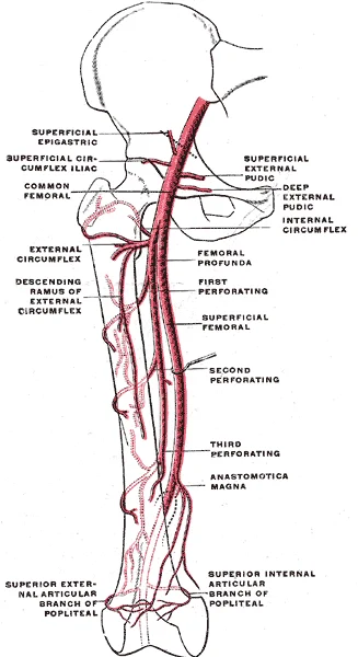
Within the triangle, the femoral artery descends vertically, lying on the psoas major muscle, then the pectineus, and finally the adductor longus. It is covered by fascia lata and skin, and its pulsation is palpable just inferior to the mid-inguinal point. The artery gives off several branches within the triangle. The most important is the profunda femoris (deep femoral) artery, which arises from the lateral side of the femoral artery. The profunda fe
Other smaller branches from the femoral artery within the triangle include the superficial epigastric artery (to the lower abdominal wall), the superficial circumflex iliac artery (to the skin and fascia of the groin), and the superficial external pudendal artery (to the external genitalia). These are clinically minor but are named in standard descriptions Mend. The femoral artery itself leaves the triangle by passing through the adductor hiatus (an opening in the adductor magnus tendon) to become the popliteal artery in the popliteal fossa.
Figure 6Gray548
Long description
Scheme of the femoral artery. (Poirier and Charpy.)

Figure 6: Scheme of the femoral artery (Poirier and Charpy.).
The femoral artery is the central vertical vessel; the profunda femoris branches laterally and then descends deep; the medial and lateral circumflex femoral arteries arise from it near its origin; the superficial branches (epigastric, circumflex iliac, external pudendal) are shown leaving the femoral artery superiorly. The femoral vein lies medial, and the femoral nerve lateral, to the artery.
Figure 7Gray549
Long description
The left femoral triangle. Labelled illustration from Wikimedia Commons, used under its open licence.

Figure 7: The left femoral triangle.
The venous drainage of the femoral triangle is dominated by the great saphenous vein and the femoral vein, which together collect blood from the superficial and deep structures of the lower limb.
It is positioned between the femoral artery laterally and the femoral canal medially. The vein receives several tributaries within the triangle, including the profunda femoris vein, which drains the deep thigh muscles, and the great saphenous vein. The femoral vein then passes beneath the inguinal ligament through
It ascends along the medial side of the leg and knee, then passes through the saphenous opening in the fascia lata to enter the femoral triangle. It terminates by draining into the femoral vein, approximately 3–4 cm below the inguinal ligament. Before its termination, it receives several tributaries: the superficial external pudendal, superficial epigastric, and superficial circumflex iliac veins, which drain the lower abdominal wall and external genitalia, plus the accessory saphenous vein when present. [2]
The venous pattern here is variable, as systemic veins show more variation than arteries. [2]
| Tributary | Origin | Course within triangle | Termination |
|---|---|---|---|
| Profunda femoris vein | Deep thigh muscles | Runs deep to femoral artery, joins posteriorly | Femoral vein |
| Great saphenous vein | Medial foot arch | Superficial, crosses anterior to femoral vessels | Femoral vein, 3–4 cm below inguinal ligament |
| Superficial external pudendal vein | External genitalia | Ascends medially, joins great saphenous | Great saphenous vein |
| Superficial epigastric vein | Lower abdominal wall | Ascends from umbilicus region, joins great saphenous | Great saphenous vein |
| Superficial circumflex iliac vein | Lateral thigh/iliac region | Runs laterally, joins great saphenous | Great saphenous vein |
The femoral triangle contains the major nerves that supply the anterior thigh. The most important of these is the femoral nerve, which arises from the dorsal divisions of the anterior rami of L2, L3 and L4. It enters the triangle by passing beneath the inguinal ligament, lying lateral to the femoral artery and femoral vein. Within the triangle, the nerve breaks up into its terminal branches: motor branches to the quadriceps femoris, sartorius and pectineus muscles, and cutaneous branches that supply the skin of the anterior and medial thigh.
The obturator nerve (L2-L4) also contributes to the region, though it passes through the obturator foramen rather than the triangle itself. It supplies the adductor muscles of the medial thigh and gives a cutaneous branch to the medial thigh.
Figure 8Femoral-triangle-diagram
Long description
An anatomical area bordered by the sartorius muscle laterally, the adductor longus medially, and the femoral ring superiorly, that includes the vessels of the deep femoral vessels, arteries, and nerves

Figure 8: An anatomical area bordered by the sartorius muscle laterally, the adductor longus medially, and the femoral ring superiorly, that includes the vessels of the deep femoral vessels, arteries, and nerves.
| Nerve | Motor supply (muscles) | Cutaneous supply (skin area) |
|---|---|---|
| Femoral nerve (L2-L4) | Quadriceps femoris (rectus femoris, vastus lateralis, vastus medialis, vastus intermedius), sartorius, pectineus, iliopsoas (via branches) | Anterior thigh, medial leg and foot (via saphenous nerve) |
| Obturator nerve (L2-L4) | Adductor longus, adductor brevis, adductor magnus (adductor part), gracilis, obturator externus | Medial thigh |
| Lateral femoral cutaneous nerve (L2-L3) | None (pure sensory) | Lateral thigh |
The saphenous nerve is the longest cutaneous branch of the femoral nerve. It descends through the triangle alongside the femoral artery, then passes into the adductor canal and emerges on the medial side of the knee. It supplies the skin of the medial leg and foot as far as the first metatarsophalangeal joint. The nerve to pectineus arises from the femoral nerve just below the inguinal ligament and enters the deep surface of the pectineus muscle.
Motor loss from a femoral nerve lesion (e.g., from a psoas haematoma or during hip surgery) results in weakness of knee extension and an absent knee jerk. Sensory loss affects the anterior thigh and the medial leg and foot. An obturator nerve lesion causes weakness of hip adduction and a small patch of sensory loss on the medial thigh. The lateral femoral cutaneous nerve is vulnerable to compression at the inguinal ligament, producing meralgia paraesthetica—numbness and tingling over the lateral thigh without motor deficit.
The lymphatic drainage of the femoral triangle is a critical pathway for immune surveillance and potential metastatic spread from the lower limb and anterior abdominal wall. Understanding this route requires a clear grasp of the anatomical boundaries and the specific nodes involved. The femoral triangle is bounded superiorly by the inguinal ligament, laterally by the sartorius muscle, and medially by the adductor longus muscle. Within this space, the deep inguinal lymph nodes play a pivotal role [1][2]
DefinitionDeep Inguinal Nodes
A group of lymph nodes located deep to the fascia lata, medial to the femoral vein, and inferior to the inguinal ligament.
These nodes receive lymph from the deep structures of the lower limb, including the muscles and bones, as well as from the superficial inguinal nodes. The superficial inguinal nodes are divided into horizontal and vertical groups. The vertical group drains the lower limb, excluding the posterior calf and lateral foot which drain to the popliteal nodes.
The femoral nerve is located lateral to the femoral artery, and the femoral sheath encloses the artery, vein, and canal.
The deep inguinal nodes ultimately drain into the external iliac lymph nodes. This pathway is significant in the context of malignancies, such as melanoma or squamous cell carcinoma of the lower limb or perineum. Early detection of enlarged nodes in the femoral triangle can indicate underlying pathology. Clinicians must palpate this area carefully, noting any tenderness, size, or fixation of the nodes. The femoral canal, being a potential site for femoral hernias, is also a common location for metastatic deposits. [2]
Exam trapFemoral Hernia Risk
Femoral hernias occur through the femoral canal, which is medial to the femoral vein. This is more common in women due to the wider pelvic anatomy.
In summary, the lymphatic drainage of the femoral triangle involves a complex network of superficial and deep nodes. The superficial nodes drain the skin and subcutaneous tissues, while the deep nodes drain the deeper structures and receive input from the superficial nodes. The deep inguinal nodes, particularly Cloquet's node, are key landmarks in this region. Awareness of this anatomy is essential for accurate diagnosis and management of conditions affecting the lower limb and perineum.
Its boundaries are the inguinal ligament superiorly, the sartorius muscle laterally, and the adductor longus muscle medially. The triangle is clinically important because it transmits the major neurovascular bundle to the lower limb.
The contents of the femoral triangle, from lateral to medial, are the femoral nerve, femoral artery, femoral vein, and the femoral canal. This arrangement is memorised by the mnemonic "NAVY" (Nerve, Artery, Vein, empty space for the canal). The femoral nerve lies lateral to the artery and divides early into anterior and posterior divisions. The femoral artery enters the triangle beneath the inguinal ligament at the mid-inguinal point (midway between the anterior superior iliac spine and the pubic symphysis) and is the main arterial supply to the lower limb. The fem
It encloses the femoral artery, femoral vein, and femoral canal, but not the femoral nerve)Skip. The sheath ends by fusing with the adventitia of the vessels about 4 cm below the inguinal ligament.
Within the triangle it gives off the superficial epigastric, superficial circumflex iliac, and superficial external pudendal arteries, plus the deep external pudendal and profunda femoris arteries. The profunda femoris is the main branch, and gives rise to the medial and lateral circumflex femoral arteries. The femoral artery then passes through the adductor canal to become the popliteal artery behind the knee. [2]
It passes beneath the ligament to become the external iliac vein. [2]
Clinical pearlClinical pearl
The femoral pulse is palpated just below the mid-inguinal point, where the artery is superficial. Femoral venous access is performed medial to the artery, and femoral nerve block is placed lateral to it.
The femoral triangle is a key anatomical region in the anterior thigh, often visualised using various imaging modalities. Understanding its radiological appearance is crucial for diagnosing conditions such as femoral hernias, vascular access complications, and soft tissue masses.
Ultrasound is the primary modality for evaluating the femoral triangle due to its real-time capability and lack of ionising radiation. The femoral vein, artery, and nerve are typically visualised in a transverse plane. The femoral sheath encloses the vein and artery, while the nerve lies outside the sheath. Compression with the ultrasound probe is used to distinguish the compressible femoral vein from the non-compressible femoral artery. [2]
Computed tomography (CT) provides detailed cross-sectional images of the femoral triangle. In axial CT scans, the femoral vessels and nerve are clearly delineated. The iliopsoas muscle forms the floor of the triangle, while the inguinal ligament forms the superior border. CT angiography can be used to assess the patency of the femoral vessels and detect any occlusions or aneurysms.
Magnetic resonance imaging (MRI) offers superior soft tissue contrast, making it useful for evaluating muscle and nerve pathology within the femoral triangle. T1-weighted images provide good anatomical detail, while T2-weighted images highlight fluid and oedema. The femoral nerve appears as a round or oval structure with intermediate signal intensity on T1-weighted images. The femoral vessels show flow voids on both T1 and T2-weighted images. MRI is particularly valuable for assessing nerve compression or entrapment syndromes.
Plain X-rays are not typically used to visualise the structures within the femoral triangle directly. However, they can be useful for identifying bony abnormalities or foreign bodies in the region. The femoral head, neck, and shaft are visible on standard hip and femur X-rays. Calcifications within the femoral vessels or surrounding soft tissues may also be detected on X-rays.
The femoral sheath encloses the vein and artery, while the nerve lies outside the sheath.
The femoral triangle is a clinically vital region because it transmits the main neurovascular bundle to the lower limb and is a common site for surgical access. Its boundaries are the inguinal ligament superiorly, the sartorius laterally, and the adductor longus medially; the floor is formed by the iliopsoas and pectineus. The femoral artery is the key surface landmark: it is palpated midway between the anterior superior iliac spine and the pubi
Femoral hernia occurs through the femoral ring, the medial compartment of the femoral sheath, and is more common in women due to a wider pelvis. The hernia emerges below and lateral to the pubic tubercle, distinguishing it from an inguinal hernia which emerges above and medial to it. Because the femoral ring is narrow and rigid, femoral hernias have a high risk of strangulation and obstruction, requiring urgent surgical repair. The lacunar ligament forms the medial boundary of the femoral ring and is the structure that may need to be divided to release a strangulated hernia, but care is needed because an aberrant obturator artery may pass close to it. [3]
Femoral nerve injury results from penetrating trauma, hip surgery, or prolonged lithotomy positioning. The nerve supplies the iliacus, pectineus, sartorius, and quadriceps femoris, and gives cutaneous branches to the anterior thigh and saphenous nerve to the medial leg. Injury causes weakness of knee extension (quadriceps) and hip flexion (iliopsoas), loss of the patellar reflex, and sensory loss over the anterior thigh and medial leg. The patient has difficulty climbing stairs and the knee may give way. Because the femoral nerve lies outside the femoral sheath, it is not affected by a femoral hernia, but it can be compressed by a psoas abscess or retroperitoneal haematoma. [3] [2]
Procedures in the triangle include femoral artery catheterisation for angiography and cardiac interventions, and femoral nerve block for anaesthesia of the anterior thigh and knee. The femoral vein is used for dialysis catheters and for emergency venous access in cardiac arrest. The femoral artery is also the site for measuring arterial pressure in shock. The pulse is felt just below the inguinal ligament, and compression here can control haemorrhage from the lower limb. The femoral triangle is also the site for surgical exposure of the femoral vessels for bypass grafting or embolectomy.
The femoral head and neck lie deep to the vessels, and the pubic tubercle marks the medial limit of the canal)Skip the rest.
- The femoral triangle is bounded by the inguinal ligament superiorly, the sartorius laterally, and the adductor longus medially; its floor is formed by the iliopsoas and pectineus muscles. The roof is fascia lata and cribriform fascia.
The femoral nerve lies outside the femoral sheath; the artery, vein, and canal are inside it.
-
The femoral artery is the continuation of the external iliac artery, entering the triangle deep to the inguinal ligament at the mid-inguinal point (midpoint between the anterior superior iliac spine and the pubic symphysis). It is a common trap to confuse this with the femoral pulse point, which is at the mid-inguinal point, not the femoral head.
-
The femoral vein lies medial to the artery within the sheath; it becomes the external iliac vein as it passes deep to the inguinal ligament. The femoral canal, the most medial compartment, contains lymphatics and the deep inguinal lymph node of Cloquet; it is the site of a femoral hernia.
-
The femoral nerve (L2–L4) lies lateral to the sheath and divides into anterior and posterior divisions; it supplies the sartorius, quadriceps femoris, pectineus, and iliacus, and gives the saphenous nerve, the longest cutaneous branch in the body.
-
The femoral artery gives off the profunda femoris (deep femoral artery) about 3–5 cm below the inguinal ligament, which is the main supply to the thigh; the profunda gives medial and lateral circumflex femoral arteries and perforating arteries. [2]
-
The femoral sheath is a funnel-shaped continuation of the transversalis and iliac fascia; it ends about 3–4 cm below the inguinal ligament, where its contents exit to continue into the thigh. [2]
-
The femoral triangle is a common site for femoral artery catheterisation; the artery is punctured just distal to the inguinal ligament, and the vein lies medial, so a misplaced puncture can enter the vein or the femoral nerve laterally.
-
A femoral hernia passes through the femoral ring into the femoral canal, deep to the inguinal ligament and medial to the femoral vein; it is more common in women and is a surgical emergency due to the narrow neck. [2]
-
The lymph nodes of the triangle (superficial and deep inguinal groups) drain the lower limb, external genitalia, and lower abdominal wall; the deep nodes lie medial to the femoral vein and receive from the superficial group. [2]
-
The femoral artery is palpable just below the mid-inguinal point; compression against the superior pubic ramus can control haemorrhage from the lower limb. The femoral pulse is absent in proximal arterial occlusion, but the femoral nerve is spared because it is lateral. [2]
-
A common exam trap: the femoral nerve is not within the femoral sheath; it is outside, lateral to the artery, and is not compressed by a femoral hernia. Another trap: the femoral canal is medial to the vein, not lateral. [2]
Confusing the femoral triangle with the femoral canal or femoral sheath. The femoral triangle is a surface landmark in the upper medial thigh, bounded by the inguinal ligament, the sartorius, and the adductor longus. The femoral sheath is a funnel-shaped fascial tube inside the triangle that contains the femoral artery, vein, and canal; the canal is the medial compartment of the sheath. Students often use these terms interchangeably, but the triangle is the region, the sheath is the fascial investment, and the canal is a potential space for hernias.
Misidentifying the order of contents from lateral to medial. The mnemonic "NAVEL" (Nerve, Artery, Vein, Empty space, Lymphatics) is frequently misapplied because the femoral nerve lies outside the femoral sheath, lateral to the artery. Within the sheath, the correct order from lateral to medial is: femoral artery, femoral vein, femoral canal (containing lymphatics). The nerve is not inside the sheath at all, and the "empty space" is the canal, not a gap between the vein and lymphatics. [2]
Forgetting that the femoral artery is the common femoral artery proximally. The common femoral artery enters the triangle beneath the inguinal ligament at the mid-inguinal point (midway between the anterior superior iliac spine and the pubic symphysis). It gives off the profunda femoris (deep femoral artery) within the triangle, then continues as the superficial femoral artery. Students often label the entire vessel "femoral artery" without distinguishing the profunda branch, which is the main supply to the thigh muscles and the source of the medial and lateral circumflex femoral arteries.
Placing the femoral vein medial to the artery at the wrong level. At the inguinal ligament, the vein lies medial to the artery, but as it descends through the triangle, it gradually moves posteriorly to lie behind the artery at the adductor hiatus. This change in relation is clinically important for central venous access: the vein is medial at the groin but becomes deeper and more posterior distally. Students who draw the vein as strictly medial throughout the triangle lose marks in spot tests.
Ignoring the floor and roof of the triangle. The floor is formed by the iliopsoas (laterally) and pectineus (medially), while the roof is fascia lata with the cribriform fascia and the saphenous opening. The great saphenous vein pierces the cribriform fascia to join the femoral vein about 3–4 cm below the inguinal ligament. Students often omit the pectineus from the floor or forget that the roof is not a single layer but has a perforated region for the saphenous vein. [2]
Confusing the boundaries with those of the femoral ring. The femoral ring is the abdominal opening of the femoral canal, bounded anteriorly by the inguinal ligament, posteriorly by the pectineal ligament (Cooper's), medially by the lacunar ligament, and laterally by the femoral vein. The triangle's boundaries are entirely different (inguinal ligament, sartorius, adductor longus). Mixing these up leads to errors in describing femoral hernia anatomy.
Forgetting that the femoral nerve is not a content of the sheath but is in the triangle. The nerve lies lateral to the artery, on the iliacus muscle, and divides into anterior and posterior divisions within the triangle. It supplies the sartorius, pectineus, and quadriceps, and gives the saphenous nerve. Students often draw the nerve inside the sheath or omit its division, which matters for nerve blocks and for understanding femoral neuropathy.
At the bedside: femoral pulse and blood pressure. The femoral artery is palpated at the mid-inguinal point to assess the lower limb circulation. In cardiac catheterisation, the common femoral artery is the standard access site for coronary angiography and intervention. The pulse is felt just below the inguinal ligament, and the vessel is compressed against the head of the femur for haemostasis after the procedure. The femoral vein, lying medial to the artery, is used for central venous cannulation when the internal jugular or subclavian routes are contraindicated; the vein is accessed 1–2 cm medial to the arterial pulse.
In the operating theatre: femoral hernia repair and vascular access. A femoral hernia emerges through the femoral ring into the femoral canal, medial to the femoral vein. Surgeons must identify the lacunar ligament medially and the pectineal ligament posteriorly to reduce the hernia safely and avoid injuring an aberrant obturator artery, which may run along the lacunar ligament in about 25% of individuals. In vascular surgery, the common femoral artery is exposed for endarterectomy or for bypass grafting; the profunda femoris is preserved because it is the main collateral to the lower limb when the superficial femoral artery is occluded.
On imaging: ultrasound-guided cannulation and CT angiography. Ultrasound is used to locate the femoral vein before cannulation; the vein is compressible and non-pulsatile, whereas the artery is pulsatile and non-compressible. The vein lies medial to the artery at the groin, but the operator must note that the vein becomes posterior to the artery more distally, so the needle should enter at a steep angle near the inguinal ligament. CT angiography of the lower limbs shows the femoral artery and its profunda branch; the profunda is the key vessel for collateral flow in peripheral arterial disease, and its patency determines whether a distal bypass is feasible.
In examinations: surface anatomy and nerve blocks. In OSCEs, students are asked to mark the femoral triangle, identify the femoral pulse, and describe the boundaries and contents. The femoral nerve block, used for analgesia in femoral fractures, is performed by injecting local anaesthetic lateral to the femoral artery just below the inguinal ligament; the nerve lies outside the sheath, so the injection is placed superficial and lateral to the artery. The saphenous nerve, a branch of the femoral nerve, supplies the medial leg and foot and is blocked at the adductor canal for ankle surgery.
In clinical reasoning: femoral neuropathy and compartment syndrome. The femoral nerve can be compressed by a psoas haematoma in anticoagulated patients, causing weakness of knee extension and loss of the knee jerk. The femoral artery can be injured in pelvic fractures, leading to a cold, pulseless limb. The triangle's contents are also relevant to the "femoral nerve stretch test" for lumbar radiculopathy, where hip extension with knee flexion reproduces pain. The relationship of the vein to the artery is used in the Seldinger technique for arterial cannulation, where the vein is deliberately avoided by staying lateral.
Femoral triangle contents
- Femoral nerve (outside sheath)
- Femoral artery (inside sheath)
- Femoral vein (inside sheath)
- Femoral canal (contains lymphatics)
- The femoral triangle is bounded by the inguinal ligament, sartorius, and adductor longus.
- The femoral nerve lies outside the femoral sheath, lateral to the artery.
- Within the sheath, order is artery, vein, canal from lateral to medial.
- The profunda femoris artery arises within the triangle and is the main thigh supply.
- The femoral vein moves posterior to the artery as it descends.
- The femoral canal is the site of femoral hernias, medial to the vein.
- The great saphenous vein joins the femoral vein through the cribriform fascia.
- The floor is iliopsoas and pectineus; the roof is fascia lata with the saphenous opening.
Answer each one in your head first, then open the answer.
What is the order of contents within the femoral sheath from lateral to medial?
Show answerHide answer
Femoral artery, femoral vein, femoral canal.Which muscle forms the lateral floor of the femoral triangle?
Show answerHide answer
Iliopsoas (specifically the psoas major and iliacus).Where does the femoral nerve lie relative to the femoral sheath?
Show answerHide answer
Lateral to the sheath, outside it, on the iliacus muscle.What is the main branch of the femoral artery given off inside the triangle?
Show answerHide answer
The profunda femoris (deep femoral artery).Which structure pierces the cribriform fascia to join the femoral vein?
Show answerHide answer
The great saphenous vein.
- Betts JG, Young KA, Wise JA, et al. Appendicular Muscles of the Pelvic Girdle and Lower Limbs. OpenStax, Anatomy and Physiology 2e (CC BY-NC-SA 4.0). Accessed .
- Betts JG, Young KA, Wise JA, et al. Circulatory Pathways. OpenStax, Anatomy and Physiology 2e (CC BY-NC-SA 4.0). Accessed .
- Betts JG, Young KA, Wise JA, et al. Bones of the Lower Limb. OpenStax, Anatomy and Physiology 2e (CC BY-NC-SA 4.0). Accessed .
Figure credits
- Figure 1: Henry Vandyke Carter, via Wikimedia Commons, Public domain, Public domain.
- Figure 2: Henry Vandyke Carter, via Wikimedia Commons, Public domain, Public domain.
- Figure 3: Henry Vandyke Carter, via Wikimedia Commons, Public domain, Public domain.
- Figure 4: Henry Vandyke Carter, via Wikimedia Commons, Public domain, Public domain.
- Figure 5: Henry Vandyke Carter, via Wikimedia Commons, Public domain, Public domain.
- Figure 6: Henry Vandyke Carter, via Wikimedia Commons, Public domain, Public domain.
- Figure 7: Henry Vandyke Carter, via Wikimedia Commons, Public domain, Public domain.
- Figure 8: Craig Hacking, via Wikimedia Commons, CC BY-SA 4.0, CC BY-SA 4.0.
Educational content for medical students and doctors. It is not a substitute for clinical judgement or local guidelines.