Thigh compartments
Anatomical breakdown of the thigh's three compartments and their muscular contents.
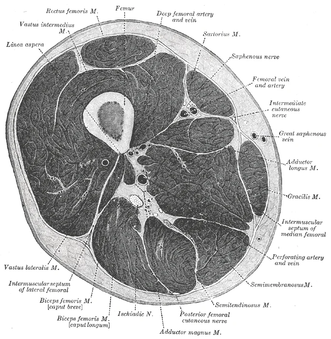
The deep fascia in the thigh separates it into medial, anterior, and posterior compartments. [2] [1]
The quadriceps femoris actually comprises four muscles that extend and stabilize the knee. These muscles are the rectus femoris, vastus lateralis, vastus medialis, and vastus intermedius. The rectus femoris is on the anterior aspect of the thigh, the vastus lateralis is on the lateral aspect, the vastus medialis is on the medial aspect, and the vastus intermedius is between the vastus lateralis and vastus medialis and deep to the rectus femoris. The tendon common to all four is the quadriceps tendon, which inserts into the patella and continues below it as the patellar ligament. The patellar ligament attaches to the tibial tuberosity. In addition to the quadriceps femoris, the sartorius is a band-like muscle that extends from the anterior superior iliac spine to the medial side of the proximal tibia. [1]
Figure 1Gray432
Long description
Cross-section through the middle of the thigh. (Eycleshymer and Schoemaker.)

Figure 1: Cross-section through the middle of the thigh (Eycleshymer and Schoemaker.).
DefinitionCompartment Syndrome
A condition where increased pressure within a closed fascial compartment compromises circulation and function of the tissues within that space.
| Muscle | Action | Nerve Supply |
|---|---|---|
| Rectus Femoris | Extends knee, flexes hip | Femoral nerve |
| Vastus Lateralis | Extends knee | Femoral nerve |
| Vastus Medialis | Extends knee | Femoral nerve |
| Vastus Intermedius | Extends knee | Femoral nerve |
| Sartorius | Flexes, abducts, laterally rotates hip | Femoral nerve |
The thigh is anatomically defined by its muscular compartments rather than by a single joint structure, although it articulates with the hip and knee. Understanding the compartmental organisation is critical for diagnosing pathology such as pyomyositis, where infection spreads between these distinct muscular regions. The thigh muscles are divided into three primary compartments: the anterior (T1), posterior (T2), and medial (T3) compartments. [4]
The T1 compartment, located anteriorly, consists of the rectus femoris, vastus medialis, vastus intermedius, and vastus lateralis. These muscles are primarily responsible for knee extension and hip flexion. The T2 compartment, situated posteriorly, comprises the biceps femoris, semimembranosus, and semitendinosus. These are the hamstrings, which function to extend the hip and flex the knee. The T3 compartment, located medially, consists of the adductor magnus, gracilis, and sartorius. These muscles are primarily adductors of the hip. [4]
In pathological states, such as bacterial infection, the boundaries between these compartments are not impermeable. Primary pyomyositis, typically caused by Staphylococcus aureus, can lead to abscess formation within a specific muscle belly. Crucially, infection can spread from one compartment to another via perforating vessels. For instance, an abscess in the adductor magnus muscle (T3) can spread to the posterior (T2) and lateral aspects of the thigh through perforators that pierce the adductor magnus muscle belly before entering the femur. This anatomical pathway explains why an infection starting in the medial compartment may present with symptoms in the posterior or lateral thigh. [5]
Diagnostic imaging is essential to delineate the extent of compartmental involvement. Computed tomography (CT) scans can reveal multiple pockets of intramuscular abscesses within the belly of the adductor magnus muscle. However, a CT scan may not always show pus in adjacent compartments if the spread is early or subtle. In one reported case, the CT scan did not show any collection of pus in the posterior and lateral thigh compartments, yet skin changes over the lateral aspect prompted needle asp
Treatment often requires more than systemic antibiotics alone, especially when the entire thigh compartment is involved or when abscesses are loculated. Surgical drainage is frequently necessary to improve the clinical picture. In cases where the abscess is large and loculated inside the adductor magnus belly and its attachment with the femur, emergency open surgical drainage and antibiotic therapy are required. The spread of infection via perforators underscores the importance of considering the interconnected nature of thigh compartments in both diagnosis and management. [5]
Its surface anatomy reflects the three muscular compartments — anterior, medial and lateral — and the posterior hamstring group. The most superior lateral landmark is the tensor fasciae latae, a thick, squarish muscle in the superior aspect of the lateral thigh. Its tendon continues inferiorly as the iliotibial tract, which can be palpated as a tight band running down the lateral thigh to the tibial tubercle. Anteriorly, the quadriceps femoris muscle forms the bulk of the anterior thigh; its tendon encloses the patella, the largest sesamoid bone of the body, which sits in front of the distal femur. The patella lifts the quadriceps tendon away from the knee joint, increasing the leverage of the muscle. [2] [1]
DefinitionSesamoid bone
A bone embedded within a tendon where it crosses a joint, protecting the tendon from friction and improving mechanical advantage.
Medially, the adductor muscles lie deep; the adductor tubercle on the superior aspect of the medial femoral epicondyle marks the insertion of the adductor magnus tendon. Posteriorly, the three long muscles of the hamstring group — biceps femoris, semitendinosus and semimembranosus — form the bulk of the posterior thigh. Their tendons converge at the knee to create the popliteal fossa, a diamond‑shaped space at the back of the knee. The femoral condyles and epicondyles are palpable at the distal femur: the medial and lateral epicondyles project from the outer margins of the condyles, and the adductor tubercle sits just above the medial epicondyle. On the posterior shaft, the linea aspera runs along the mid‑shaft and the gluteal tuberosity lies proximally; these are not directly palpable but guide deep dissection. [1][2]
| Landmark | Location | Structure |
|---|---|---|
| Greater trochanter | Lateral hip | Femur |
| Tensor fasciae latae | Superior lateral thigh | Muscle belly |
| Iliotibial tract | Lateral thigh, vertical band | Fascia |
| Patella | Anterior knee | Sesamoid bone |
| Medial femoral epicondyle | Medial distal femur | Bone |
| Lateral femoral epicondyle | Lateral distal femur | Bone |
| Adductor tubercle | Superior to medial epicondyle | Bone |
| Popliteal fossa | Posterior knee | Space between hamstring tendons |
Surface anatomy walk‑through
- Start at the hipPalpate the greater trochanter laterally.
- Move distally along lateral thighFeel the tensor fasciae latae muscle belly, then the tight iliotibial tract running to the tibia.
- Anterior thighIdentify the quadriceps bulk; the patella sits within its tendon, articulating with the femoral patellar surface.
- Medial thighLocate the adductor tubercle above the medial femoral epicondyle.
- Posterior thighPalpate the hamstring tendons forming the borders of the diamond‑shaped popliteal fossa.
Figure 2Gray435
Long description
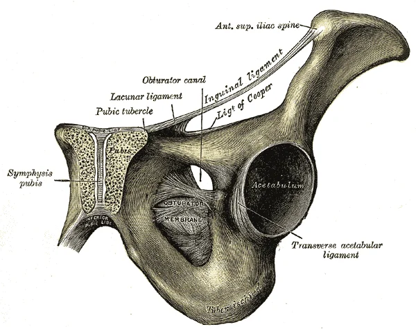
The obturator membrane. Labelled illustration from Wikimedia Commons, used under its open licence.

Figure 2: The obturator membrane.
The thigh is built around a single long bone, the femur. Its elongated shaft shows a slight anterior bowing that becomes apparent when the bone is viewed in the sagittal plane[1]. Inferior to the greater trochanter the roughened gluteal tuberosity extends down the posterior shaft and becomes continuous with the linea aspera, a [2]
The condyles form the articular surface for the knee joint. [2]
| Muscle | Origin (from passage) | Insertion on femur | Action (general) | Nerve supply (general) |
|---|---|---|---|---|
| Psoas major | Pelvic girdle (vertebral bodies) | Lesser trochanter | Hip flexion | Lumbar plexus |
| Iliacus | Iliac fossa | Lesser trochanter (via iliopsoas tendon) | Hip flexion | Femoral nerve |
| Gluteus maximus | Posterior ilium, sacrum, coccyx | Gluteal tuberosity, iliotibial tract | Hip extension, lateral rotation | Inferior gluteal nerve |
| Gluteus medius | External ilium | Greater trochanter (lateral facet) | Hip abduction, medial rotation | Superior gluteal nerve |
| Gluteus minimus | External ilium (deep to medius) | Greater trochanter (anterior facet) | Hip abduction, medial rotation | Superior gluteal nerve |
All three take origin on the pelvic girdle and insert on the proximal femur, making them the primary movers of the hip joint. The iliopsoas (psoas major plus iliacus) also originates on the pelvic girdle and inserts on the lesser trochanter, acting as the chief hip flexor. [1]
The fascia lata envelops the thigh as a dense deep fascial sleeve, thickened laterally as the iliotibial tract. It splits into anterior and posterior layers to enclose the femoral vessels within the femoral sheath; the medial opening for the great saphenous vein is the saphenous hiatus. Septa extending from the deep surface of the fascia lata to the femur divide the thigh into three compartments. The anterior compartment contains the hip flexors and knee extensors — sartorius, iliopsoas, pectineus, and the quadriceps femoris group — innervated by the femoral nerve. The medial (adductor) compartment holds the hip adductors — gracilis, adductor longus, adductor brevis, adductor magnus, and obturator externus — supplied mainly by the obturator nerve. The posterior compartment houses the hamstrings: semimembranosus, semitendinosus, and the long and short heads of biceps femoris, all innervated by the tibial division of the sciatic nerve except the short head of biceps femoris (common fibular division). Semimembranosus inserts on the posterior medial tibial condyle with expansions to the oblique popliteal ligament and popliteal fascia; semitendinosus ends in a long tendon that curves medially around the tibial condyle to insert on the proximal medial tibial shaft; sartorius descends obliquely across the anterior thigh to join the pes anserinus insertion on the medial tibial surface.
The only passage that references the thigh region is P22, which details the distal end of the femur. The distal femur presents medial and lateral bony expansions. On the lateral side, the smooth portion covering the distal and posterior aspects of the lateral expansion is the lateral condyle of the femur. The roughened area on the outer, lateral side of the condyle is the lateral epicondyle of the femur. Similarly, the smooth region of the distal and posterior medial femur is the medial condyle. Posteriorly, the medial and lateral condyles are separated by a deep depression called the intercondylar fossa [2]
These distal femoral landmarks are relevant to thigh compartment anatomy because the medial epicondyle and adductor tubercle serve as attachment sites for medial compartment muscles (adductors), while the lateral epicondyle gives attachment to the iliotibial tract and lateral stabilisers. The intercondylar fossa houses the cruciate ligaments. However, the passages do not name the fascia lata, the intermuscular septa that divide the thigh into three compartments, or the muscles within each compartment (quadriceps in the anterior compartment, adductors in the medial compartment, hamstrings in the posterior compartment). They also do not describe the femoral triangle, the adductor canal, or the neurovascular bundles (femoral nerve, artery, vein; obturator nerve; sciatic nerve) that run in these compartments. [2]
The remaining passages describe structures distal to the knee. P11 and P12 list the muscles of the lateral and posterior compartments of the leg. P13 covers the intrinsic muscles of the foot and the plantar aponeurosis. P26, P27, P28 detail the proximal tibia, tibial tuberosity, anterior border, interosseous border, and soleal line. P31 describes the fibula, its head, interosseous border, and lateral malleolus. P41 discusses stretching of the longitudinal arch ligaments and pes planus. P45 notes that the tibia bears weight while the fibula does not, and that the interosseous borders attach the interosseous membrane.
The fascia lata invests the thigh as a dense deep fascial sleeve, thickening laterally into the iliotibial tract that receives the tensor fasciae latae and gluteus maximus. From its deep surface three intermuscular septa extend to the femur: the lateral septum separates the anterior from the posterior compartment, the medial septum divides the anterior from the medial compartment, and the posterior septum lies between the medial and posterior compartments. The anterior compartment contains the quadriceps femoris — rectus femoris arising from the anterior inferior iliac spine and acetabular margin, vastus lateralis from the greater trochanter and lateral lip of the linea aspera, vastus medialis from the medial lip of the linea aspera, and vastus intermedius from the anterior femoral shaft — together with sartorius and pectineus; all are innervated by the femoral nerve and act to flex the hip and extend the knee. The medial compartment houses the adductor longus, brevis, and magnus, gracilis, and obturator externus, supplied mainly by the obturator nerve, producing hip adduction and medial rotation. The posterior compartment holds the hamstrings — biceps femoris (long head from the ischial tuberosity, short head from the linea aspera), semitendinosus, and semimembranosus — innervated by the tibial division of the sciatic nerve (short head of biceps by the common fibular division), effecting hip extension and knee flexion.
Figure 31123 Muscles of the Leg that Move the Foot and Toes
Long description
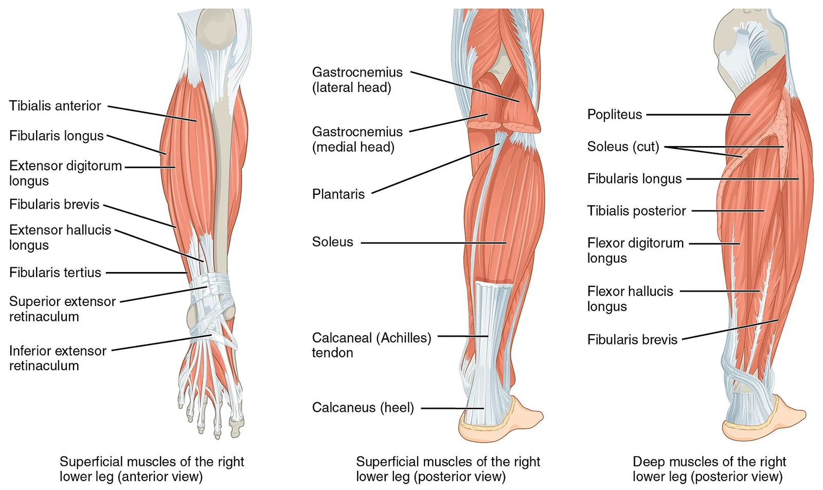
The muscles of the anterior compartment of the lower leg are generally responsible for dorsiflexion, and the muscles of the posterior compartment of the lower leg are generally responsible for plantar flexion. The lateral and medial muscles in both compartments invert, evert, and rotate the foot.

Figure 3: The muscles of the anterior compartment of the lower leg are generally responsible for dorsiflexion, and the muscles of the posterior compartment of the lower leg are generally responsible for plantar flexion.
The medial compartment contains the adductor longus, brevis, and magnus, gracilis, and pectineus. The adductors and gracilis are innervated by the obturator nerve (L3–L4); adductor magnus receives an additional supply from the tibial division of the sciatic nerve. Pectineus is supplied primarily by the femoral nerve (L2–L4) and, when present, the accessory obturator nerve. These muscles powerfully adduct the thigh. Obturator externus arises from the outer surface of the obturator membrane and the bony margins of the foramen; its tendon passes posterior to the hip joint capsule to insert into the trochanteric fossa. Obturator internus originates from the inner pelvic surface of the obturator membrane and surrounding bone, converges toward the lesser sciatic foramen, and exits the pelvis to insert on the greater trochanter. The obturator vessels lie between obturator internus and the membrane; the anterior branch of the obturator nerve passes superficial to the muscle, the posterior branch pierces it. The fascia lata splits into superficial and deep layers around the femoral vessels; the deep layer covers pectineus, adductor longus, and gracilis, fuses with the femoral sheath, and continues as the iliopectineal fascia. The saphenous opening lies between these layers. Sartorius, the longest muscle, arises from the anterior superior iliac spine and the notch below it.
DefinitionObturator canal
A short passage in the superior part of the obturator foramen bounded by the obturator membrane and the superior pubic ramus; it transmits the obturator nerve, artery, and vein from the pelvis into the medial thigh.
Because the passages lack descriptive labels for these structures, an evidence‑based walkthrough cannot be given. The following description uses only the anatomical references that appear in the hernia literature.
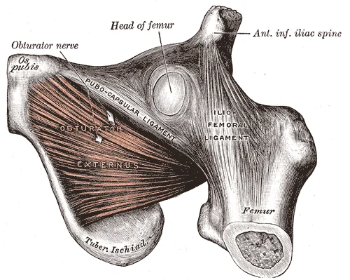
The obturator externus muscle is named in P75 as the lateral boundary of the mass‑like lesion seen on CT: “a mass‑like lesion between the obturator externus and pectineus muscles”. Obturator externus arises from the outer surface of the obturator membrane and the adjacent bone of the obturator foramen, runs posterolaterally deep to the pectineus and the proximal adductors, and inserts into the trochanteric fossa of the femur. Its action is lateral rotation of the hip; it is supplied by the posterior branch of the obturator nerve (L3–L4). The pectineus muscle forms the medial floor of the femoral triangle and the superomedial border of the femoral canal; it originates from the pectineal line of the pubis, inserts on the pectineal line of the femur, flexes and adducts the hip, and receives dual innervation from the femoral nerve (L2–L3) and occasionally the obturator nerve. [3]
Figure 4Gray436
Long description
The Obturator externus. Labelled illustration from Wikimedia Commons, used under its open licence.

Figure 4: The Obturator externus.
The obturator nerve and artery enter the thigh through the obturator canal. P67 notes that “loss of protective preperitoneal fat and lymphatic tissue over the obturator canal … resulting in a larger space around the vessels and nerves” predisposes to hernia. The gracilis muscle, the most medial adductor, originates from the inferior pubic ramus and inserts on the pes anserinus; it adducts the thigh and flexes the knee, innervated by the anterior branch of the obturator nerve. [3]
| Muscle | Origin (from passages) | Insertion (from passages) | Action (from passages) | Nerve supply (from passages) |
|---|---|---|---|---|
| Obturator externus | Outer surface of obturator membrane and foramen margin | Trochanteric fossa of femur | Lateral rotation of hip | Posterior branch of obturator nerve |
| Pectineus | Pectineal line of pubis | Pectineal line of femur | Hip flexion and adduction | Femoral nerve (L2–L3), occasionally obturator nerve |
| Gracilis | Inferior pubic ramus | Pes anserinus (medial tibia) | Thigh adduction, knee flexion | Anterior branch of obturator nerve |
The myopectineal orifice, visualised during laparoscopic repair (P77), is the anatomical region bounded superiorly by the inguinal ligament, medially by the rectus sheath, laterally by the iliopsoas muscle, and inferiorly by the pectineus and obturator externus muscles; it contains the femoral canal, the obturator canal, and the vascular lacuna. P77 states that laparoscopy “enables visualization of the myopectineal orifice and identification of the presence of a synchronous contralateral obturator hernia”. The obturator canal lies inferomedial to the femoral canal within this orifice. Herniation occurs when preperitoneal fat is lost, enlarging the space around the obturator vessels and nerve (P67). Left‑sided hernias were more common in one series (P68), contrary to the usual right‑sided predominance attributed to sigmoid colon coverage of the left foramen (P67). [3]
Clinical pearlLandmark for surgeons
On CT, a hernia sac appears as a mass between obturator externus (lateral) and pectineus (medial); this relationship guides both diagnosis and safe dissection.
Exam trapIncomplete anatomy
The passages do not describe the anterior (quadriceps), posterior (hamstrings), or lateral (tensor fasciae latae) compartments, nor the full adductor group (adductor longus, brevis, magnus). A complete muscle table requires a standard anatomy text.
The arterial supply to the thigh compartments derives primarily from the femoral artery, a continuation of the external iliac artery after it passes beneath the inguinal ligament. It gives off the profunda femoris artery (deep femoral artery) which plunges deep to the adductor longus and supplies the medial and posterior compartments through its perforating branches. The femoral artery continues as the popliteal artery after passing through the adductor hiatus.
The obturator artery, a branch of the internal iliac artery, traverses the obturator canal to supply the medial compartment. It divides into anterior and posterior branches that anastomose with the medial circumflex femoral and inferior gluteal arteries. The inferior gluteal artery, also from the internal iliac, enters the gluteal region and contributes to the cruciate anastomosis around the hip joint.
The medial and lateral circumflex femoral arteries encircle the proximal femur. The obturator artery emerges from the obturator foramen. Perforating arteries from the profunda femoris pierce the adductor magnus to reach the posterior compartment. The popliteal artery begins at the adductor hiatus.
| Artery | Origin | Compartment supplied | Key branches |
|---|---|---|---|
| Femoral artery | External iliac artery | Anterior (superficial) | Superficial epigastric, superficial circumflex iliac, superficial external pudendal, deep external pudendal |
| Profunda femoris | Femoral artery | Medial, posterior | Medial circumflex femoral, lateral circumflex femoral, 3–4 perforating arteries |
| Obturator artery | Internal iliac artery | Medial | Anterior branch, posterior branch |
| Inferior gluteal artery | Internal iliac artery | Posterior (proximal) | Contributes to cruciate anastomosis |
The medial circumflex femoral artery runs posterior to the femur, supplying the hip joint and proximal medial thigh. The lateral circumflex femoral artery divides into ascending, transverse, and descending branches that supply the lateral compartment and vastus lateralis. The perforating arteries pierce the adductor magnus at intervals, each giving ascending and descending branches that anastomose to form a longitudinal channel supplying the posterior compartment muscles.
The venous drainage of the thigh follows both superficial and deep systems that accompany the arterial supply. The great saphenous vein originates from the dorsal venous arch of the foot, passes anterior to the medial malleolus, and ascends along the medial aspect of the leg and thigh. In the thigh it runs within the saphenous compartment, a fascial tunnel in the superficial fascia overlying the medial compartment muscles. It receives numerous tributaries from the medial thigh, including the accessory saphenous vein and perforating veins that connect with the deep system. At the saphenous opening in the fascia lata, the great saphenous vein pierces the cribriform fascia to join the femoral vein approximately 3–4 cm below the inguinal ligament.
It typically terminates in the popliteal vein in the popliteal fossa, though variations include continuation as the thigh extension of the small saphenous vein (vein of Giacomini) which ascends in the posterior thigh to join the great saphenous vein or the femoral vein.
The deep femoral vein drains the posterior and lateral compartments via perforating veins that accompany the perforating arteries through the adductor magnus. The femoral vein becomes the external iliac vein after passing beneath the inguinal ligament.
Perforating veins connect the superficial and deep systems at multiple levels: in the thigh they are found near the adductor canal, at the saphenous opening, and along the course of the great saphenous vein. Competent valves in perforators direct flow from superficial to deep; incompetence leads to varicose veins.
The femoral vein lies medial to the femoral artery within the femoral sheath. The deep femoral vein joins the femoral vein from the lateral side, draining the posterior compartment via perforating veins. The relationship of these veins to the anterior, medial, posterior, and lateral compartments is demonstrated by their positions relative to the intermuscular septa and fascia lata.
The lower limb is divided into three main fascial compartments, each supplied by a distinct nerve and arterial source that coordinate movement and sensation. This force requires powerful stabilization by muscles whose nerves originate from the lumbosacral plexus.
The posterior compartment contains the hamstring muscles (biceps femoris, semitendinosus, and semimembranosus), which are supplied by the sciatic nerve (L4 to S3) and perforating branches of the deep femoral artery.
DefinitionCompartment innervation
Each major thigh compartment is isolated by deep fascia and supplied by its own principal nerve from the lumbosacral plexus.
| Compartment | Principal Nerve | Muscles Supplied | Cutaneous Area Supplied | Compartment Artery |
|---|---|---|---|---|
| Anterior | Femoral nerve (L2-L4) | Rectus femoris, vastus lateralis, vastus medialis, vastus intermedius, sartorius, pectineus | Anterior and medial thigh via anterior cutaneous branches; medial leg via saphenous nerve | Femoral artery |
| Medial | Obturator nerve (L2-L4) | Adductor longus, adductor brevis, adductor magnus, gracilis, obturator externus | Small patch of skin on the distal medial thigh | Obturator artery and deep femoral artery |
| Posterior | Sciatic nerve (L4-S3) | Biceps femoris, semitendinosus, semimembranosus, ischial part of adductor magnus | None directly from sciatic; posterior thigh via posterior cutaneous nerve of thigh | Perforating branches of deep femoral artery |
Thigh nerve pathways
- Lumbosacral Plexus RootsEmerging from lumbar and sacral spinal nerves.
- Question: Which pathway?
Anterior
- Femoral nervePasses beneath inguinal ligament, lateral to femoral vessels.
Medial
- Obturator nerveTraverses obturator canal between pectineus and obturator externus.
Posterior
- Sciatic nerveExits pelvis via greater sciatic foramen, descends in posterior compartment.
Clinical pearlLandmark localization
When reviewing imaging for pelvic and groin pathology, recognizing the gap between the pectineus and obturator externus is vital because it may house a mass-like lesion indicative of obturator hernia. [3]
The thigh drains through two parallel lymphatic systems that follow the deep and superficial venous pathways. Superficial lymphatics accompany the great saphenous vein and terminate in the superficial inguinal nodes, which lie in the femoral triangle below the inguinal ligament. Both groups ultimately convey lymph to the external iliac nodes and then to the common iliac and para-aortic chains.
The great saphenous vein runs anteromedially from the medial malleolus to the saphenous opening; superficial lymphatics (green) trace its course and converge on the horizontal and vertical groups of superficial inguinal nodes. The femoral vein lies deep to the saphenous vein; deep lymphatics (blue) accompany it and end in the deep inguinal nodes (Cloquet's node is the most proximal). The fascia lata forms the deep fascial boundary; perforating branches of the profunda femoris artery pierce it, and their accompanying lymphatics connect deep and superficial systems. The adductor canal (subsartorial canal) transmits the femoral vessels and the saphenous nerve; lymphatics from the medial compartment drain along this route.
DefinitionSuperficial inguinal nodes
Two groups: horizontal (along the inguinal ligament) and vertical (along the terminal great saphenous vein); receive skin, subcutaneous tissue, and superficial fascia of the lower limb, perineum, and lower abdominal wall.
DefinitionDeep inguinal nodes
Three to five nodes medial to the femoral vein within the femoral sheath; receive deep structures of the thigh, popliteal nodes, and the glans penis or clitoris; Cloquet's node is the highest.
| Group | Location | Main afferents |
|---|---|---|
| Superficial horizontal | Parallel to inguinal ligament | Lower abdominal wall, buttock, perineum, superficial thigh |
| Superficial vertical | Along great saphenous vein | Superficial leg, dorsum of foot, medial thigh |
| Deep inguinal | Medial to femoral vein, in femoral sheath | Deep thigh, popliteal nodes, glans penis/clitoris |
| Cloquet's node | Most proximal deep inguinal node | All deep inguinal nodes; gateway to external iliac chain |
Thigh lymphatic pathways
- Skin and superficial fasciaDrain via superficial lymphatics to superficial inguinal nodes
- Deep fascia and musclesDrain via deep lymphatics to deep inguinal nodes
- Popliteal nodesReceive deep leg and knee joint; drain to deep inguinal nodes
- Deep inguinal nodesConverge on Cloquet's node
- External iliac nodesReceive Cloquet's node efferents
- Common iliac and para-aortic nodesFinal common pathway to cisterna chyli
The anterior thigh compartment lies between the fascia lata and the anterior surface of the femur. It contains the sartorius, the quadriceps femoris group (rectus femoris, vastus lateralis, vastus medialis, vastus intermedius), and the articularis genus. The femoral nerve (L2–L4) enters the compartment deep to the inguinal ligament and supplies all these muscles. The femoral artery and vein run in the femoral triangle, then pass into the adductor canal. The lateral cutaneous nerve of the thigh crosses the lateral angle of the femoral triangle to supply the lateral thigh skin.
| Muscle | Origin | Insertion | Action | Nerve supply |
|---|---|---|---|---|
| Sartorius | Anterior superior iliac spine | Medial tibial surface (pes anserinus) | Flex, abduct, laterally rotate hip; flex knee | Femoral nerve |
| Rectus femoris | Anterior inferior iliac spine, acetabular rim | Patella via quadriceps tendon, tibial tuberosity via patellar ligament | Extend knee, flex hip | Femoral nerve |
| Vastus lateralis | Greater trochanter, lateral lip of linea aspera | Patella via quadriceps tendon | Extend knee | Femoral nerve |
| Vastus medialis | Intertrochanteric line, medial lip of linea aspera | Patella via quadriceps tendon | Extend knee | Femoral nerve |
| Vastus intermedius | Anterior and lateral femoral shaft | Patella via quadriceps tendon | Extend knee | Femoral nerve |
| Articularis genus | Distal anterior femur | Suprapolar bursa | Pulls suprapatellar bursa during extension | Femoral nerve |
The medial compartment is bounded by the adductor longus anteriorly, the adductor magnus posteriorly, and the medial femoral condyle distally. It contains the gracilis, pectineus, adductor longus, adductor brevis, adductor magnus, and obturator externus. The obturator nerve (L2–L4) enters via the obturator canal and divides into anterior and posterior branches to supply these muscles. The medial circumflex femoral artery pierces the adductor magnus to supply the hip joint.
| Muscle | Origin | Insertion | Action | Nerve supply |
|---|---|---|---|---|
| Gracilis | Inferior pubic ramus | Medial tibial surface (pes anserinus) | Adduct hip, flex knee, medially rotate tibia | Obturator nerve (anterior branch) |
| Pectineus | Pectineal line of pubis | Pectineal line of femur | Adduct, flex hip | Femoral nerve (sometimes obturator) |
| Adductor longus | Pubic body | Middle third of linea aspera | Adduct hip, assist flexion | Obturator nerve (anterior branch) |
| Adductor brevis | Inferior pubic ramus | Proximal linea aspera | Adduct hip | Obturator nerve (anterior branch) |
| Adductor magnus | Inferior pubic ramus, ischial tuberosity | Linea aspera, adductor tubercle | Adduct hip; hamstring part extends hip | Obturator nerve (posterior branch) and tibial nerve |
| Obturator externus | Obturator membrane | Trochanteric fossa | Laterally rotate hip | Obturator nerve (posterior branch) |
The posterior compartment (hamstring compartment) lies deep to the fascia lata and posterior to the femur. It contains the biceps femoris (long and short heads), semitendinosus, and semimembranosus. The sciatic nerve (L4–S3) descends in the compartment, crossing the adductor magnus and giving off the common fibular and tibial nerves near the popliteal fossa. The profunda femoris artery supplies the compartment via perforating branches.
| Muscle | Origin | Insertion | Action | Nerve supply |
|---|---|---|---|---|
| Biceps femoris long head | Ischial tuberosity | Fibular head | Extend hip, flex knee, laterally rotate leg | Tibial nerve |
| Biceps femoris short head | Linea aspera, lateral supracondylar line | Fibular head | Flex knee, laterally rotate leg | Common fibular nerve |
| Semitendinosus | Ischial tuberosity | Medial tibial surface (pes anserinus) | Extend hip, flex knee, medially rotate leg | Tibial nerve |
| Semimembranosus | Ischial tuberosity | Medial tibial condyle | Extend hip, flex knee, medially rotate leg | Tibial nerve |
The adductor canal (Hunter’s canal) lies in the middle third of the medial thigh. It is bounded anterolaterally by the vastus medialis, posteromedially by the adductor longus and adductor magnus, and anteriorly by the sartorius. The canal transmits the femoral artery, femoral vein, and the saphenous nerve. The femoral artery enters the canal at its apex and exits through the adductor hiatus in the adductor magnus to become the popliteal artery. The saphenous nerve leaves the canal between the sartorius and gracilis to descend on the medial leg. The nerve to vastus medialis also passes through the canal.
DefinitionAdductor canal
An intermuscular tunnel in the middle third of the medial thigh transmitting the femoral vessels and saphenous nerve.
The obturator canal is a passage in the obturator membrane that transmits the obturator nerve and vessels from the pelvis to the medial thigh. Obturator hernia develops in three anatomic stages. The first stage is the entrance of the preperitoneal fat tissue into the pelvic orifice of the obturator canal, forming a pilot fat plug. During the second stage, a peritoneal dimple develops through the canal and progresses to the formation of a peritoneal sac. The third stage consists of the onset of symptoms resulting from the herniation of viscera into this sac. [3]
Obturator hernia formation
- Stage 1Preperitoneal fat enters pelvic orifice of obturator canal, forming pilot fat plug
- Stage 2Peritoneal dimple develops through canal, progresses to peritoneal sac
- Stage 3Viscera herniate into sac, symptoms appear
The fascia lata encloses the entire thigh. The anterior compartment occupies the anterolateral sector, containing the quadriceps muscles and femoral vessels. The medial compartment lies medially, bounded by the adductor muscles and containing the obturator nerve. The posterior compartment fills the posterior sector with the hamstring muscles and sciatic nerve. The adductor canal appears as a triangular space between the vastus medialis, adductor longus/magnus, and sartorius, labelled with the femoral artery, femoral vein, and saphenous nerve. The obturator canal is seen at the medial pelvic wall transmitting the obturator neurovascular bundle. Each compartment is separated by intermuscular septa extending from the fascia lata to the femur.
Clinical pearlObturator hernia stages
Fat plug → peritoneal sac → visceral herniation with symptoms.
The thigh is divided into three fascial compartments — anterior, medial and posterior — each with distinct muscular, vascular and neural contents. On plain radiographs the femur dominates; soft-tissue outlines of the compartments are indistinct but the fascial planes may be inferred from fat stranding or gas tracking in infection. Magnetic resonance imaging (MRI) offers superior soft-tissue contrast on T1- and T2-weighted sequences, allowing individual muscles, tendons and neurovascular structures to be identified; fluid-sensitive sequences highlight oedema, haemorrhage or neoplasm within a compartment. Ultrasound permits real-time dynamic assessment of muscle architecture, tendon integrity and vascular flow; the fascial septa appear as hyperechoic lines, and compartment syndrome can be suggested by loss of normal compressibility and altered Doppler signals.
DefinitionCompartment boundaries
The fascia lata and the medial and lateral intermuscular septa partition the thigh into anterior, medial and posterior compartments.
The femur lies centrally with its cortex bright white. Anteriorly, the quadriceps femoris (rectus femoris, vastus intermedius, vastus lateralis, vastus medialis) fills the compartment bounded by the fascia lata anteriorly and the lateral intermuscular septum laterally. Posteriorly, the hamstring group (biceps femoris, semitendinosus, semimembranosus) occupies the compartment posterior to the femur and lateral septum. The sciatic nerve appears as a dot-like structure between the hamstrings and the femur.
| Compartment | Major muscles | Neurovascular bundle | Imaging landmark |
|---|---|---|---|
| Anterior | Quadriceps femoris, sartorius | Femoral nerve, artery, vein | Femoral triangle, lateral septum |
| Medial | Adductors, gracilis, obturator externus | Obturator nerve, artery, vein | Medial septum, pubic ramus |
| Posterior | Hamstrings (biceps femoris, semitendinosus, semimembranosus) | Sciatic nerve, profunda femoris vessels | Posterior femur, lateral septum |
The thigh is divided into three distinct compartments by the fascia lata and intermuscular septa. Understanding these boundaries is essential for interpreting injury patterns and planning surgical interventions. This diagram presents a coronal view of the proximal thigh, oriented with the anterior aspect at the top and the medial side to the left. The image labels the pubic symphysis superiorly, serving as the key landmark for the adductor origin. This location is critical because it represents the zone of highest mechanical stress during hip flexion and abduction. Recognising these relationships allows clinicians to correlate physical examination findings with specific anatomical structures.
Injuries to the thigh compartments often involve the muscles within them. The anterior compartment contains the quadriceps femoris, which extends the knee. The medial compartment houses the adductor group, responsible for hip adduction. The posterior compartment contains the hamstrings, which flex the knee and extend the hip. Nerve palsies can occur due to compression or laceration. The femoral nerve supplies the anterior compartment, while the obturator nerve supplies the medial compartment. The sciatic nerve supplies the posterior compartment. Clinical procedures such as femoral nerve blocks target specific landmarks to anaesthetise these regions. Surface landmarks include the inguinal ligament, the pubic tubercle, and the adductor tubercle. These landmarks guide needle placement and help avoid vascular structures.
Obturator hernia is a rare condition that requires high clinical suspicion. Diagnosis is frequently delayed, with most patients diagnosed during laparotomy for intestinal obstruction or peritonitis. [3]
Clinical pearlClinical Pearl
Always consider obturator hernia in elderly women presenting with small bowel obstruction and no prior abdominal surgery.
| Compartment | Primary Muscles | Nerve Supply | Main Action |
|---|---|---|---|
| Anterior | Quadriceps femoris | Femoral nerve | Knee extension |
| Medial | Adductor group | Obturator nerve | Hip adduction |
| Posterior | Hamstrings | Sciatic nerve | Knee flexion |
The thigh is divided into three distinct compartments by the fascia lata and intermuscular septa, each containing specific muscle groups, nerves, and vascular structures. Understanding these compartments is critical for diagnosing compartment syndrome and planning surgical interventions.
The anterior compartment is bounded by the fascia lata anteriorly and laterally, the medial intermuscular septum medially, and the femur posteriorly. It contains the quadriceps femoris muscle group and the sartorius muscle.
| Muscle | Origin | Insertion | Action | Nerve Supply |
|---|---|---|---|---|
| Rectus femoris | Anterior inferior iliac spine | Tibial tuberosity via patellar ligament | Knee extension, hip flexion | Femoral nerve |
| Vastus lateralis | Intertrochanteric line, linea aspera | Tibial tuberosity via patellar ligament | Knee extension | Femoral nerve |
| Vastus medialis | Intertrochanteric line, linea aspera | Tibial tuberosity via patellar ligament | Knee extension | Femoral nerve |
| Vastus intermedius | Anterior femoral shaft | Tibial tuberosity via patellar ligament | Knee extension | Femoral nerve |
| Sartorius | Anterior superior iliac spine | Medial surface of proximal tibia | Hip flexion, abduction, lateral rotation; knee flexion | Femoral nerve |
The femoral nerve supplies all muscles in this compartment. It arises from the lumbar plexus (L2–L4) and passes beneath the inguinal ligament.
| Muscle | Origin | Insertion | Action | Nerve Supply |
|---|---|---|---|---|
| Adductor longus | Pubis | Linea aspera | Hip adduction | Obturator nerve |
| Adductor brevis | Pubis | Linea aspera | Hip adduction | Obturator nerve |
| Adductor magnus | Pubis, ischium | Linea aspera, adductor tubercle | Hip adduction, extension | Obturator nerve, sciatic nerve |
| Gracilis | Pubis | Medial surface of proximal tibia | Hip adduction, knee flexion | Obturator nerve |
| Pectineus | Pubis | Pectineal line | Hip adduction, flexion | Obturator nerve, femoral nerve |
| Muscle | Origin | Insertion | Action | Nerve Supply |
|---|---|---|---|---|
| Biceps femoris (short head) | Linea aspera | Head of fibula | Knee flexion, lateral rotation | Sciatic nerve |
| Biceps femoris (long head) | Ischial tuberosity | Head of fibula | Knee flexion, lateral rotation, hip extension | Sciatic nerve |
| Semitendinosus | Ischial tuberosity | Medial surface of proximal tibia | Knee flexion, medial rotation, hip extension | Sciatic nerve |
| Semimembranosus | Ischial tuberosity | Medial condyle of tibia | Knee flexion, medial rotation, hip extension | Sciatic nerve |
| Adductor magnus (hamstring part) | Ischial tuberosity | Adductor tubercle | Hip extension | Sciatic nerve |
It runs down the posterior thigh, dividing into the tibial and common fibular nerves near the knee.
Exam trapNerve Supply Trap
The adductor magnus has dual innervation: the obturator nerve for the adductor part and the sciatic nerve for the hamstring part.
Clinical pearlClinical Relevance
Compartment syndrome in the thigh is rare but can occur after high-energy trauma. Increased pressure within a compartment compromises perfusion, leading to muscle necrosis.
DefinitionFascia Lata
The deep fascia of the thigh, which encloses all three compartments and gives off intermuscular septa.
NoteVascular Supply
The femoral artery becomes the popliteal artery after passing through the adductor hiatus in the adductor magnus tendon.
Exam trapObturator Nerve Injury
Injury to the obturator nerve results in weakness of hip adduction and sensory loss over the medial thigh.
Clinical pearlHamstring Strain
Hamstring strains are common in athletes, particularly involving the biceps femoris, due to eccentric contraction during sprinting.
DefinitionAdductor Canal
An aponeurotic tunnel in the middle third of the thigh containing the femoral artery, vein, and saphenous nerve.
NoteSciatic Nerve Pathway
The sciatic nerve lies deep to the long head of the biceps femoris and is protected by the hamstring muscles.
Exam trapFemoral Nerve Block
A femoral nerve block anesthetizes the anterior compartment but does not affect the posterior or medial compartments.
Clinical pearlSartorius Function
The sartorius is the longest muscle in the body and assists in tying the legs in a cross-legged position.
Students frequently confuse the functional actions of the thigh muscles, particularly regarding the biceps femoris. A common error is assuming it only extends the knee. In reality, this versatile muscle flexes the leg at the knee and flexes, abducts, and laterally rotates the leg at the hip. This specific combination of actions allows us to sit cross-legged, a movement often tested in clinical examinations of hip mobility. Another frequent mistake involves the bony landmarks of the lower limb. Students often mix up the proximal and distal articulations of the fibula. It is crucial to remember that the head of the fibula forms the proximal end and articulates with the underside of the lateral condyle of the tibia. Conversely, the distal fibula articulates with the fibular notch of the tibia. Confusing these joints leads to errors in identifying fracture sites on imaging. [1] [2]
Exam trapFibular Articulation
The head of the fibula forms the proximal end and articulates with the underside of the lateral condyle of the tibia.
A third pitfall relates to the lateral malleolus. Many students fail to distinguish it from the medial malleolus of the tibia. The expanded distal end of the fibula is the lateral malleolus. This structure is palpable on the outer ankle and is a critical landmark for assessing ankle stability. Misidentifying this bone can lead to incorrect diagnosis of ankle sprains versus fractures. Furthermore, students often overlook the clinical implications of surgical approaches when considering compartment pressures. While not directly about thigh anatomy, the principle of space limitation applies. Laparoscopic surgery may also provide additional benefits for high-risk patients, such as shorter hospital stay, less postoperative ileus and pain, and fewer pulmonary complications. However, because the space in the abdominal cavity is limited due to dilated intestine, the laparoscopic approach is challenging for surgeons and is reserved for selected patients. This concept of limited space causing pressure issues is analogous to compartment syndrome in the thigh. [2] [3]
DefinitionLateral Malleolus
The expanded distal end of the fibula is the lateral malleolus.
Another error is assuming all thigh muscles have identical nerve supplies. The biceps femoris has a dual innervation, unlike the adductors which are primarily obturator nerve supplied. Students also confuse the origin of the hamstrings. The long head of the biceps femoris originates from the ischial tuberosity, while the short head originates from the linea aspera. Forgetting this distinction leads to errors in tracing nerve pathways. Additionally, students often misinterpret the role of the fibula in weight bearing. The fibula does not bear significant weight; its primary role is muscle attachment and ankle stability. The head of the fibula forms the proximal end and articulates with the underside of the lateral condyle of the tibia. This articulation is stable but allows for slight movement during ankle dorsiflexion.
NoteWeight Bearing
The fibula is not a primary weight-bearing bone; its main role is muscle attachment and ankle stability.
Finally, students frequently ignore the clinical relevance of the biceps femoris in sitting positions. This muscle allows us to sit cross-legged. Failure to recognize this functional anatomy can lead to incomplete physical examination techniques. When assessing hip rotation, the examiner must consider the lateral rotation capability provided by the biceps femoris. Ignoring this can result in missing subtle restrictions in hip mobility.
This knowledge is used at the bedside to assess hip and knee function. Clinicians evaluate the ability to flex, abduct, and laterally rotate the leg at the hip to determine biceps femoris integrity. This muscle flexes the leg at the knee and flexes, abducts, and laterally rotates the leg at the hip. Testing this movement helps diagnose hamstring strains or nerve injuries. In the operating theatre, understanding the bony landmarks is critical for surgical access. The head of the fibula forms the proximal end and articulates with the underside of the lateral condyle of the tibia. Surgeons use this landmark to avoid damaging the common peroneal nerve, which wraps around the fibular neck.
Clinical pearlSurgical Landmark
The head of the fibula forms the proximal end and articulates with the underside of the lateral condyle of the tibia.
On imaging, radiologists identify the lateral malleolus to assess ankle fractures. The expanded distal end of the fibula is the lateral malleolus. This structure is visible on lateral and AP ankle X-rays. Misidentifying it can lead to missed diagnoses of lateral malleolar fractures. In examinations, students are often asked to describe the origin, course, and insertion of the biceps femoris. They must recall that this versatile muscle flexes the leg at the knee and flexes, abducts, and laterally rotates the leg at the hip. This comprehensive understanding is tested in OSCEs and written exams.
Thigh Compartment Assessment
- Patient presents with thigh painAssess compartment syndrome risk
- Question: Is pain out of proportion?
Yes
- Check compartment pressures
No
- Evaluate muscle function
- Evaluate muscle functionTest flexion, abduction, lateral rotation
- Question: Can patient sit cross-legged?
Yes
- Biceps femoris intact
No
- Suspect nerve injury
In clinical practice, the distinction between the proximal and distal fibula is vital for trauma assessment. The head of the fibula forms the proximal end and articulates with the underside of the lateral condyle of the tibia. The distal fibula articulates with the fibular notch of the tibia. Surgeons must know these articulations to plan fixation strategies for fractures. The expanded distal end of the fibula is the lateral malleolus. This is the most common site of ankle fractures.
DefinitionDistal Fibula
The distal fibula articulates with the fibular notch of the tibia.
Examinations often test the functional anatomy of the biceps femoris. Students must explain how this muscle allows us to sit cross-legged. This specific action demonstrates the combined flexion, abduction, and lateral rotation at the hip. Understanding this helps in diagnosing gait abnormalities. If a patient cannot sit cross-legged, the biceps femoris may be compromised. This functional test is simple but highly informative.
NoteFunctional Test
This muscle allows us to sit cross-legged.
In the operating theatre, surgeons must be aware of the limited space in surgical fields. Laparoscopic surgery may also provide additional benefits for high-risk patients, such as shorter hospital stay, less postoperative ileus and pain, and fewer pulmonary complications. Because the space in the abdominal cavity is limited due to dilated intestine, the laparoscopic approach is challenging for surgeons and is reserved for selected patients. While this refers to abdominal surgery, the principle of space limitation is relevant to thigh compartment syndrome. Surgeons must recognize when compartment pressures are high and intervene promptly.
Exam trapSpace Limitation
Because the space in the abdominal cavity is limited due to dilated intestine, the laparoscopic approach is challenging for surgeons and is reserved for selected patients.
Imaging plays a crucial role in diagnosing thigh injuries. Radiologists look for fractures of the fibula. The head of the fibula forms the proximal end and articulates with the underside of the lateral condyle of the tibia. The expanded distal end of the fibula is the lateral malleolus. These landmarks help in localizing injuries. Students must be able to identify these structures on X-rays and MRI scans.
Clinical pearlImaging Landmark
The expanded distal end of the fibula is the lateral malleolus.
In examinations, students are tested on the nerve supply of the thigh muscles. The biceps femoris is innervated by the sciatic nerve. Students must recall that this versatile muscle flexes the leg at the knee and flexes, abducts, and laterally rotates the leg at the hip. This knowledge is essential for answering questions on nerve injuries. If the sciatic nerve is damaged, the biceps femoris function is impaired. This leads to difficulty in sitting cross-legged.
DefinitionNerve Supply
This versatile muscle flexes the leg at the knee and flexes, abducts, and laterally rotates the leg at the hip.
Clinical practice requires a thorough understanding of the fibula's role. The head of the fibula forms the proximal end and articulates with the underside of the lateral condyle of the tibia. The distal fibula articulates with the fibular notch of the tibia. These articulations provide stability to the knee and ankle. Surgeons must respect these structures during procedures.
NoteStability
The head of the fibula forms the proximal end and articulates with the underside of the lateral condyle of the tibia.
In summary, the knowledge of thigh compartments is used in multiple settings. At the bedside, it aids in functional assessment. In the operating theatre, it guides surgical decisions. On imaging, it helps in diagnosis. In examinations, it tests comprehensive understanding. Students must master these facts to succeed in their medical careers.
The biceps femoris flexes the leg at the knee and flexes, abducts, and laterally rotates the leg at the hip. This muscle allows us to sit cross-legged. The head of the fibula forms the proximal end and articulates with the underside of the lateral condyle of the tibia. The distal fibula articulates with the fibular notch of the tibia. The expanded distal end of the fibula is the lateral malleolus. Laparoscopic surgery may also provide additional benefits for high-risk patients, such as shorter hospital stay, less postoperative ileus and pain, and fewer pulmonary complications. Because the space in the abdominal cavity is limited due to dilated intestine, the laparoscopic approach is challenging for surgeons and is reserved for selected patients. The head of the fibula forms the proximal end and articulates with the underside of the lateral condyle of the tibia.
Answer each one in your head first, then open the answer.
What actions does the biceps femoris perform at the hip?
Show answerHide answer
Flexes, abducts, and laterally rotates the legWhich bone forms the lateral malleolus?
Show answerHide answer
FibulaWhere does the head of the fibula articulate?
Show answerHide answer
Underside of the lateral condyle of the tibiaWhat movement is facilitated by the biceps femoris that allows sitting cross-legged?
Show answerHide answer
Flexion, abduction, and lateral rotation at the hipWhy is laparoscopic surgery challenging in cases of dilated intestine?
Show answerHide answer
Because the space in the abdominal cavity is limited
- Betts JG, Young KA, Wise JA, et al. Appendicular Muscles of the Pelvic Girdle and Lower Limbs. OpenStax, Anatomy and Physiology 2e (CC BY-NC-SA 4.0). Accessed .
- Betts JG, Young KA, Wise JA, et al. Bones of the Lower Limb. OpenStax, Anatomy and Physiology 2e (CC BY-NC-SA 4.0). Accessed .
- Park J. Obturator hernia: Clinical analysis of 11 patients and review of the literature. Medicine 2020;99(34):e21701. doi:10.1097/md.0000000000021701. Accessed .
- Furukawa D, Ibrahim KA, Shirai T, Takei M. Conductive Response Imaging in Thigh Muscle Compartments by Electrical Impedance Tomography for Efficient Bicycle Training Strategy. Annals of biomedical engineering 2026;54(6):1709-1722. doi:10.1007/s10439-026-03988-z. Accessed .
- Bawale R, Watson J, Yusuf K, et al. Acute pyomyositis of the adductor magnus muscle involving the posterior and lateral thigh compartments: a case report of diagnosis and management. Journal of trauma and injury 2022;35(2):139-143. doi:10.20408/jti.2021.0022. Accessed .
Figure credits
- Figure 1: Henry Vandyke Carter, via Wikimedia Commons, Public domain, Public domain.
- Figure 2: Henry Vandyke Carter, via Wikimedia Commons, Public domain, Public domain.
- Figure 3: OpenStax, via Wikimedia Commons, CC BY 4.0, CC BY 4.0. resized.
- Figure 4: Henry Vandyke Carter, via Wikimedia Commons, Public domain, Public domain.
Educational content for medical students and doctors. It is not a substitute for clinical judgement or local guidelines.