Hip joint
Multiaxial ball-and-socket joint between the femoral head and acetabulum providing stability.
The hip joint is a multiaxial ball-and-socket joint. It is formed by the articulation between the head of the femur and the acetabulum of the hip bone. The hip carries the weight of the body and thus requires strength and stability during standing and walking. For these reasons, its range of motion is more limited than at the shoulder joint. Movements at the hip joint are restricted by strong ligaments, which reduce its range of motion but confer stability during standing and weight bearing. The acetabulum is deepened by the acetabular labrum. [1]
DefinitionJoint Classification
The hip is a multiaxial ball-and-socket synovial joint characterized by a fluid-filled joint cavity allowing smooth movement.
| Muscle | Origin | Insertion | Action | Nerve Supply |
|---|---|---|---|---|
| Iliopsoas | Lumbar vertebrae and iliac fossa | Lesser trochanter of femur | Flexion | Femoral nerve |
| Gluteus maximus | Ilium, sacrum, and coccyx | Iliotibial tract and gluteal tuberosity | Extension and lateral rotation | Inferior gluteal nerve |
| Gluteus medius | Outer surface of ilium | Greater trochanter of femur | Abduction and internal rotation | Superior gluteal nerve |
| Adductor longus | Body of pubis | Linea aspera of femur | Adduction | Obturator nerve |
The abductors of the hip move the lower limb away from the midline. The primary abductors are the gluteus medius and gluteus minimus, assisted by the tensor fasciae latae. These muscles originate from the outer surface of the ilium and insert onto the greater trochanter of the femur.
The adductors bring the lower limb back toward the midline. This group includes the adductor longus, adductor brevis, adductor magnus, gracilis, and pectineus. They originate mainly from the pubis and ischium and insert along the linea aspera of the femur.
The extensors straighten the hip joint, moving the thigh posteriorly. The primary extensor is the gluteus maximus, aided by the hamstring muscles (semitendinosus, semimembranosus, and biceps femoris long head).
The flexors bring the thigh forward toward the trunk. The major flexor is the iliopsoas muscle, supported by the rectus femoris, sartorius, and pectineus.
Internal rotation turns the anterior surface of the thigh medially. This movement is performed by the anterior fibers of the gluteus medius, gluteus minimus, and the tensor fasciae latae.
External rotation turns the anterior surface of the thigh laterally. This is executed by the deep lateral rotators, including the piriformis, obturator internus, obturator externus, superior gemellus, inferior gemellus, and quadratus femoris, along with the gluteus maximus.
Hip Joint Mechanics
- Weight bearingFemoral head articulates with acetabulum.
- Question: Movement type?
Flexion/Extension
- Sagittal plane motion
Abduction/Adduction
- Coronal plane motion
The hip joint is a synovial ball-and-socket joint, functionally classified as a diarthrosis. This classification indicates that the bones move freely within a joint cavity, allowing for a large range of motion while maintaining stability. The articulation occurs between the head of the femur and the acetabulum of the hip bone. The articular surfaces are covered by hyaline cartilage, which acts like a low-friction coating, allowing smooth movement without damaging the underlying bone. Unlike cartilaginous joints, the cartilage on the femoral head and the acetabulum are not continuous with each [1][2]
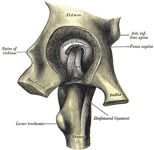
It is formed by the articulation of the head of the femur with the acetabulum of the pelvis. The acetabulum is a deep cup-shaped cavity located on the lateral aspect of the hip bone, formed by the fusion of the ilium, ischium, and pubis. [1]
Figure 1Gray341
Long description
Left hip-joint, opened by removing the floor of the acetabulum from within the pelvis.

Figure 1: Left hip-joint, opened by removing the floor of the acetabulum from within the pelvis.
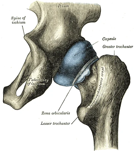
This structure increases the surface area for articulation and helps seal the joint cavity. The articular capsule is strong, with several thickened areas forming intrinsic ligaments. These ligaments arise from the hip bone, at the margins of the acetabulum, and attach to the femur at the base of the neck. They are tightened by extension at the hip, thus pulling the head of the femur tightly into the acetabulum when in the upright, standing position. This mechanism provides significant stability during weight-bearing activities. [1]
Figure 2Gray343
Long description
Capsule of hip-joint (distended). Posterior aspect.

Figure 2: Capsule of hip-joint (distended) (posterior aspect).
Synovial joints are strengthened by the presence of ligaments, which hold the bones together and resist excessive or abnormal movements of the joint. Ligaments are classified as extrinsic ligaments if they are located outside of the articular capsule, intrinsic ligaments if they are fused to the wall of the articular capsule, or intracapsular ligaments if they are located inside the articular capsule. They limit the range of motion, preventing excessive or abnormal joint movements. The synovial membrane lines the inner surface of the articular capsule and secretes synovial fluid. This fluid provides lubrication to further reduce friction between the bones of the joint. It also provides nourishment to the articular cartilage, which does not contain blood vessels. [2]
Clinical pearlStability Mechanism
The hip joint achieves stability through a combination of deep bony articulation, a strong capsule, and ligaments that tighten during extension.
The large range of motions at the shoulder joint is provided by the articulation of the large, rounded humeral head with the small and shallow glenoid cavity, which is only about one third of the size of the humeral head. In contrast, the hip joint socket is deep, providing greater stability. The acetabulum is the socket portion of the hip joint. This space is deep and has a large articulation area for the femoral head, thus giving stability and weight bearing ability to the joint. The surrounding articular capsule is strong, with several thickened areas forming intrinsic ligaments. These ligaments arise from the hip bone, at the margins of the acetabulum, and attach to the femur at the base of the neck. The ligaments are tightened by extension at the hip, thus pulling the head of the femur tightly into the acetabulum when in the upright, standing position. [1]
| Ligament | Location | Function |
|---|---|---|
| Iliofemoral | Anterior | Limits extension |
| Pubofemoral | Anterior-Inferior | Limits abduction and extension |
| Ischiofemoral | Posterior | Limits internal rotation |
The ability of the bones to move smoothly against each other within the joint cavity, and the freedom of joint movement this provides, means that each synovial joint is functionally classified as a diarthrosis. The articular cartilage acts like a Teflon® coating over the bone surface, allowing the articulating bones to move smoothly against each other without damaging the underlying bone tissue. Lining the inner surface of the articular capsule is a thin synovial membrane. The cells of this membrane secrete synovial fluid (synovia = “a thick fluid”), a thick, slimy fluid that provides lubrication to further reduce friction between the bones of the joint. This fluid also provides nourishment to the articular cartilage, which does not contain blood vessels. [2]
Outside of their articulating surfaces, the bones are connected together by ligaments, which are strong bands of fibrous connective tissue. These strengthen and support the joint by anchoring the bones together and preventing their separation. Ligaments allow for normal movements at a joint, but limit the range of these motions, thus preventing excessive or abnormal joint movements. Ligaments are classified based on their relationship to the fibrous articular capsule. An extrinsic ligament is located outside of the articular capsule, an intrinsic ligament is fused to or incorporated into the wall of the articular capsule, and an intracapsular ligament is located inside of the articular capsule.
DefinitionAcetabular Labrum
A fibrocartilage lip attached to the outer margin of the acetabulum that deepens the socket.
Synovial joints are the most common type of joints in the body. They are characterized by the presence of a joint cavity, inside of which the bones of the joint articulate with each other. The articulating surfaces of the bones at a synovial joint are not directly connected to each other by connective tissue or cartilage, which allows the bones to move freely against each other. The walls of the joint cavity are formed by the articular capsule. Friction between the bones is reduced by a thin layer of articular cartilage covering the surfaces of the bones, and by a lubricating synovial fluid, which is secreted by the synovial membrane. [2]
Exam trapCartilage Continuity
Articular cartilages of each bone are not continuous with each other in synovial joints.
Some synovial joints also have an articular disc (meniscus), which can provide padding between the bones, smooth their movements, or strongly join the bones together to strengthen the joint. However, the hip joint does not typically have an articular disc. Muscles and their tendons acting across a joint can also increase their contractile strength when needed, thus providing indirect support for the joint. The hip joint relies heavily on its bony architecture and ligamentous support for stability. [2]
The articular capsule is a strong and dense fibrous connective tissue structure that encloses the hip joint. It is attached proximally to the bony rim of the acetabulum and the transverse acetabular ligament, and it attaches distally to the neck of the femur. [1]
The synovial membrane lines the inner surface of the articular capsule and reflects onto the femoral neck up to the articular margin of the head of the femur. It secretes synovial fluid to lubricate the articulating surfaces within the joint cavity.
The stability of the hip joint is significantly enhanced by its surrounding anatomical structures. Very little additional extension of the thigh is permitted beyond the vertical position, and these ligaments stabilize the hip joint to allow an upright standing position with only minimal muscle contraction. [1]
This intracapsular ligament is normally slack and does not provide any significant joint support, but it does provide a pathway for an important artery that supplies the head of the femur. [1]
Surrounding the joint, the strong articular capsule encloses the articulation, reinforced by its named ligaments. The synovial membrane lies deep to the fibrous capsule, lining the internal joint cavity.
DefinitionIntracapsular ligament
The ligament of the head of the femur is an intracapsular structure that transmits a vital blood vessel to the femoral head despite offering minimal mechanical support.
| Structure | Proximal/Origin Attachment | Distal/Insertion Attachment | Main Function |
|---|---|---|---|
| Articular Capsule | Acetabular rim and transverse ligament | Intertrochanteric line and femoral neck | Encloses joint and resists excessive movement |
| Ligament of Head of Femur | Acetabular notch and transverse ligament | Fovea capitis on femoral head | Transports nutrient artery to femoral head |
| Synovial Membrane | Inner surface of fibrous capsule | Articular margins of femoral head | Secretes synovial fluid for joint lubrication |
Hip capsule anatomy
- Articular CapsuleEncloses joint from acetabular rim to femoral neck.
- Question: Intracapsular structures present?
Yes
- Ligamentum teresSpans from acetabular notch to fovea capitis.
No
- Synovial liningContinues internally to lubricate articulating surfaces.
The hip joint is a ball-and-socket joint, functionally classified as multiaxial, allowing for a wide range of motion including flexion, extension, abduction, adduction, and rotation. This structural design provides significant mobility but relies heavily on strong ligamentous support to maintain stability during weight-bearing activities. The primary ligaments reinforcing the hip joint capsule are the iliofemoral, pubofemoral, and ischiofemoral ligaments. These structures prevent excessive movem [2]
Figure 3Gray339
Long description
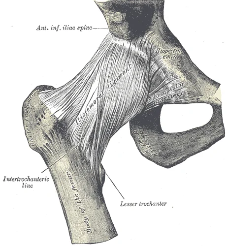
Right hip-joint from the front. (Spalteholz.)

Figure 3: Right hip-joint from the front (Spalteholz.).
The most prominent structure is the iliofemoral ligament, often described as Y-shaped. Its superior limb attaches to the anterior inferior iliac spine, while its inferior limb attaches to the intertrochanteric line of the femur. This ligament lies anterior to the joint capsule and is the strongest ligament in the body, preventing hyperextension. Lateral to the iliofemoral ligament is the pubofemoral ligament. It limits excessive abduction and extension. [1]
Figure 4Gray342
Long description
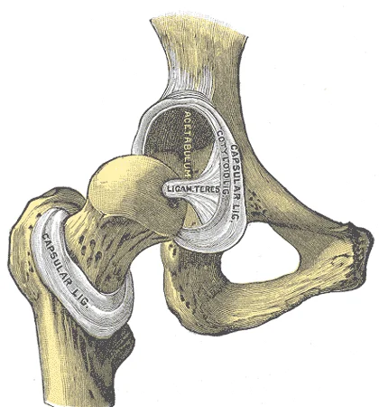
Hip-joint, front view. The capsular ligament has been largely removed.

Figure 4: Hip-joint, front view.
Figure 5Gray340
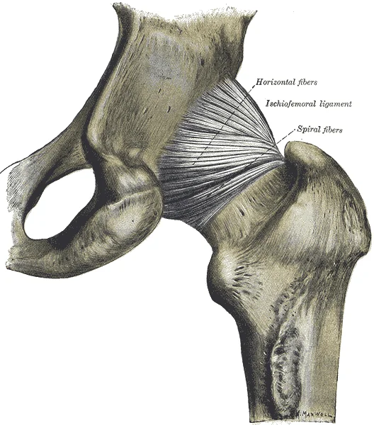
Long description
The hip-joint from behind. (Quain.). Labelled illustration from Wikimedia Commons, used under its open licence.

Figure 5: The hip-joint from behind (Quain.).
| Ligament | Attachments | Function |
|---|---|---|
| Iliofemoral | Anterior inferior iliac spine to intertrochanteric line | Prevents hyperextension; strongest ligament |
| Pubofemoral | Superior pubic ramus to femoral neck | Limits excessive abduction and extension |
| Ischiofemoral | Ischium to femoral neck | Reinforces posterior capsule; limits internal rotation |
A common injury in elderly individuals, particularly those with weakened bones due to osteoporosis, is a “broken hip,” which is actually a fracture of the femoral neck. This may result from a fall, or it may cause the fall.
Clinical pearlClinical Pearl
Fractures of the femoral neck can disrupt blood supply, leading to avascular necrosis of the femoral head.
The stability of the hip is further enhanced by the acetabular labrum, a fibrocartilaginous rim that deepens the socket. While not a ligament in the strict sense, it works in concert with the capsular ligaments to maintain joint congruence. The tension in these ligaments varies with joint position; they are tautest during extension, which helps maintain upright posture with minimal muscular effort. This passive stability is crucial for efficient bipedal locomotion.
Understanding the hip requires examining its stabilising structures, particularly the bursae and intra-articular components, which facilitate smooth movement and reduce friction.
Bursae are small, fluid-filled sacs that act as cushions between moving structures. They contain a lubricating fluid that serves to reduce friction between structures. In the hip region, bursae are categorised by their anatomical position relative to muscles and bones. Subcutaneous bursae prevent friction between the skin and an underlying bone. Submuscular bursae protect muscles from rubbing against a bone or another muscle. A subtendinous bursa prevents friction between bone and a muscle tendon. [2]
Within the joint capsule, the hip joint contains specific structures that enhance stability and nutrition.
In adults, its vascular contribution is minimal, but it remains a key landmark for identifying the centre of the femoral head.
The joint capsule itself is a strong fibrous structure. It attaches proximally to the acetabular rim and the transverse acetabular ligament.
DefinitionSynovial Joint
A joint type where articulating bones are separated by a fluid-filled cavity, allowing for free movement.
The synovial membrane lines the inner surface of the joint capsule, excluding the articular cartilage and the labrum. It secretes synovial fluid, which nourishes the articular cartilage and reduces friction. The hip joint is a multiaxial joint, allowing for flexion, extension, abduction, adduction, and rotation. This wide range of motion is supported by the strong ligaments and the deep socket of the acetabulum.
| Bursa Type | Location | Function |
|---|---|---|
| Trochanteric | Between greater trochanter and gluteus maximus | Prevents friction during hip movement |
| Iliopsoas | Between iliopsoas tendon and hip capsule | Lubricates tendon crossing anterior joint |
| Subcutaneous | Between skin and underlying bone | Protects skin from pressure and friction |
Injuries to the hip can lead to significant morbidity. Hip fractures are associated with increased rates of morbidity and mortality[1]. Surgery for a hip fracture followed by prolonged bed rest may lead to life-threatening complications, including pneumonia, infection of pressure ulcers (bedsores), and thrombophlebitis (deep vein thrombosis; blood clot formation) that can result in a pulmonary embolism (blood clot within the lung)[1]. Understanding the anatomy of the bursae and intra-articular structures is crucial for diagnosing and treating these conditions.
The hip joint is a synovial ball-and-socket joint, classified as a multiaxial joint. This structural classification allows for movement in all three anatomical planes: sagittal, frontal, and transverse. The robust nature of the lower limb joints provides the stability required to support body weight during standing, walking, and running, contrasting with the greater mobility of the upper limb joints. [1]
The muscles are grouped by their primary action. The abductors, including the gluteus medius and minimus, lie on the lateral aspect. The adductors, such as the adductor longus and brevis, are located on the medial thigh. The flexors, including the iliopsoas, are anterior. The extensors, primarily the gluteus maximus, are posterior. The rotators, both internal and external, are situated deep to the gluteal muscles.
| Muscle Group | Primary Action | Nerve Supply |
|---|---|---|
| Abductors | Abduction | Superior gluteal nerve |
| Adductors | Adduction | Obturator nerve |
| Flexors | Flexion | Femoral and lumbosacral plexus |
| Extensors | Extension | Inferior gluteal nerve |
| Rotators | Rotation | Superior and inferior gluteal nerves |
Flexion and extension occur in the sagittal plane. Extension is the reverse movement, powered by the gluteus maximus and the hamstring muscles. Similarly, the hip requires coordinated muscle action to initiate movement. [1]
Abduction and adduction occur in the frontal plane. Abduction moves the limb away from the midline, primarily performed by the gluteus medius and minimus. Adduction brings the limb toward the midline, executed by the adductor group. These movements are crucial for maintaining balance during gait.
Rotation occurs in the transverse plane. Internal rotation is produced by the gluteus medius and minimus (anterior fibres) and the tensor fasciae latae. External rotation is performed by the deep lateral rotators, including the piriformis, obturator internus, and gemelli. The hip joint allows for a significant range of motion, enabling actions such as kicking or climbing.
Figure 6Gray344
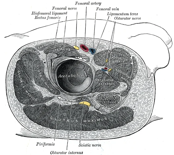
Long description
Structures surrounding right hip-joint. Labelled illustration from Wikimedia Commons, used under its open licence.

Figure 6: Structures surrounding right hip-joint.
Bursae reduce friction during these movements. A large submuscular bursa, the trochanteric bursa, is found at the lateral hip, between the greater trochanter of the femur and the overlying gluteus maximus muscle. This structure prevents rubbing of the muscle during movements. Understanding the actions of hip muscles is essential for diagnosing gait abnormalities and planning surgical interventions. [2]
DefinitionHip joint stability
The stability of the hip joint is achieved through a combination of deep bony congruity, a strong capsuloligamentous complex, and surrounding musculature.
The hip joint is a ball-and-socket synovial joint formed by the articulation of the spherical femoral head with the cup-shaped acetabulum of the pelvis.
The acetabulum deepens the socket to enclose more than half of the femoral head, while the labrum further increases the articular surface area and creates a suction seal that resists distraction forces.
The joint capsule is reinforced by three powerful extracapsular ligaments that twist during extension to lock the joint and economise muscular effort.
| Ligament | Attachment proximal | Attachment distal | Primary function |
|---|---|---|---|
| Iliofemoral ligament | Anterior inferior iliac spine | Intertrochanteric line | Resists hyperextension and external rotation |
| Pubofemoral ligament | Iliopubic eminence | Inferior aspect of intertrochanteric line | Resists abduction and hyperextension |
| Ischiofemoral ligament | Ischial wall of acetabulum | Greater trochanter | Resists internal rotation and extension |
Muscular support is critical for dynamic stability. The surrounding muscles act as compressive stabilisers that press the femoral head firmly into the acetabulum across all ranges of motion.
| Muscle | Origin | Insertion | Action | Nerve supply |
|---|---|---|---|---|
| Gluteus medius | Outer surface of ilium | Greater trochanter | Abduction and internal rotation | Superior gluteal nerve |
| Iliopsoas | T12 to L5 vertebrae and iliac fossa | Lesser trochanter | Flexion of the hip | Femoral nerve and direct branches |
| Rectus femoris | Anterior inferior iliac spine and acetabular rim | Patella via patellar tendon | Flexion of hip and extension of knee | Femoral nerve |
Stability mechanisms of the hip joint
- Static factorsBony congruity of the acetabulum and femoral head combined with the acetabular labrum.
- Question: Capsular tension?
Yes
- Iliofemoral, pubofemoral and ischiofemoral ligaments tighten in extension.
No
- Ligaments relax in flexion; dynamic stabilisers compensate.
The blood supply to the hip joint is derived mainly from branches of the femoral and obturator arteries, with key contributions from the medial femoral circumflex and lateral femoral circumflex arteries. These vessels form an extracapsular arterial ring at the base of the femoral neck. From this ring, ascending branches travel along the neck beneath the synovial membrane to form the retinacular blood supply, which is critical for nourishing the femoral head and neck. Disruption of these vessels, for example during a transcervical femoral neck fracture, carries a high risk of avascular necrosis of the femoral head.
Retinacular blood supply appears as ascending channels running along the femoral neck beneath the capsule.
According to Hilton's law, the nerves supplying a joint also supply the muscles moving that joint and the skin covering the insertions of those muscles.
Irritation of these articular branches within the capsule leads to nerve supply and referred pain patterns that frequently project to the knee or anteromedial thigh rather than the hip itself. This phenomenon explains why hip pathology often presents as isolated knee discomfort in clinical practice.
DefinitionHilton's law
The nerve supplying a joint also supplies the muscles moving that joint and the skin covering their insertions.
| Nerve | Origin | Joint innervation area | Referred pain distribution |
|---|---|---|---|
| Femoral nerve | L2-L4 | Anterior capsule | Anterior thigh and knee |
| Obturator nerve | L2-L4 | Inferomedial capsule | Medial knee |
| Nerve to quadratus femoris | L4-S1 | Posterior capsule | Gluteal region |
| Superior gluteal nerve | L4-S1 | Superior capsule | Lateral hip |
Hip pain evaluation
- Presenting complaintPatient reports joint discomfort.
- Question: Is pain referred from the hip?
Yes
- Examine hip ROMCheck internal rotation.
No
- Examine local knee structureAssess menisci and ligaments.
Understanding the hip joint on imaging requires a systematic approach to identifying osseous landmarks, soft tissue interfaces, and joint spaces. The primary modalities are plain radiography and magnetic resonance imaging (MRI). Plain films provide an overview of bone integrity and alignment, while MRI offers superior soft tissue contrast for cartilage, labrum, and marrow pathology.
The femoral head is the rounded superior portion of the femur that articulates with the acetabulum. In the AP view, it appears as a smooth, spherical structure. It is covered by articular cartilage, which is radiolucent and thus not directly visible on X-ray but inferred by the uniform joint space. On the AP view, the neck appears foreshortened due to its anterior angulation. The greater trochanter is the large, lateral prominence where several abductor muscles attach. It lies lateral to the femoral neck and head.
The acetabulum is the cup-shaped socket of the pelvis. It is formed by the fusion of the ilium, ischium, and pubis. The acetabular fossa is the non-articular central depression. The lunate surface is the C-shaped articular area covered by cartilage. The acetabular labrum is a fibrocartilaginous rim that deepens the socket. It is not visible on plain X-ray but is critical for joint stability. [1]
The joint space is the radiolucent gap between the femoral head and the acetabulum. It represents the articular cartilage and synovial fluid. A normal joint space is approximately 3–4 mm wide. Narrowing suggests cartilage loss, as seen in osteoarthritis.
The iliopsoas tendon runs along the anterior aspect of the hip joint. It is not directly visible on plain X-ray but may cause a soft tissue shadow anterior to the joint. The gluteal muscles surround the posterior and lateral aspects.
MRI is the gold standard for evaluating soft tissue structures. It visualizes the labrum, ligamentum teres, and bone marrow. On T1-weighted images, fat is bright, and fluid is dark. On T2-weighted images, fluid is bright, aiding in the detection of edema or effusion. The marrow within the femoral head and neck shows high signal intensity on T1-weighted images due to fatty content.
DefinitionJoint Space
The radiolucent gap between the femoral head and acetabulum on plain radiographs, representing articular cartilage and synovial fluid.
Exam trapFemoral Neck Foreshortening
The femoral neck appears shorter on AP views due to its anterior angulation; true length is best assessed on cross-sectional imaging or lateral views.
| Structure | Location | Appearance on AP X-ray |
|---|---|---|
| Femoral Head | Superior, medial | Smooth sphere |
| Femoral Neck | Anterior, medial | Foreshortened cylinder |
| Greater Trochanter | Lateral | Large prominence |
| Lesser Trochanter | Medial, posterior | Small profile |
| Acetabulum | Pelvic bone | Cup-shaped socket |
Hip Imaging Selection
- Suspected Hip PathologyInitial assessment
- Question: Trauma or suspected fracture?
Yes
- Plain Radiography (AP & Lateral)
No
- Soft tissue or marrow concern?
- Question: Soft tissue or marrow concern?
Yes
- MRI Hip
No
- CT for bony detail
The hip joint is a synovial ball-and-socket joint formed by the articulation of the head of the femur with the acetabulum of the pelvis. This structure allows for a wide range of motion, including flexion, extension, abduction, adduction, and rotation. The stability of the joint is derived from the deep socket of the acetabulum, the strong surrounding ligaments, and the powerful muscles that cross the joint. [1]
Dislocation of the hip is a serious injury often resulting from high-energy trauma, such as a motor vehicle accident. Posterior dislocations are the most common type, where the femoral head is displaced posteriorly out of the acetabulum. This injury pattern is frequently associated with fractures of the posterior wall of the acetabulum. Anterior dislocations are less common and may be associated with fractures of the anterior column. [1]
Neck of femur fractures are common in the elderly population, particularly those with osteoporosis. These fractures are classified as intracapsular or extracapsular. Intracapsular fractures, such as femoral neck fractures, carry a risk of avascular necrosis due to the disruption of the blood supply to the femoral head. Extracapsular fractures, including intertrochanteric fractures, generally have a better prognosis for healing. [1]
Clinical examination of the hip involves assessing range of motion, strength, and specific signs of pathology. The log roll test is used to detect intra-articular pathology; pain during passive internal and external rotation of the hip suggests joint involvement. The Trendelenburg test assesses the strength of the gluteus medius and minimus muscles; a positive test, where the pelvis drops on the contralateral side, indicates weakness or nerve injury.
Hip joint aspiration is performed to diagnose septic arthritis or crystal arthropathy. The anterior approach is commonly used, with the needle inserted just lateral to the femoral artery and below the inguinal ligament. Ultrasound guidance can improve accuracy and reduce complications.
Injury patterns to the hip often involve complex fractures of the pelvic ring. Acetabular fractures can result from axial loading forces, such as falling from a height or dashboard injuries in car crashes. These fractures may involve the weight-bearing dome of the acetabulum, requiring surgical intervention to restore joint congruity.
Clinical pearlClinical Pearl
Posterior hip dislocation presents with the leg shortened, internally rotated, and adducted.
| Muscle | Origin | Insertion | Action | Nerve Supply |
|---|---|---|---|---|
| Gluteus Maximus | Ilium, Sacrum | Iliotibial Tract | Extension, Lateral Rotation | Inferior Gluteal Nerve |
| Gluteus Medius | Ilium | Greater Trochanter | Abduction, Medial Rotation | Superior Gluteal Nerve |
| Iliopsoas | Lumbar Vertebrae, Ilium | Lesser Trochanter | Flexion, Lateral Rotation | Femoral Nerve, Lumbosacral Plexus |
It is formed by the articulation of the head of the femur with the acetabulum of the pelvis. The acetabulum is a deep cup-shaped cavity located on the lateral aspect of the hip bone, formed by the fusion of the ilium, ischium, and pubis. [1]
It is formed by the articulation of the head of the femur with the acetabulum of the pelvis. The acetabulum is a deep cup-shaped cavity located on the lateral aspect of the hip bone, formed by the fusion of the ilium, ischium, and pubis. [1]
It is formed by the articulation of the head of the femur with the acetabulum of the pelvis. The acetabulum is a deep cup-shaped cavity located on the lateral aspect of the hip bone, formed by the fusion of the ilium, ischium, and pubis. [1]
The iliofemoral ligament, also known as the Y-ligament of Bigelow, is a strong ligament. It prevents hyperextension of the hip and is crucial for maintaining upright posture with minimal muscular effort. The pubofemoral ligament limits excessive abduction and extension, while the ischiofemoral ligament spirals around the neck and limits internal rotation. [1]
Blood supply to the femoral head is critical. The artery of the ligamentum teres provides supplementary blood flow, particularly in children. Disruption of this supply, such as in femoral neck fractures, can lead to avascular necrosis. [1]
Nerve supply to the hip joint is derived from the femoral, obturator, and sciatic nerves. This innervation pattern explains why hip pathology can present as referred pain to the knee. [1]
Movements at the hip joint include flexion, extension, abduction, adduction, and rotation. [1]
Common clinical traps include confusing the blood supply sources. Students often mistake the lateral circumflex femoral artery as the main supply, whereas it primarily supplies the lateral aspect of the neck and shaft. Another trap is misidentifying the attachment of the capsule posteriorly; it attaches to the neck, not the trochanters. [1]
Fractures of the femoral neck are a common injury in elderly individuals, particularly those with weakened bones due to osteoporosis. [1]
Exam trapBlood Supply
The medial circumflex femoral artery is the main supplier to the femoral head, not the lateral circumflex femoral artery.
Clinical pearlLigament Strength
The iliofemoral ligament is the strongest ligament in the human body, preventing hyperextension.
| Ligament | Location | Function |
|---|---|---|
| Iliofemoral | Anterior | Prevents hyperextension |
| Pubofemoral | Anterior-Inferior | Limits abduction and extension |
| Ischiofemoral | Posterior | Limits internal rotation |
Exam trapSocket depth misconception
Mistaking the depth of the hip socket for a shallow articulation; in truth, the socket is deepened by a fibrocartilaginous collar.
A frequent mistake is viewing the hip joint as a simple ball-and-socket structure without appreciating the deepened stability provided by the acetabular labrum.
Another error involves confusing the main intracapsular ligament with superficial ligaments; the ligament of the head of femur transmits a small branch of the obturator artery.
Students often misattribute the primary blood supply to the femoral head entirely to the ligamentum teres, overlooking the critical ascending branches of the medial circumflex femoral artery.
A common oversight in cross-sectional imaging is failing to identify the iliopsoas bursa, which communicates directly with the synovial cavity of the hip joint in a significant proportion of individuals.
Another pitfall is misidentifying the axis of inclination of the femoral neck, leading to errors in understanding conditions such as coxa vara and coxa valga.
Confusing the attachments of the strong extracapsular ligaments—namely the iliofemoral, pubofemoral, and ischiofemoral ligaments—frequently leads to misunderstandings regarding which movements are limited by hyperextension.
Finally, students frequently assume that all surrounding bursae are isolated entities, forgetting that inflammation of the trochanteric or iliopsoas bursae can present with overlapping clinical signs of deep hip pain.
At the bedside, clinical examination of the hip involves assessing the range of flexion, extension, abduction, adduction, internal rotation, and external rotation to detect early signs of osteoarthritis or femoroacetabular impingement.
In the operating theatre, orthopaedic surgeons approach the hip joint through various surgical intervals, such as the anterolateral or posterolateral approaches, to perform total hip arthroplasty while protecting the surrounding neurovascular structures.
On imaging, plain radiographs, computed tomography, and magnetic resonance arthrography are utilised to evaluate joint space narrowing, subchondral sclerosis, osteophyte formation, and labral tears.
In examinations, students must recall the exact anatomical relations of the femoral nerve, femoral artery, and femoral vein lying anterior to the joint capsule within the femoral triangle.
Hip Assessment Pathway
- Bedside ExaminationAssess range of motion and pain provocation tests.
- Question: Structural abnormality suspected?
Yes
- Request cross-sectional imaging (MRI or CT).
No
- Monitor conservatively.
- The hip joint is a multiaxial ball-and-socket synovial articulation specialised for both stability and weight-bearing.
- The acetabulum is deepened by the fibrocartilaginous acetabular labrum.
- The joint capsule is reinforced by the iliofemoral, pubofemoral, and ischiofemoral ligaments. [1]
- The iliofemoral ligament is the strongest ligament in the body and prevents hyperextension during standing.
- The main arterial supply to the femoral head is derived from the medial circumflex femoral artery.
- The ligament of the head of the femur carries a small branch of the obturator artery.
- Clinical assessment requires thorough evaluation of rotational and flexion movements.
Answer each one in your head first, then open the answer.
What is the primary function of the iliofemoral ligament during standing
Show answerHide answer
It prevents hyperextension of the hip joint.Which artery provides the main blood supply to the head of the femur
Show answerHide answer
The medial circumflex femoral artery.What structure deepens the acetabular socket
Show answerHide answer
The acetabular labrum.Which ligament contains a small branch of the obturator artery
Show answerHide answer
The ligament of the head of the femur.What type of synovial joint is the hip joint
Show answerHide answer
A multiaxial ball-and-socket joint.
- Betts JG, Young KA, Wise JA, et al. Anatomy of Selected Synovial Joints. OpenStax, Anatomy and Physiology 2e (CC BY-NC-SA 4.0). Accessed .
- Betts JG, Young KA, Wise JA, et al. Synovial Joints. OpenStax, Anatomy and Physiology 2e (CC BY-NC-SA 4.0). Accessed .
Figure credits
- Figure 1: Henry Vandyke Carter, via Wikimedia Commons, Public domain, Public domain.
- Figure 2: Henry Vandyke Carter, via Wikimedia Commons, Public domain, Public domain.
- Figure 3: Henry Vandyke Carter, via Wikimedia Commons, Public domain, Public domain.
- Figure 4: Henry Vandyke Carter, via Wikimedia Commons, Public domain, Public domain.
- Figure 5: Henry Vandyke Carter, via Wikimedia Commons, Public domain, Public domain.
- Figure 6: Henry Vandyke Carter, via Wikimedia Commons, Public domain, Public domain.
Educational content for medical students and doctors. It is not a substitute for clinical judgement or local guidelines.