Gluteal region
The gluteal region is the posterior buttock area of the hip, bounded by bone and filled by the gluteal muscles.
The gluteal region is the posterior part of the hip, forming the buttock. Its skeletal walls are the ilium above, the ischium below and behind, and the femur laterally. Most muscles that insert on the femur and move it originate on the pelvic girdle.
The gluteal muscles form the bulk of the region. They are among the largest and most powerful muscles in the body. Three muscles are stacked in layers: the gluteus maximus is the largest, the gluteus medius lies deep to it, and the gluteus minimus, the smallest of the trio, lies deepest. [1]
DefinitionGluteal group
The gluteal group consists of the gluteus maximus, gluteus medius and gluteus minimus, arranged from superficial to deep.
Superiorly, the ilium forms a fan-like region that is the largest part of the hip bone and is united to the sacrum at the sacroiliac joint[1]. Inferiorly, the ischium forms the posteroinferior part of the hip bone and supports the body when sitting[1]. The ischial tuberosities are farther apart in the female pelvis, which increases the size of the pelvic outlet[1]. Anteriorly, the femur gives attachment to muscles acting across the hip joint. [2][3]
Clinical pearlInjection site
An intramuscular injection is given into the upper outer quadrant of the buttock, away from the sciatic nerve that runs through the lower medial part of the region. This is textbook anatomy teaching, not a trial result.
Exam trapPiriformis relations
The piriformis runs across the greater sciatic foramen and separates the structures above it from those below it. The superior gluteal vessels and nerve pass above the piriformis; the inferior gluteal vessels and nerve and the sciatic nerve pass below it. This is why an injection placed low and medial risks the sciatic nerve.
The piriformis is the key landmark of the region because it divides the neurovascular structures passing through the greater sciatic foramen into two groups. Injections placed above and lateral to the piriformis, in the upper outer quadrant, avoid the sciatic nerve and are the textbook safe site.
| Muscle | Position | Key feature |
|---|---|---|
| Gluteus maximus | Most superficial | The largest gluteal muscle |
| Gluteus medius | Deep to maximus | Middle layer |
| Gluteus minimus | Deepest | The smallest of the trio |
Gluteal region: boundaries and safe injection
- Identify the regionThe buttock, bounded by ilium above, ischium below and femur laterally.
- Question: Which layer?
Superficial
- Gluteus maximusThe largest gluteal muscle.
Middle
- Gluteus mediusDeep to the maximus.
Deepest
- Gluteus minimusThe smallest of the trio.
- Question: Injection site
Upper outer quadrant
- SafeAbove and lateral to the piriformis, clear of the sciatic nerve.
Lower inner quadrant
- AvoidSciatic nerve and inferior gluteal vessels pass below the piriformis.
Palpable landmarks of the gluteal region are all derived from the hip bone and the proximal femur. The iliac crest forms the superior boundary; run your fingers laterally along it and you will meet the anterior superior iliac spine (ASIS) at its front end and the posterior superior iliac spine (PSIS) at its back end. The PSIS is often visible as a small dimple over the sacroiliac joint, which lies just deep to it. Inferior to the ASIS, the anterior inferior iliac spine is less easily felt but ma [2]
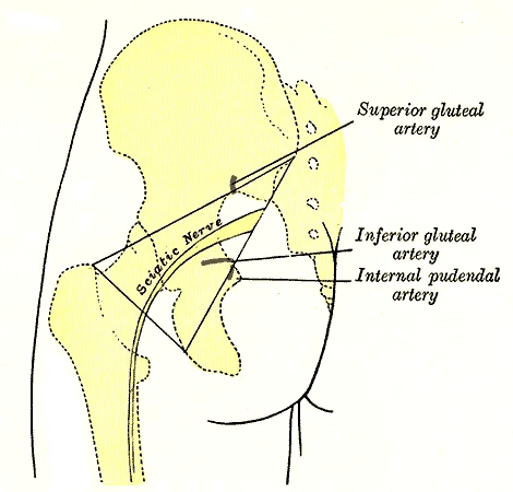
Surface markings of the gluteal region are used clinically to locate deep structures. The sciatic nerve, the largest nerve in the body, emerges from the greater sciatic foramen below the piriformis muscle. The superior gluteal artery and nerve exit above the piriformis, marked by
Figure 1Gray1244
Long description
Left gluteal region, showing surface markings for arteries and sciatic nerve.

Figure 1: Left gluteal region, showing surface markings for arteries and sciatic nerve.
Identify the iliac crest running horizontally at the top; the PSIS as a dimple just medial to it; the greater trochanter laterally at the level of the pubic symphysis; the ischial tuberosity in the lower medial buttock; and the natal cleft in the midline. The sciatic nerve marking is drawn as a curved line from the PSIS to the midpoint between the ischial tuberosity and greater trochanter, passing deep to gluteus maximus. The piriformis muscle is outlined deep to the gluteus maximus, with the sciatic nerve emerging below it and the superior gluteal vessels above it. [1]
The gluteal region overlies the buttock; its framework is the hip bone and the bony pelvis. The pelvic girdle (hip girdle) is formed by a single bone on each side, the hip bone or coxal bone (coxal = "hip"), which serves as the attachment point for each lower limb. Each hip bone is firmly joined to the axial skeleton through its attachment to the sacrum of the vertebral column, and the right and left hip bones converge anteriorly to attach to each other. The bony pelvis is the whole structure fo [2]
This framework is built for rigidity. Unlike the pectoral girdle, whose bones are highly mobile for upper limb movement, the pelvic bones are strongly united to form a largely immobile, weight-bearing structure. Body weight thus transfers laterally from the vertebral column through the pelvic girdle and hip joints into whichever limb bears weight, so the pelvis is a strong foundation for the upper body. [2]
Each adult hip bone is formed by three separate bones that fuse together during the late teenage years: the ilium, ischium and pubis; these names define the three regions of the adult hip bone. [2]
Anteriorly, the pubic body is joined to the pubic body of the opposite hip bone by the pubic symphysis. The inferior pubic ramus extends downward and laterally from the body, then joins the ischial ramus to form the single ischiopubic ramus, running from the pubic body to the ischial tuberosity. The pubic arch is formed by the pubic symphysis and the bodies and inferior pubic rami of the adjacent pubic bones; the inverted V of the two ischiopubic rami at the symphysis is the subpubic angle. [2]
On each side identify the ilium above, the ischium posteroinferiorly and the pubis anteriorly, the three fusing in the late teenage years. In the anterior midline is the pubic symphysis. Follow the inferior pubic ramus down to its junction with the ischial ramus; the ischiopubic ramus runs back to the ischial tuberosity. The two ischiopubic rami make the inverted V of the subpubic angle below the symphysis. Posteriorly the sacrum lies between the hip bones, with the coccyx inferiorly. [2]
The gluteal region is the thick, muscular mass that forms the buttock and the upper lateral thigh. Its deep fascia, the fascia lata, is a strong, dense sheet that invests the muscles of the thigh and gluteal region. In the gluteal area, the fascia lata splits to enclose the gluteus maximus and the tensor fascia latae, and it thickens laterally to form the iliotibial tract (band). This tract is a strong, fibrous band that runs down the lateral side of the thigh from the iliac crest to the lateral tibial condyle (Gerdy's tubercle). It is made taut by the tensor fascia latae, which pulls on it to help stabilise the lateral aspect of the knee. The fascia lata also sends deep septa between muscles, dividing the thigh into medial, anterior, and posterior compartments; the gluteal muscles themselves lie superficial to these septa but are enclosed by the same deep fascia. [1]
The skin and superficial fascia have been removed to reveal the deep fascia (fascia lata) as a white, encircling line. On the lateral side, the fascia is thickened into the iliotibial tract. Deep to the fascia, the large gluteus maximus lies posteriorly, its fibres running obliquely downwards and laterally. Anterior to it, the gluteus medius is partly covered by the gluteus maximus, and the tensor fascia latae lies anterolaterally, embedded within the fascia.
The deep fascia of the gluteal region does not create true osteofascial compartments like those of the thigh or leg; instead, the muscles are arranged in layers. The superficial layer contains the gluteus maximus and tensor fascia latae. The middle layer contains the gluteus medius, piriformis, obturator internus, and the gemelli. The deep layer contains the gluteus minimus and quadratus femoris. This layered arrangement is clinically important because infections or haematomas can track along fascial planes, and the sciatic nerve, which emerges from the greater sciatic foramen below the piriformis, lies between the deep and middle layers. [1] [3]
| Muscle | Origin | Insertion | Action | Nerve supply |
|---|---|---|---|---|
| Gluteus maximus | Posterior ilium, sacrum, coccyx | Iliotibial tract, gluteal tuberosity of femur | Extends and laterally rotates thigh | Inferior gluteal nerve |
| Tensor fascia latae | Anterior iliac crest | Iliotibial tract | Stabilises knee | Superior gluteal nerve |
| Gluteus medius | External ilium | Greater trochanter | Abducts thigh; stabilises pelvis | Superior gluteal nerve |
| Gluteus minimus | External ilium | Greater trochanter | Abducts thigh; stabilises pelvis | Superior gluteal nerve |
| Piriformis | Sacrum | Greater trochanter | Laterally rotates thigh | Nerve to piriformis |
| Obturator internus | Inner obturator membrane | Greater trochanter | Laterally rotates thigh | Nerve to obturator internus |
| Superior gemellus | Ischial spine | Greater trochanter | Laterally rotates thigh | Nerve to obturator internus |
| Inferior gemellus | Ischial tuberosity | Greater trochanter | Laterally rotates thigh | Nerve to quadratus femoris |
| Quadratus femoris | Ischial tuberosity | Intertrochanteric crest | Laterally rotates thigh | Nerve to quadratus femoris |
The deep group of short lateral rotators—piriformis, obturator internus, superior and inferior gemelli, and quadratus femoris—all lie deep to the gluteus maximus and act together to laterally rotate the femur at the hip. The obturator externus, though originating in the medial thigh, also joins this group functionally. The piriformis is a key landmark: the sciatic nerve, the inferior gluteal nerve and vessels, and the pudendal nerve all pass through the greater sciatic foramen in relation to it, most commonly inferior to the muscle belly. [1]
The gluteal region contains several important muscles that are organized into compartments. The gluteal muscles include the gluteus maximus, medius, and minimus, along with other deep muscles such as piriformis and obturator internus.
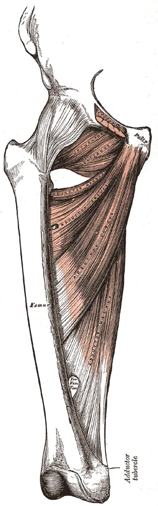
Figure 2Gray433
Long description
Deep muscles of the medial femoral region.

Figure 2: Deep muscles of the medial femoral region.
| Muscle | Origin | Insertion | Action | Nerve Supply |
|---|---|---|---|---|
| Gluteus maximus | Posterior gluteal line, iliac crest, sacrum, coccyx, sacrotuberous ligament | Iliotibial tract, gluteal tuberosity | flexes the leg at the knee and flexes, abducts, and laterally rotates the leg at the hip | Inferior gluteal nerve [1] |
| Gluteus medius | Outer surface of ilium, between anterior and posterior gluteal lines | Greater trochanter | Abducts thigh, medially rotates thigh | Superior gluteal nerve |
| Gluteus minimus | Outer surface of ilium, between anterior and posterior gluteal lines | Greater trochanter | Abducts thigh, medially rotates thigh | Superior gluteal nerve |
| Piriformis | Anterior surface of sacrum, greater sciatic foramen | Greater trochanter | Laterally rotates thigh | Nerve to piriformis |
| Obturator internus | Inner surface of obturator membrane, surrounding bone | Greater sciatic foramen | Greater trochanter | Laterally rotates thigh |
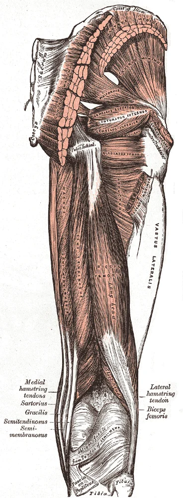
The sciatic nerve is the largest nerve in the body and passes through the gluteal region. It emerges from the greater sciatic foramen, inferior to piriformis muscle. The sciatic nerve then courses inferiorly through the posterior thigh, where it divides into the tibial nerve and common fibular nerve at the level of the popliteal fossa.
Figure 3Gray434
Long description
Muscles of the gluteal and posterior femoral regions.

Figure 3: Muscles of the gluteal and posterior femoral regions.
The gluteal muscles perform several important actions. The gluteus maximus extends and laterally rotates the thigh, and helps us stand up from a sitting position. The gluteus medius and minimus abduct and medially rotate the thigh, stabilizing the pelvis during walking. Piriformis laterally rotates the thigh.
The superior gluteal nerve supplies gluteus medius and minimus, while the inferior gluteal nerve supplies gluteus maximus. These nerves emerge from the greater sciatic foramen, superior and inferior to piriformis respectively.
The gluteus maximus is the most superficial muscle, forming the bulk of the buttock. Deep to it are the gluteus medius and minimus, which are partially covered by the gluteus maximus. The piriformis muscle is located deep in the gluteal region, passing through the greater sciatic foramen.
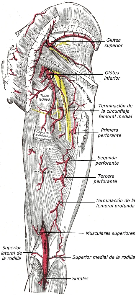
Figure 4Gray544transespa
Long description
The arteries of the gluteal and posterior femoral regions.

Figure 4: The arteries of the gluteal and posterior femoral regions.
The arterial supply of the gluteal region is derived primarily from the internal iliac artery, the main artery of the pelvis. The blood flow is distributed through two major vessels that exit the pelvic cavity to reach the buttock and the proximal thigh.
The superior gluteal artery is the largest branch of the internal iliac artery. It originates from the posterior division of the internal iliac artery. It follows a course that takes it out of the pelvis through the greater sciatic foramen, specifically passing through the suprapiriform space, which is the gap located above the piriformis muscle.
Once in the gluteal region, it runs laterally between the gluteus medius and gluteus minimus muscles. Its primary function is to provide oxygenated blood to the abductor mechanism of the hip. It gives off numerous muscular branches to the gluteus medius, gluteus minimus, and the tensor fasciae latae. It also provides branches to the joint capsule of the hip.
The inferior gluteal artery also originates from the posterior division of the internal iliac artery. Its course differs from the superior gluteal artery as it exits the pelvis through the greater sciatic foramen in the infrapiriform space, which is the area below the piriformis muscle.
It descends in the gluteal region, passing deep to the gluteus maximus. It provides the primary blood supply to the gluteus maximus muscle and the short lateral rotators of the hip. Additionally, it provides essential nutrient branches to the sciatic nerve.
| Vessel | Primary Target Structures | Key Relations |
|---|---|---|
| Superior gluteal artery | Gluteus medius, gluteus minimus, tensor fasciae latae | Above piriformis muscle |
| Inferior gluteal artery | Gluteus maximus, short lateral rotators, sciatic nerve | Below piriformis muscle |
The gluteal region is characterised by a rich anastomotic network. This ensures that if one major vessel is compressed or blocked, blood can still reach the tissues. The superior and inferior gluteal arteries anastomose with each other and with the medial and lateral circumflex femoral arteries. This network forms a crucial collateral circle around the hip joint, linking the internal iliac system with the femoral system.
The buttock is drained by veins that accompany its arteries and carry blood into the true pelvis. Almost all of them leave through the greater sciatic foramen and end in the internal iliac vein. Because each vein is a companion of an artery, the map of the veins repeats the map of the arteries; learn one and the other follows.
These are the venae comitantes of the superior gluteal artery. They begin in the buttock, taking tributaries from gluteus maximus, gluteus medius, gluteus minimus and tensor fasciae latae. The paired veins climb behind the artery, leave the pelvis through the greater sciatic foramen and cross above piriformis together with the superior gluteal nerve. Behind the iliac bone they join into one vessel, which opens into the internal iliac vein.
These accompany the inferior gluteal artery. They gather blood from gluteus maximus, the tissues resting on it and the back of the thigh. They pass upward through the greater sciatic foramen below piriformis, with the inferior gluteal nerve and the sciatic nerve close behind. In the pelvis they usually fuse into a single trunk, which may join the internal pudendal vein before draining into the internal iliac vein. [1]
This vein begins in the perineum, formed by the deep dorsal vein of the penis or clitoris with veins leaving the erectile tissue and the perineal muscles. It tracks the internal pudendal artery out of the pelvis through the greater sciatic foramen below piriformis, crosses the ischial spine, re-enters through the lesser sciatic foramen, and ends in the internal iliac vein.
Small paired veins run with the lateral sacral arteries on the front of the sacrum. They enter the pelvis through the anterior sacral foramina, drain the sacral bone and its attached muscles, and end in the internal iliac vein. Across the midline they meet the median sacral vein, linking the two sides.
The gluteal veins join freely with one another and with the medial and lateral circumflex femoral veins. Through perforating veins they also communicate with the femoral vein and its branches. This web gives alternative routes when the external iliac or femoral vein is narrowed, and blood may leave the buttock by a deep path through the internal iliac vein or a superficial path down the thigh.
DefinitionVena comitans
A vena comitans is a vein, usually one of a pair, that runs alongside an artery. Every vein in this region is a companion of an artery.
Clinical pearlAbove or below piriformis
The superior gluteal vessels pass above piriformis and the inferior gluteal vessels pass below it. That single border sorts the whole region.
Exam trapInternal pudendal vein
The internal pudendal vein is a perineal vein, not a buttock vein. It only crosses the greater and lesser sciatic foramina on its way to the perineum.
NoteWhy the connections matter
The gluteal veins link the internal iliac and femoral systems, so a clot forming in a thigh vein can extend upwards and infection of the perineum can track into the pelvis.
| Vein | Companion artery | Key relation | Termination |
|---|---|---|---|
| Superior gluteal | Superior gluteal artery | Greater sciatic foramen, above piriformis | Internal iliac vein |
| Inferior gluteal | Inferior gluteal artery | Greater sciatic foramen, below piriformis | Internal iliac vein |
| Internal pudendal | Internal pudendal artery | Ischial spine, then lesser sciatic foramen | Internal iliac vein |
| Lateral sacral | Lateral sacral artery | Anterior sacral foramina | Internal iliac vein |
At the top the superior gluteal vein crosses above piriformis with the superior gluteal artery and nerve, having just left the greater sciatic foramen, and lies deep to gluteus medius. Below piriformis the inferior gluteal vein emerges with the inferior gluteal artery, while the sciatic nerve rests medial to them. Medial to that foramen the internal pudendal vein curves round the ischial spine, the point between the greater and lesser sciatic notches, and slips forward into the lesser sciatic foramen. On the sacral surface the lateral sacral veins accompany the lateral sacral arteries beside the midline median sacral vein, and all drain medially into the internal iliac vein, which lies deep in the pelvis. The cut edges of gluteus maximus frame the field laterally.
Tracing gluteal venous drainage
- Start: buttock and perineumBlood collects in tributaries of the gluteal veins and in the internal pudendal vein.
- Question: Which vein is followed?
Superior gluteal
- Leave the pelvis through the greater sciatic foramen above piriformisTravel with the superior gluteal artery and nerve.
- End in the internal iliac veinThe paired veins unite behind the ilium.
Inferior gluteal
- Pass through the greater sciatic foramen below piriformisThe sciatic nerve lies close behind.
- End in the internal iliac veinOften shares a trunk with the internal pudendal vein.
Internal pudendal
- Hook around the ischial spineExit through the greater, then re-enter through the lesser sciatic foramen.
- End in the internal iliac veinCarries blood from the perineum into the pelvis.
The gluteal region is supplied by nerves that arise from the sacral plexus, a network formed by the anterior rami of spinal nerves L4–S4. These nerves leave the pelvis through the greater sciatic foramen, a large opening bounded by the sacrum, ilium, and sacrospinous ligament. The two principal nerves of the region are the superior gluteal nerve and the inferior gluteal nerve, both of which are motor nerves to the gluteal muscles, with no cutaneous branches. Cutaneous supply to the skin of the buttock comes instead from the posterior rami of the upper lumbar and sacral spinal nerves, as well as from branches of the sacral plexus such as the posterior femoral cutaneous nerve. [2]
The superior gluteal nerve (L4, L5, S1) exits the pelvis through the greater sciatic foramen above the piriformis musclecars. It then runs laterally between the gluteus medius and gluteus minimus muscles, supplying both of these abductors of the thighcars. It also innervates the tensor fasciae latae, a small muscle that stabilises the pelvis during walking. The inferior gluteal nerve (L5, S1, S2) leaves the pelvis through the greater sciatic foramen below the piriformis muscle and immediately en [1]
The piriformis muscle itself lies centrally, dividing the greater sciatic foramen into a suprapiriform and infrapiriform part, which is the key landmark for locating the two nerves.
| Nerve | Origin | Motor supply (muscles) | Cutaneous supply | Key relations |
|---|---|---|---|---|
| Superior gluteal nerve | L4, L5, S1 | Gluteus medius, gluteus minimus, tensor fasciae latae | None | Exits above piriformis; runs between gluteus medius and minimus |
| Inferior gluteal nerve | L5, S1, S2 | Gluteus maximus | None | Exits below piriformis; enters deep surface of gluteus maximus |
| Posterior femoral cutaneous nerve | S1, S2, S3 | None | Skin of posterior thigh and lower buttock | Runs below piriformis, deep to gluteus maximus |
| Posterior rami of L1–S3 | Spinal nerves | None | Skin of medial buttock and sacral area | Pierce the thoracolumbar fascia directly |
The gluteal muscles themselves act on the hip joint, which is stabilised by the pelvic girdle. The pelvic girdle has very little movement because of its connection with the sacrum at the base of the axial skeleton, and it was designed to stabilise and support the body[1]. The leg muscles therefore make up in size and power what they lack in range of motion, facilitating posture and movement[1]. The gluteus maximus is the largest muscle of the region and is the chief extensor [2]
Clinically, the sciatic nerve is at risk during gluteal injections, which should be placed in the upper outer quadrant of the buttock to avoid it.
Understanding this drainage requires a clear distinction between superficial and deep lymphatic vessels, as their destinations differ significantly.
The vessels converge towards the inguinal lymph nodes. Specifically, the lateral vessels drain into the superficial inguinal nodes, while medial vessels may drain into the deep inguinal nodes or directly into the external iliac nodes.
The superficial lymphatic vessels accompany the superficial veins. They drain the skin and subcutaneous tissue of the gluteal region. Most of these vessels drain into the superficial inguinal lymph nodes. These nodes are located in the upper part of the femoral triangle, just below the inguinal ligament. The superficial inguinal nodes are arranged in two groups: horizontal and vertical. The horizontal group receives lymph from the lower abdominal wall, the perineum, and the external genitalia. The vertical group receives lymph from the lower limb and the gluteal region.
Some lymphatic vessels from the upper lateral part of the gluteal region may drain directly into the deep inguinal lymph nodes or even the external iliac lymph nodes. This direct drainage is clinically significant because it bypasses the superficial inguinal nodes. The deep inguinal lymph nodes are located deep to the fascia lata, medial to the femoral vein. They receive lymph from the deep structures of the lower limb and the gluteal region.
The internal iliac lymph nodes are located in the pelvis, along the internal iliac artery. They receive lymph from the pelvic viscera, the perineum, and the gluteal muscles.
Clinical relevance is paramount in this region. Infections or malignancies of the gluteal skin can spread to the inguinal lymph nodes. For example, squamous cell carcinoma of the buttock skin may metastasize to the superficial inguinal nodes. Enlargement of these nodes can be palpated in the groin. It is essential to examine the inguinal region when assessing patients with gluteal skin lesions.
Clinical pearlClinical Pearl
Always palpate the inguinal lymph nodes when evaluating gluteal skin lesions, as they are the primary drainage site for most of the region.
The lymphatic drainage pattern is not always uniform. Anatomical variations exist, and some vessels may cross the midline. This variability must be considered in surgical planning and staging of diseases.
| Structure | Primary Drainage Site | Secondary Drainage Site |
|---|---|---|
| Gluteal Skin (Superficial) | Superficial Inguinal Nodes | Deep Inguinal Nodes |
| Gluteal Muscles (Deep) | Internal Iliac Nodes | External Iliac Nodes |
| Upper Lateral Gluteal Skin | Deep Inguinal Nodes | External Iliac Nodes |
Understanding these pathways is essential for diagnosing and managing conditions affecting the gluteal region. The lymphatic system provides a route for immune cells to monitor for pathogens and abnormal cells. Disruption of this system can lead to lymphoedema, although this is rare in the gluteal region unless there is significant surgical intervention or radiation therapy.
The gluteal region is compartmentalised by two fibro-osseous spaces: the greater sciatic foramen and the lesser sciatic foramen. These are formed by the sacrospinous ligament (running from sacrum to ischial spine) and the sacrotuberous ligament (from sacrum to ischial tuberosity). The sacrospinous ligament converts the greater sciatic notch into the greater foramen; the sacrotuberous ligament, together with the sacrospinous ligament, creates the lesser foramen below the ischial spine. The greater foramen is the main gateway between the pelvis and the gluteal region; the lesser foramen connects the gluteal region to the perineum. [2]
Contents of the greater sciatic foramen. The piriformis muscle passes through its centre, dividing the foramen into a suprapiriform and an infrapiriform part. Above piriformis pass the superior gluteal nerve (L4–S1) and superior gluteal vessels (branches of the internal iliac artery and vein). Below piriformis pass, from lateral to medial: the sciatic nerve (L4–S3), the posterior femoral cutaneous nerve, the inferior gluteal nerve (L5–S2) and **inferior gluteal ve
Contents of the lesser sciatic foramen. This transmits the tendon of obturator internus as it turns sharply around the ischium, the nerve to obturator internus (which supplies the muscle and also the superior gemellus), and the pudendal nerve with the internal pudendal vessels as they pass from the gluteal region into the perineal pouch.
Surface markings of the gluteal region are used clinically to locate deep structures. The sciatic nerve, the largest nerve in the body, emerges from the greater sciatic foramen below the piriformis muscle. The superior gluteal artery and nerve exit above the piriformis, marked by
Bursae and fascial spaces. Three clinically important bursae lie in the region: the trochanteric bursa (between gluteus maximus and the greater trochanter), the ischial bursa (over the ischial tuberosity, under gluteus maximus), and the gluteofemoral bursa (between the tendon of gluteus maximus and the vastus lateralis). The iliotibial tract receives the insertion of gluteus maximus and tensor fasciae latae and passes down the lateral thigh.
In the figure, the piriformis is seen emerging from the greater sciatic foramen; the sciatic nerve passes below it, crossing the superior gemellus, the tendon of obturator internus, and the inferior gemellus, then lies on quadratus femoris before disappearing under the lower border of gluteus maximus. The greater trochanter is lateral, the ischial tuberosity medial, and the sacrotuberous ligament forms the medial boundary of the foramen. The superior gluteal vessels are seen above piriformis, and the inferior gluteal vessels below it, with the pudendal nerve curving around the ischial spine toward the lesser foramen.
The inferior cluneal nerves, three or four in number, curve upward around the lower border of gluteus maximus to supply the skin over its lower and lateral portion, while perineal branches innervate the skin on the upper medial thigh. Gluteus maximus originates from the posterior gluteal line of the ilium, the adjacent rough bone including the iliac crest, the posterior surfaces of the lower sacrum and lateral coccyx, the sacrospinalis aponeurosis, the sacrotuberous ligament, and the gluteal aponeurosis covering gluteus medius. Its fibers run obliquely inferolaterally; the upper and larger portion, with superficial fibers of the lower portion, forms a thick tendinous lamina crossing the greater trochanter to insert into the iliotibial band, while deeper lower fibers insert into the gluteal tuberosity between vastus lateralis and adductor magnus. Three bursae typically relate to its deep surface. Gluteus medius and minimus converge onto radiated aponeuroses ending in tendons inserting on the anterior greater trochanter, with expansions to the hip capsule. Between these two muscles run the deep branches of the superior gluteal vessels and the superior gluteal nerve. The deep surface of gluteus minimus contacts the reflected tendon of rectus femoris and the hip capsule. The sacral plexus lies on the posterior pelvic wall between piriformis and the pelvic fascia; anterior to it are the hypogastric vessels, ureter, and sigmoid colon. The superior gluteal vessels pass between the lumbosacral trunk and first sacral nerve, while the inferior gluteal vessels run between the second and third sacral nerves.
Plain films, computed tomography (CT), magnetic resonance imaging (MRI) and ultrasound each display a different component of the gluteal region. Cortical bone is radiopaque and appears white on radiographs and CT, whereas muscle, fat and neural tissue differ mainly in their soft-tissue density or signal. The gluteal muscles themselves are seldom the primary target of imaging; they are seen because they surround the bony pelvis and the sciatic nerve.
On an anteroposterior (AP) radiograph of the pelvis the gluteal region is framed by the iliac wings superiorly and laterally, the sacrum and coccyx posteriorly, and the ischium and pubis inferiorly. The iliac crest, anterior superior iliac spine, iliac wing, sacroiliac joint, sacral ala, coccyx, acetabulum, femoral head and neck, greater and lesser trochanters, ischial spine, ischial tuberosity, obturator foramen and pubic symphysis can all be identified. The gluteal soft-tissue shadow is faint,
Axial CT slices through the greater trochanter show, from posterior to anterior, the subcutaneous fat, gluteus maximus, the plane containing the sciatic nerve, gluteus medius and minimus on the lateral surface of the ilium, and iliopsoas anteriorly. Medial to the sciatic nerve lie the obturator internus and gemelli, with the ischial tuberosity below and the acetabulum medially. Bone windows show cortical and trabecular detail; soft-tissue windows show the muscles and fat planes. Coronal and sagittal reformats display the full extent of the gluteal muscles and their relation to the hip joint.
On T1-weighted images fat is bright and muscle is of intermediate signal; on T2-weighted and fat-suppressed images fluid and oedema are bright. Gluteus maximus, medius and minimus, tensor fasciae latae and the iliotibial tract are well seen, as is the piriformis with the sciatic nerve passing below it. Marrow signal within the ilium, sacrum and proximal femur is assessed on the same sequences.
With the patient prone or side-lying, the probe shows the layered gluteus maximus, medius and minimus, the echogenic iliotibial tract, the ischial tuberosity and the sciatic nerve as a honeycomb structure. Ultrasound can demonstrate a hip effusion and guide a gluteal or peri-sciatic injection.
Named labels include the iliac crest, iliac wing, anterior superior iliac spine, sacroiliac joint, sacrum, coccyx, acetabulum, femoral head and neck, greater trochanter, ischial spine, ischial tuberosity, obturator foramen and pubic symphysis; the gluteal soft-tissue shadow overlies the iliac wings and the lateral aspect of the sacrum.
The gluteal region is a frequent site for intramuscular injections and is susceptible to nerve injuries due to the proximity of major neurovascular structures to the skin.
To avoid injury to the sciatic nerve, injections are typically administered in the upper outer quadrant of the gluteal region. This area ensures the needle enters the gluteus medius rather than the deeper structures.
Damage to the nerves of the gluteal region often results from iatrogenic causes, such as improper injection technique, or traumatic causes, such as posterior hip dislocations.
| Nerve | Cause of Lesion | Clinical Presentation |
|---|---|---|
| Superior Gluteal Nerve | Iatrogenic or trauma | Weakness in hip abduction; Trendelenburg sign |
| Inferior Gluteal Nerve | Iatrogenic or trauma | Difficulty climbing stairs or rising from a chair |
The Trendelenburg sign is a clinical indicator of weakness in the hip abductors, primarily the gluteus medius and gluteus minimus. These muscles are supplied by the superior gluteal nerve.
When a patient stands on one leg, the abductors of the weight-bearing hip must contract to keep the pelvis level. If the superior gluteal nerve is damaged or the muscles are weak, the pelvis drops toward the unsupported side. This is known as a positive Trendelenburg sign.
Clinicians use palpable landmarks to navigate the region. The posterior superior iliac spine (PSIS) and the greater trochanter of the femur are key points for locating the sciatic nerve.
The nerve descends deep to the gluteus maximus and superficial to the quadratus femoris. The superior gluteal nerve is positioned superior to the piriformis muscle, while the inferior gluteal nerve emerges inferior to the piriformis. [2]
- Another trap is assuming the sacroiliac joint is freely movable; it is largely immobile, with only slight gliding movements. [2]
Exam trapLigament vs Foramen
The sacrospinous ligament forms the inferior border of the greater sciatic foramen; the sacrotuberous ligament forms the inferior border of the lesser sciatic foramen.
Students repeatedly confuse the posterior landmarks of the hip bone and the ligaments that convert the sciatic notches into foramina. These are the corrections examiners test.
The ischial spine is the sharp projection on the posterior margin of the ischium; it separates the greater and lesser sciatic notches. The ischial tuberosity is the expanded part of the ischium that supports body weight when sitting. Swapping them also hides the fact that the sacrotuberous ligament attaches to the tuberosity, not the spine. [2]
The posterior margin of the ischium has the shallow lesser sciatic notch and the ischial spine. The spine is the divider, so the two notches are never continuous; they are separated by bone. [2]
The sacrospinous ligament attaches to the ischial spine, whereas the sacrotuberous ligament attaches to the ischial tuberosity. Both run from the sacrum to the hip bone, so the distal attachment is the only reliable way to tell them apart. [2]
The sacrospinous and sacrotuberous ligaments contribute to the formation of the greater and lesser sciatic foramina. [2]
The sacroiliac joint is supported by the anterior and posterior sacroiliac ligaments. The sacrospinous and sacrotuberous ligaments are different structures: they join the sacrum to the hip bone and help form the sciatic foramina.
The acetabulum is the cup-like socket on the lateral side of the hip bone and is part of the hip joint. The obturator foramen is the large anterior opening. Placing the socket anteriorly reverses the whole bone. [2]
The ischial ramus projects anteriorly and superiorly. Drawing it backwards displaces the pubic portion of the hip bone and turns the obturator foramen the wrong way. [2]
Clinical pearlOne sentence to remember
The spine divides the notches, and the tuberosity takes the weight.
Identifying the posterior landmarks of the hip bone
- Posterior margin of the ischiumTwo notches and a spine are felt or seen here.
- Question: Is the projection the sharp spine?
Yes
- Ischial spineSeparates the greater and lesser sciatic notches and gives attachment to the sacrospinous ligament.
No
- Question: Is the mass the expanded weight-bearing part?
Yes
- Ischial tuberositySupports body weight when sitting and gives attachment to the sacrotuberous ligament.
No
- Re-check the ischial ramusIt projects anteriorly and superiorly, so it is not a posterior landmark.
The ischial tuberosity supports body weight when sitting, so the skin and soft tissue over it carry the full load in a seated patient. In a patient who cannot move, the load is sustained for hours and the tuberosity becomes a typical site of pressure injury; regular repositioning relieves it. The ischial spine is the landmark a clinician seeks when locating the sacrospinous ligament and the lesser sciatic notch.
The surgeon working on the posterior pelvis uses the ischial spine to find the sacrospinous ligament and the greater sciatic foramen, because the sacrospinous and sacrotuberous ligaments contribute to the formation of the greater and lesser sciatic foramina. The obturator foramen is the large anterior opening, and the structures that cross it must be protected during pelvic and hip operations. The cup-like acetabulum is part of the hip joint, so the hip surgeon checks its rim before and after any procedure on the joint.
On a plain radiograph of the pelvis, the acetabulum, the obturator foramen, the ischial spine and the ischial tuberosity are all seen. The cup-like acetabulum is part of the hip joint, so its outline is checked for fracture and for joint disease. Computed tomography shows the notches and the ligaments that convert them into foramina. The ischial spine is the divider to find first, because it separates the two notches.
Students are asked to palpate the ischial tuberosity and to state what it does, and to name the ligament that attaches to the ischial spine and the one that attaches to the ischial tuberosity. A common viva question is how a notch becomes a foramen: the answer is that the sacrospinous and sacrotuberous ligaments bridge the notch.
DefinitionSciatic foramen
A sciatic foramen is the opening that remains when the sacrospinous and sacrotuberous ligaments bridge a sciatic notch; the greater and lesser sciatic foramina are formed in this way.
| Ligament | Key relation | Role |
|---|---|---|
| Anterior sacroiliac | Supports the sacroiliac joint | Stabilises the sacroiliac joint |
| Posterior sacroiliac | Supports the sacroiliac joint | Stabilises the sacroiliac joint |
| Sacrospinous | Attaches to the ischial spine | Contributes to the greater and lesser sciatic foramina |
| Sacrotuberous | Attaches to the ischial tuberosity | Contributes to the greater and lesser sciatic foramina |
- The ischial tuberosity is the expanded part of the ischium that supports body weight when sitting.
- The ischial spine separates the greater and lesser sciatic notches.
- The ischial ramus projects anteriorly and superiorly.
- The sacrospinous ligament attaches to the ischial spine; the sacrotuberous ligament attaches to the ischial tuberosity.
- These two ligaments contribute to the formation of the greater and lesser sciatic foramina.
- The anterior and posterior sacroiliac ligaments support the sacroiliac joint. [2]
- The acetabulum is the cup-like socket on the lateral side of the hip bone and forms part of the hip joint. [2]
- The obturator foramen is the large anterior opening of the hip bone.
Answer each one in your head first, then open the answer.
Which part of the ischium supports body weight when sitting?
Show answerHide answer
The expanded ischial tuberosity.Name the structure that separates the greater and lesser sciatic notches.
Show answerHide answer
The ischial spine.To which bony points do the sacrospinous and sacrotuberous ligaments attach?
Show answerHide answer
The sacrospinous ligament attaches to the ischial spine and the sacrotuberous ligament to the ischial tuberosity.Which ligaments support the sacroiliac joint?
Show answerHide answer
The anterior and posterior sacroiliac ligaments.Describe the acetabulum and the obturator foramen of the hip bone.
Show answerHide answer
The acetabulum is the cup-like socket on the lateral side that forms part of the hip joint; the obturator foramen is the large anterior opening.
- Betts JG, Young KA, Wise JA, et al. Appendicular Muscles of the Pelvic Girdle and Lower Limbs. OpenStax, Anatomy and Physiology 2e (CC BY-NC-SA 4.0). Accessed .
- Betts JG, Young KA, Wise JA, et al. The Pelvic Girdle and Pelvis. OpenStax, Anatomy and Physiology 2e (CC BY-NC-SA 4.0). Accessed .
- Betts JG, Young KA, Wise JA, et al. Bones of the Lower Limb. OpenStax, Anatomy and Physiology 2e (CC BY-NC-SA 4.0). Accessed .
Figure credits
- Figure 1: Henry Vandyke Carter, via Wikimedia Commons, Public domain, Public domain.
- Figure 2: Henry Vandyke Carter, via Wikimedia Commons, Public domain, Public domain.
- Figure 3: Henry Vandyke Carter, via Wikimedia Commons, Public domain, Public domain.
- Figure 4: Henry Gray, via Wikimedia Commons, Public domain, Public domain.
Educational content for medical students and doctors. It is not a substitute for clinical judgement or local guidelines.