Osteology of the lower limb
Comprehensive anatomical guide to the lower limb bones, joints, and ossification patterns for medical students.
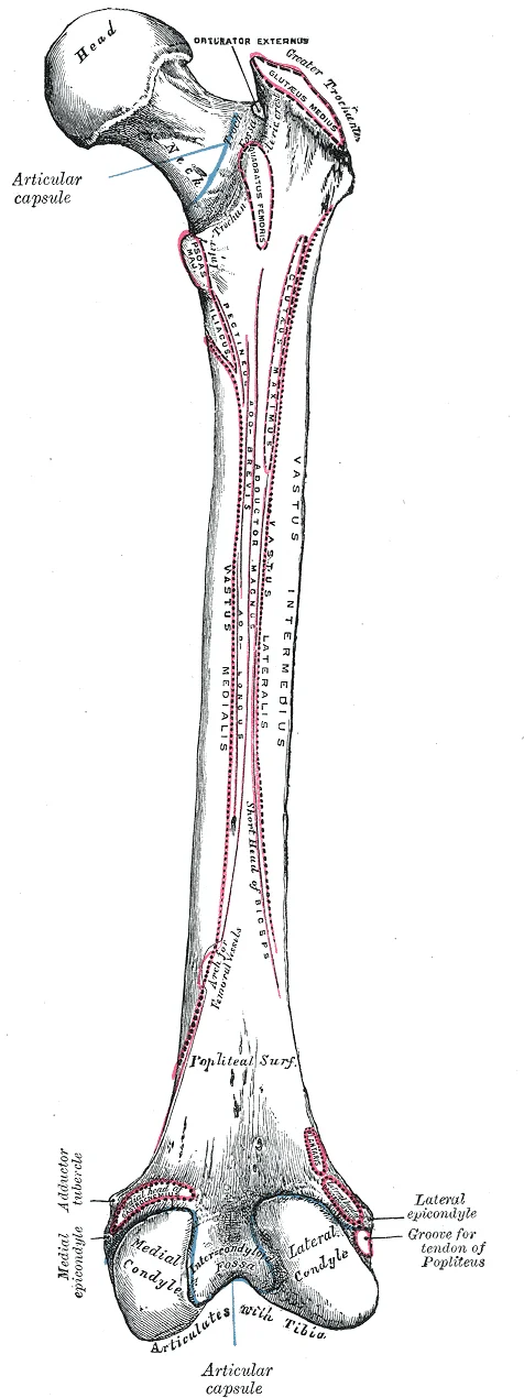
The distal femur has medial and lateral bony expansions. The lateral condyle articulates with the tibia to form the knee joint. The lateral epicondyle provides attachment for knee ligaments. Similarly, the medial condyle and medial epicondyle serve articulation and attachment functions respectively. The adductor tubercle is located at the superior margin of the medial epicondyle. Posteriorly, the condyles are separated by the intercondylar fossa. [1]
Figure 1Gray245
Long description
Right femur. Posterior surface. Labelled illustration from Wikimedia Commons, used under its open licence.

Figure 1: Right femur (posterior surface).
DefinitionHip Bone
The hip bone is formed by the fusion of the ilium, ischium, and pubis, which converge to form the acetabulum. [3]
The femur is the single bone of the thigh and is the longest and strongest bone in the body. It accounts for approximately one-quarter of a person’s total height. The proximal end features the head, which articulates with the acetabulum. This ligament carries an important artery supplying the femoral head. The neck of the femur is a narrowed region below the head and is a common site for [1]
The distal femur has medial and lateral bony expansions. The lateral condyle articulates with the tibia to form the knee joint. The lateral epicondyle provides attachment for knee ligaments. Similarly, the medial condyle and medial epicondyle serve articulation and attachment functions respectively. The adductor tubercle is located at the superior margin of the medial epicondyle. Posteriorly, the condyles are separated by the intercondylar fossa.
The lower limb can be mapped by a series of palpable landmarks that guide the identification of underlying bones. The most prominent of these are the femoral condyles, the tibial tuberosity, the malleoli, and the metatarsal heads.
The femur is the longest bone in the body and its distal end is characterised by a horseshoe‑shaped contour formed by the medial and lateral condyles and the patellar surface. Palpation of the femoral condyles is straightforward: the medial condyle lies just below the knee cap, while the lateral condyle is slightly more prominent on the outer side of the knee. [1]
The tibia, the medial bone of the leg, lies directly beneath the skin along the entire length of the medial leg, making it easily palpable. Its prominence is most noticeable at the tibial tuberosity, a bony ridge just below the knee that serves as the attachment point for the patellar tendon. [1]
At the ankle, the medial and lateral malleoli are the most obvious landmarks. The medial malleolus is the distal end of the tibia, while the lateral malleolus is the distal end of the fibula. These structures form the medial and lateral borders of the ankle joint and provide a reference for the position of the subtalar joint. [1]
The foot contains seven tarsal bones, five metatarsals and fourteen phalanges. [1]
View: lateral view of the lower limb in anatomical position. Orientation: anterior is to the left, posterior to the right. Labelled structures: femur, patella, tibia, fibula, tarsals (calcaneus, talus, navicular, cuboid, cuneiforms), metatarsals, phalanges. [1] The figure shows the femur’s condyles and patellar surface, the tibial crest and tuberosity, the malleoli, and the foot’s tarsal arrangement. The cuboid’s deep groove is highlighted, indicating the passage for the peroneus longus tendon. This arrangement allows the reader to visualise the spatial relationships between t [1]
The surface anatomy of the lower limb is therefore a practical guide for locating the femur, tibia, fibula, and foot bones, and for understanding how these structures relate to each other in the anatomical position. [1]
Figure 2Gray246
Long description
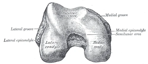
Lower extremity of right femur viewed from below.

Figure 2: Lower extremity of right femur viewed from below.
We begin proximally with the hip bone, or coxal bone, which forms the lateral and anterior aspects of the pelvic girdle.
Moving distally, we encounter the femur, the longest bone in the body. The neck of the femur is a critical region; fractures here are common in elderly patients due to osteoporosis and can compromise the blood supply to the femoral head, leading to avascular necrosis. The patella, or kneecap, sits within the tendon of the quadriceps femoris muscle[1]. It is the largest sesamoid bone in the body, a bone embedded within a tendon[1]. By articulating with the patellar surface of the femur, the patella prevents tendon rubbing and increases the leverage of the quadriceps[1]. It does not articulate with the tibia[1].
Figure 3Gray257
Long description
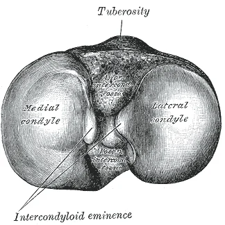
Upper surface of right tibia. Labelled illustration from Wikimedia Commons, used under its open licence.

Figure 3: Upper surface of right tibia.
Figure 4Gray252
Long description
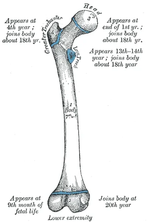
Plan of ossification of the femur. From five centers.

Figure 4: Plan of ossification of the femur (from five centers).
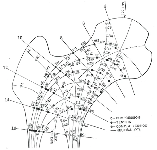
Figure 5Gray251
Long description
Intensity of the maximum tensile and compressive stresses in the upper femur. Computed for the load of 100 pounds on the right femur. Corresponds to the upper part of Fig. 250. (After Koch.)

Figure 5: Intensity of the maximum tensile and compressive stresses in the upper femur.
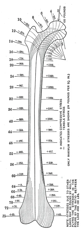
Figure 6Gray250
Long description
Diagram of the computed lines of maximum stress in the normal femur. The section numbers 2, 4, 6, 8, etc., show the positions of the transverse sections analyzed. The amounts of the maximum tensile and compressive stress at the various sections are given for a load of 100 pounds on the femur-head. F

Figure 6: Diagram of the computed lines of maximum stress in the normal femur.
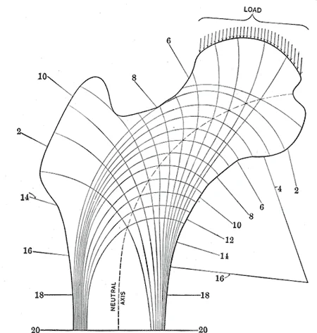
Figure 7Gray248
Long description
Diagram of the lines of stress in the upper femur, based upon the mathematical analysis of the right femur. These result from the combination of the different kinds of stresses at each point in the femur. (After Koch.)

Figure 7: Diagram of the lines of stress in the upper femur, based upon the mathematical analysis of the right femur.
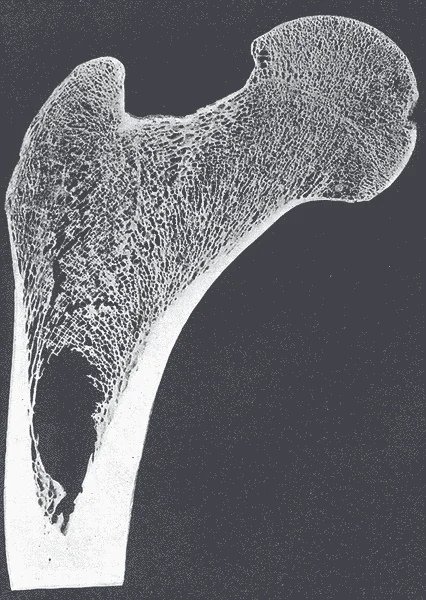
Figure 8Gray247
Long description
Frontal longitudinal midsection of upper femur.

Figure 8: Frontal longitudinal midsection of upper femur.
Figure 9Gray244
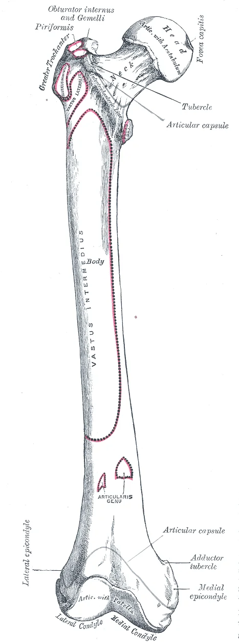
Long description
Right femur. Anterior surface. Labelled illustration from Wikimedia Commons, used under its open licence.

Figure 9: Right femur (anterior surface).
Figure 10Gray243
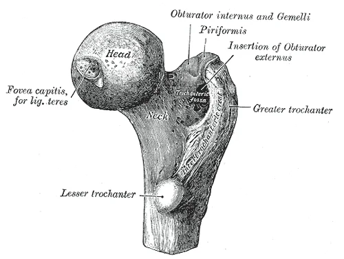
Long description
Upper extremity of right femur viewed from behind and above.

Figure 10: Upper extremity of right femur viewed from behind and above.
The leg comprises the tibia and fibula. The fibula is the thinner lateral bone. Its proximal end, the head of the fibula, articulates with the lateral tibial condyle[1]. The distal end forms the lateral malleolus, a palpable bony prominence on the lateral ankle[1]. The medial malleolus is part of the tibia. Together, these malleoli grip the talus.
Figure 11807 Pelvis
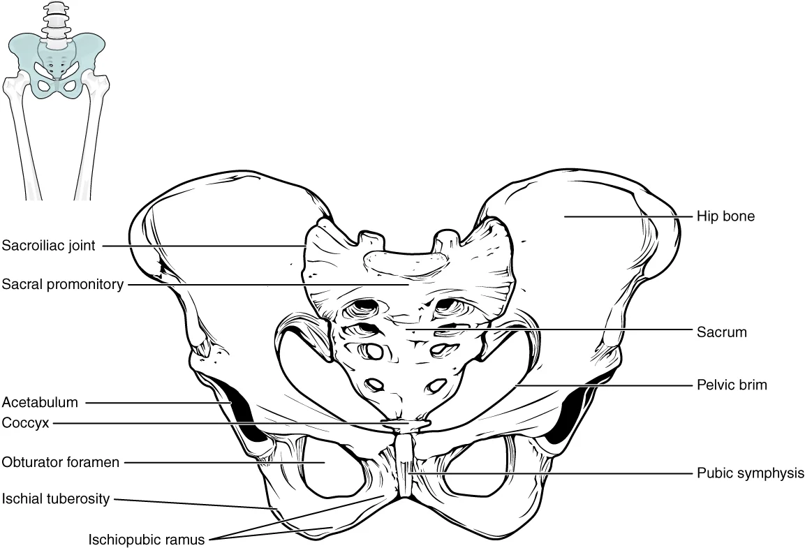
Long description
The pelvic girdle is formed by a single hip bone. The hip bone attaches the lower limb to the axial skeleton through its articulation with the sacrum. The right and left hip bones, plus the sacrum and the coccyx, together form the pelvis.

Figure 11: The pelvic girdle is formed by a single hip bone.
Figure 12Gray262
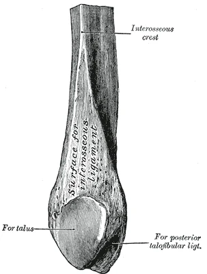
Long description
Lower extremity of right fibula. Medial aspect.

Figure 12: Lower extremity of right fibula (medial aspect).
Figure 13Gray260
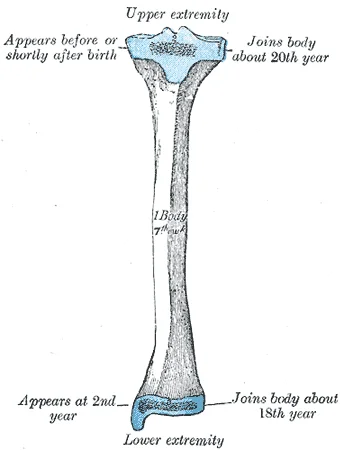
Long description
Plan of ossification of the tibia. From three centers.

Figure 13: Plan of ossification of the tibia (from three centers).
Figure 14Gray259
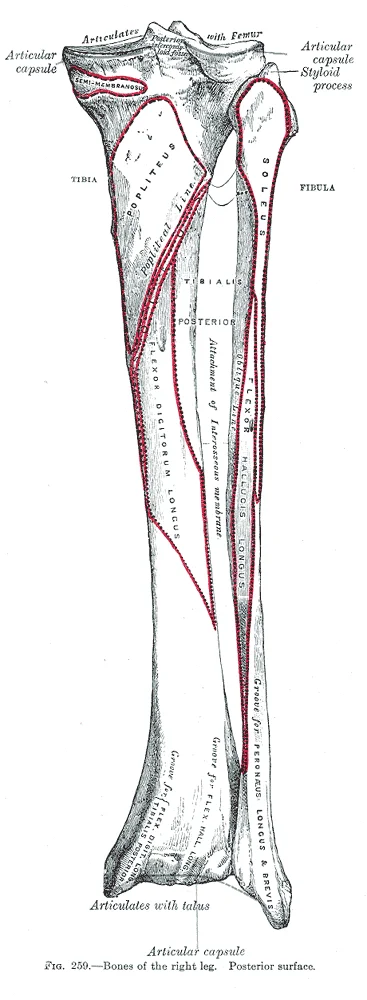
Long description
Bones of the right leg. Posterior surface.

Figure 14: Bones of the right leg (posterior surface).
Figure 15Gray258
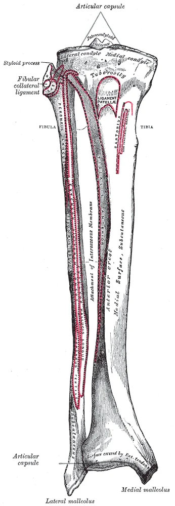
Long description
Bones of the right leg. Anterior surface.

Figure 15: Bones of the right leg (anterior surface).
The foot begins with the tarsals. The talus is the most superior tarsal bone, transmitting body weight from the tibia to the calcaneus[1]. The calcaneus is the largest foot bone, forming the heel[1]. It features the sustentaculum tali, a shelf supporting the talus medially[1]. Anterior to the tarsals lie the five metatarsals[1]. The first metatarsal is short and thick, while the second is the longest[1]. The base of the fifth metatarsal has a pro
Figure 16Gray265
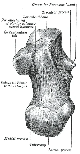
Long description
Left calcaneus, inferior surface. Labelled illustration from Wikimedia Commons, used under its open licence.

Figure 16: Left calcaneus, inferior surface.
Figure 17Gray264
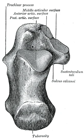
Long description
Left calcaneus, superior surface. Labelled illustration from Wikimedia Commons, used under its open licence.

Figure 17: Left calcaneus, superior surface.
The hip joint is a multiaxial ball-and-socket joint, offering extensive mobility but relying on strong ligaments for stability during weight bearing. The knee joint contains intracapsular cruciate ligaments that cross each other, forming an X-shape. The ankle joint is a hinge joint supported by strong lateral and medial ligaments. The medial side features the broad deltoid ligament, which resists excessive eversion. The lateral side has several smaller ligaments prone to injury in inversion sprains. Below the ankle, the subtalar joint lies between the talus and calcaneus, allowing for inversion and eversion. These tarsal joints are plane joints, contributing to the foot’s ability to adapt to uneven ground. [2]
The lower limb joints are specialized synovial joints, with movements determined by their structural classification. The hip joint’s range of motion is restricted by strong ligaments, conferring stability. The ankle joint allows dorsiflexion and plantar flexion while preventing abnormal side-to-side movements. The foot’s arches absorb shock, flattening under weight and rebounding to provide spring. During running, forces on the foot can reach 2.5 times body weight. Prolonged stretching of ligaments supporting the longitudinal arches can lead to pes planus, or flat foot. This condition involves the depression of the medial longitudinal arch. [2] [1]
The sacroiliac joint is largely immobile, supported by anterior and posterior sacroiliac ligaments. These structures ensure the sacrum can carry the weight of the body effectively. The joints between the tarsal bones are all plane joints, facilitating small motions that combine to produce inversion and eversion. The talocrural joint is supported by ligaments extending from the malleoli to the talus and calcaneus. These ligaments anchor the joint, ensuring proper alignment during gait. [3] [2] [1]
Clinical pearlClinical Pearl
Inversion ankle sprains commonly injure the lateral ligaments, which are smaller and more susceptible to trauma than the robust deltoid ligament on the medial side. [2]
| Structure | Location | Function |
|---|---|---|
| Deltoid Ligament | Medial ankle | Resists excessive eversion |
| Posterior Cruciate Ligament | Knee | Supports knee when flexed and weight bearing |
| Sacrospinous Ligament | Pelvis | Runs from sacrum to ischial spine |
| Sacrotuberous Ligament | Pelvis | Runs from sacrum to ischial tuberosity |
Understanding these boundaries is essential for localising pathology and planning surgical approaches.
DefinitionFascia
A dense connective tissue layer that surrounds muscles, bones, nerves, and blood vessels, providing structural support and compartmentalisation.
The lower limb is divided into distinct compartments by intermuscular septa that extend from the deep fascia to the bone. These compartments contain muscles with similar actions and neurovascular supplies. In the thigh, the femur is surrounded by the fascia lata, which is thickened laterally to form the iliotibial tract. This tract extends from the iliac tubercle to the lateral condyle of the tibia, providing lateral stability. [1]
| Compartment | Primary Muscles | Nerve Supply |
|---|---|---|
| Anterior | Quadriceps femoris | Femoral nerve |
| Medial | Adductor group | Obturator nerve |
| Posterior | Hamstrings | Sciatic nerve |
The anterior compartment contains the quadriceps femoris, which extends the knee. The medial compartment houses the adductor muscles, which primarily adduct the hip. The posterior compartment contains the hamstrings, which flex the knee and extend the hip. Each compartment is enclosed by its own fascial boundary, ensuring that swelling or infection remains contained within specific anatomical regions.
In the leg, the crural fascia encloses the muscles into anterior, lateral, and posterior compartments. The anterior compartment contains the tibialis anterior, which dorsiflexes the foot. The lateral compartment contains the peroneus longus and brevis, which evert the foot. The deep layer contains the tibialis posterior, flexor digitorum longus, and flexor hallucis longus.
Exam trapCompartment Syndrome
Increased pressure within a closed fascial compartment can compromise blood flow, leading to muscle and nerve damage. This is a surgical emergency requiring immediate fasciotomy.
The neurovascular bundles run within or between these compartments. The sciatic nerve runs through the posterior compartment. In the leg, the anterior tibial artery and deep fibular nerve supply the anterior compartment. The posterior tibial artery and tibial nerve supply the posterior compartment. Knowledge of these relationships is critical for interpreting clinical signs and managing trauma.
The lower limb musculature is organised into compartments, each with distinct neurovascular supplies. The anterior compartment of the thigh contains the quadriceps femoris, a powerful extensor of the leg at the knee. This muscle group includes the rectus femoris, vastus lateralis, vastus medialis, and vastus intermedius. They converge into the quadriceps tendon, which incorporates the patella, a sesamoid bone located within the tendon. The patella articulates with the patellar surface on the anterior side of the distal femur, thereby protecting the muscle tendon from rubbing against the femur. Continuing from the patella to the anterior tibia just below the knee is the patellar ligament. Acting via the patella and patellar ligament, the quadriceps femoris is a powerful muscle that acts to extend the leg at the knee. [1] [2]
The lateral compartment of the leg contains the fibularis longus and fibularis brevis. These muscles evert the foot and plantarflex the ankle. The fibula is the slender bone located on the lateral side of the leg. It serves primarily for muscle attachments and thus is largely surrounded by muscles. Only the proximal and distal ends of the fibula can be palpated. The head of the fibula forms the proximal end and articulates with the underside of the lateral condyl [1]
The posterior compartment of the thigh contains the hamstrings, which flex the knee and extend the hip. The medial compartment of the thigh contains the adductors, which adduct the thigh.
| Compartment | Muscle | Origin | Insertion | Action | Nerve Supply |
|---|---|---|---|---|---|
| Anterior Thigh | Quadriceps Femoris | Ilium and Femur | Tibia via Patellar Ligament | Extend Leg | Femoral Nerve |
| Lateral Leg | Fibularis Longus | Fibula | Medial Cuneiform | Evert Foot | Superficial Fibular Nerve [1] |
| Posterior Thigh | Biceps Femoris | Ischial Tuberosity | Fibular Head | Flex Knee | Sciatic Nerve |
The ligament of the head of the femur (ligamentum teres) spans between the acetabulum and femoral head. This intracapsular ligament is normally slack and does not provide any significant joint support, but it does provide a pathway for an important artery that supplies the head of the femur. This arterial supply is critical for the viability of the femoral head, particularly in cases of fracture. [2]
The arterial supply to the lower limb originates from the abdominal aorta, which bifurcates into the common iliac arteries. The external iliac artery continues into the thigh, becoming the femoral artery as it passes beneath the inguinal ligament. This vessel is the primary source of blood for the lower extremity. It travels through the femoral triangle, where it gives off several important branches. It supplies the muscles of the anterior and medial compartments of the thigh. [1]
DefinitionFemoral Triangle
A triangular space in the upper thigh bounded by the inguinal ligament, sartorius, and adductor longus, containing the femoral artery, vein, and nerve.
As the femoral artery descends, it enters the adductor hiatus, a gap in the adductor magnus muscle. Here, it becomes the popliteal artery. The popliteal artery lies deep in the popliteal fossa, behind the knee joint. It gives off genicular branches that anastomose around the knee, ensuring blood supply during flexion and extension. These branches include the superior medial and lateral genicular arteries, as well as the inferior medial and lateral genicular arteries.
Arterial Pathway
- Abdominal AortaBifurcates into common iliacs
- Question: External Iliac Artery
Passes under inguinal ligament
- Femoral Artery
Continues
- Common Iliac
The popliteal artery divides into the anterior and posterior tibial arteries. The anterior tibial artery passes through the interosseous membrane to the anterior compartment of the leg. It supplies the extensor muscles of the leg. The posterior tibial artery runs down the posterior compartment, supplying the flexor muscles. It gives off the peroneal artery, which supplies the lateral compartment.
The labels indicate the origin and course of each vessel.
| Vessel | Origin | Course |
|---|---|---|
| Femoral | External Iliac | Anterior thigh |
| Popliteal | Femoral | Popliteal fossa |
| Anterior Tibial | Popliteal | Anterior leg |
| Posterior Tibial | Popliteal | Posterior leg |
The dorsalis pedis artery is the continuation of the anterior tibial artery. It runs over the dorsum of the foot. The plantar arteries arise from the posterior tibial artery. They supply the sole of the foot. These vessels form an extensive network, ensuring adequate perfusion to the bones and soft tissues of the lower limb.
The venous drainage of the lower limb is organised into superficial and deep systems, which communicate via perforating veins. Understanding this architecture is fundamental for clinical assessments of venous insufficiency and thrombosis.
The superficial system lies within the subcutaneous tissue and does not accompany arteries. It consists primarily of the great saphenous vein and the small saphenous vein.
DefinitionGreat Saphenous Vein
The longest vein in the body, running from the foot to the groin.
The great saphenous vein originates from the medial end of the dorsal venous arch of the foot. It ascends anterior to the medial malleolus, then travels up the medial aspect of the leg and thigh. In the thigh, it lies superficial to the fascia lata. [1]
Exam trapSaphenous Nerve Relation
The saphenous nerve runs with the great saphenous vein in the leg but diverges in the thigh.
The small saphenous vein arises from the lateral end of the dorsal venous arch. It ascends posterior to the lateral malleolus and runs up the posterior aspect of the leg, between the two heads of the gastrocnemius muscle. It typically terminates by draining into the popliteal vein within the popliteal fossa. [1]
The deep venous system accompanies the major arteries of the lower limb. These veins are valve-rich and rely on skeletal muscle pumps for return flow.
The venae comitantes of the anterior and posterior tibial arteries form the proximal part of the deep system. These pairs of veins unite to form the popliteal vein behind the knee. [1]
| Structure | Origin | Termination | Key Relation |
|---|---|---|---|
| Great Saphenous Vein | Medial dorsal arch | Femoral vein | Anterior to medial malleolus |
| Small Saphenous Vein | Lateral dorsal arch | Popliteal vein | Posterior to lateral malleolus |
| Popliteal Vein | Union of tibial veins | Femoral vein | Deep to popliteal fascia |
| Femoral Vein | Adductor hiatus | Inguinal ligament | Medial to femoral artery |
Perforating veins connect the superficial and deep systems. They contain one-way valves that prevent retrograde flow from the high-pressure deep system to the low-pressure superficial system. Incompetence of these valves leads to varicose veins.
The nervous supply to the lower limb is divided into motor and cutaneous components, originating primarily from the lumbar and sacral plexuses. Understanding this supply requires tracing the path of major nerves relative to the bony landmarks of the pelvis and femur.
The sciatic nerve is a major nerve in the body, emerging from the greater sciatic notch below the piriformis muscle. It descends along the posterior thigh, lying deep to the gluteus maximus and the long head of the biceps femoris. It typically divides into the tibial and common fibular nerves near the apex of the popliteal fossa.
DefinitionSciatic Nerve
The largest nerve in the body, emerging from the greater sciatic notch and dividing into tibial and common fibular branches. [3]
The following table details the motor and cutaneous supply of the key nerves.
| Nerve | Motor Supply (Muscles) | Cutaneous Supply (Skin Area) |
|---|---|---|
| Femoral | Iliopsoas, Sartorius, Quadriceps femoris | Anterior thigh, medial leg (via saphenous) |
| Obturator | Adductor longus, Adductor brevis, Adductor magnus | Medial thigh |
| Sciatic (Tibial) | Hamstrings, All muscles below knee | Posterior thigh, sole of foot |
| Sciatic (Common Fibular) | Biceps femoris, All muscles of lateral/anterior leg | Lateral leg, dorsum of foot |
Lesions of these nerves result in specific deficits. Damage to the femoral nerve causes weakness in knee extension and loss of sensation over the anterior thigh. The common fibular nerve is susceptible to compression at the fibular neck, resulting in foot drop. The tibial nerve lesion affects plantar flexion and sensation of the sole.
| Nerve | Primary Deficit | Sensory Loss |
|---|---|---|
| Femoral | Weak knee extension | Anterior thigh |
| Common Fibular | Foot drop | Lateral leg, dorsum foot |
| Tibial | Weak plantar flexion | Sole of foot |
The relationship between nerves and bones is critical for surgical approaches. The sciatic nerve lies close to the ischial tuberosity. Knowledge of these relations prevents iatrogenic injury during hip and knee procedures.
The lymphatic drainage of the lower limb follows a predictable pattern, moving from superficial tissues to deep structures before entering the pelvic cavity. This system is crucial for immune surveillance and fluid balance. Understanding the pathways is essential for interpreting clinical signs such as lymphoedema or metastatic spread.
Deep inguinal nodes lie medial to the femoral vein and receive efferents from the superficial nodes.
DefinitionSuperficial Inguinal Nodes
A chain of lymph nodes located in the subcutaneous tissue of the upper medial thigh, receiving drainage from the lower limb and lower abdominal wall.
The superficial lymphatics accompany the great saphenous vein. They drain the skin of the foot, leg, and anterior thigh. Infections in the foot often present with swollen inguinal nodes. The deep lymphatics follow the major arteries. They drain the muscles, joints, and bones of the lower limb. The popliteal nodes are situated behind the knee. They receive lymph from the lateral side of the foot and the posterior calf. Efferents from the popliteal nodes join the deep inguinal nodes.
| Structure | Primary Drainage | Destination Nodes |
|---|---|---|
| Foot and Leg | Superficial and Deep | Popliteal and Superficial Inguinal |
| Thigh | Superficial and Deep | Superficial and Deep Inguinal |
| Buttock | Superficial | Superficial Inguinal |
| Perineum | Superficial | Superficial Inguinal |
Clinical examination of the inguinal region is vital. Palpation of the superficial inguinal nodes can detect infection or malignancy. The nodes are soft, mobile, and non-tender in health. Enlargement suggests pathology in the drainage area. The deep inguinal nodes are deeper and harder to palpate. They are assessed via imaging if clinically indicated.
Clinical pearlClinical Pearl
Always examine the inguinal nodes when a patient presents with an infection or wound on the lower limb.
The lymphatic vessels have valves to prevent backflow. They rely on muscle contraction and arterial pulsation for propulsion. Obstruction of these vessels leads to lymphoedema. This condition causes swelling and skin thickening. Early intervention is key to managing symptoms. The anatomical knowledge of these pathways guides surgical and diagnostic procedures.
The pubis forms the anterior portion of the hip bone. Located superiorly on the pubic body is a small bump called the pubic tubercle. These bony landmarks define the boundaries of the obturator foramen and the acetabulum. [3]
The acetabulum, formed by the fusion of the ilium, ischium, and pubis, articulates with the head of the femur. This ball-and-socket joint allows for a wide range of motion. The femoral head is covered with articular cartilage, except for the fovea capitis, where the ligament of the head of the femur attaches. This ligament transmits small blood vessels to the femoral head.
Anterior to the hip joint lies the femoral triangle. Its boundaries are the inguinal ligament superiorly, the sartorius muscle laterally, and the adductor longus muscle medially. The contents of the femoral triangle, from lateral to medial, are the femoral nerve, femoral artery, femoral vein, and lymphatics (NAVEL).
Posterior to the hip joint is the gluteal region. The greater sciatic foramen allows passage of the sciatic nerve, superior and inferior gluteal vessels and nerves, and the piriformis muscle. The sciatic nerve is the largest nerve in the body, emerging from the pelvis below the piriformis muscle. It divides into the tibial and common fibular nerves in the posterior thigh. [3]
The ischium forms the posteroinferior portion of the hip bone. The ischial tuberosity is the roughened area where the hamstrings originate. It bears weight when sitting. The ischial spine separates the greater and lesser sciatic notches.
DefinitionHip Bone
The hip bone is formed by the fusion of the ilium, ischium, and pubis.
| Structure | Location | Clinical Relevance |
|---|---|---|
| Pubic Tubercle | Superior pubic ramus | Attachment of inguinal ligament |
| Pectineal Line | Superior margin of superior pubic ramus | Origin of pectineus muscle |
| Ischial Tuberosity | Posteroinferior ischium | Weight bearing in sitting |
| Acetabulum | Lateral hip bone | Articulation with femoral head |
Understanding the skeletal architecture of the lower limb requires proficiency in interpreting various imaging modalities. Each technique highlights different aspects of bone integrity, joint congruity, and surrounding soft tissue relationships.
Plain radiography remains the primary initial investigation for suspected lower limb trauma. It provides a clear view of bone density, alignment, and cortical continuity. In a standard anteroposterior view, the femur, tibia, and fibula are assessed for fractures or degenerative changes. The hip joint is evaluated for the integrity of the femoral head and acetabulum. The knee joint assessment focuses on the patellofemoral relationship and tibiofemoral alignment. Ankle radiographs typically include anteroposterior, lateral, and mortise views to detect subtle fractures of the malleoli. The foot series includes dorsoplantar and lateral views to identify metatarsal or tarsal fractures. Radiographic signs of pathology include cortical disruption, joint space narrowing, or osteophyte formation. These findings guide immediate management decisions, such as the need for immobilisation or surgical intervention. [1]
Computed tomography offers superior cross-sectional detail and is invaluable for complex fractures. It allows for three-dimensional reconstruction, which is particularly useful for intra-articular fractures of the ankle, knee, or hip. CT scans can delineate fracture lines that are not visible on plain X-rays. They are essential for pre-operative planning in pelvic fractures or tibial plateau fractures. The high contrast resolution helps distinguish between bone fragments and soft tissue interposition. In the context of the lower limb, CT is often used to assess the extent of comminution and to plan internal fixation strategies. It provides precise measurements of bone loss and alignment, ensuring accurate reduction during surgery.
Magnetic resonance imaging is the gold standard for evaluating soft tissue structures and bone marrow pathology. It is particularly sensitive for detecting stress fractures, which may be occult on X-ray. MRI visualises ligaments, tendons, and menisci with high clarity. In the knee, it is used to assess cruciate ligament tears and meniscal injuries. In the hip, it can detect avascular necrosis or labral tears. Bone marrow oedema appears as high signal intensity on fluid-sensitive sequences, indicating acute injury or inflammation. MRI is also crucial for diagnosing osteomyelitis, where it can identify early bone infection before structural changes are visible on X-ray.
Ultrasound is dynamic and excellent for evaluating superficial structures. It is used to assess tendons, such as the Achilles tendon, for tears or tendinopathy. It can visualise joint effusions and guide aspiration or injection. In the lower limb, ultrasound is useful for detecting deep vein thrombosis, although this is a vascular rather than osteological assessment. It provides real-time imaging of muscle tears and can assess the integrity of the patellar tendon. While it does not penetrate bone well, it is invaluable for surrounding soft tissue pathology.
The tibia and fibula extend distally to form the ankle joint with the talus. The talus sits within the mortise formed by the tibia and fibula. The calcaneus is the large bone forming the heel.
The femur is the single bone of the thigh. Its rounded head articulates with the acetabulum of the hip bone to form the hip joint. The head has the fovea capitis for attachment of the ligament of the head of the femur. The narrow neck joins inferiorly with the greater and lesser trochanters. Passing between these bony expansions are the intertrochanteric line on the anterior femur and the larger intertrochanteric crest on the posterior femur. On the posterior shaft of the femur is the gluteal tuberosity proximally and the linea aspera in the mid-shaft region. The expanded distal end consists of three articulating surfaces: the medial and lateral condyles, and the patellar surface. The outside margins of the condyles are the medial and lateral epicondyles. The adductor tubercle is on the superior aspect of the medial epicondyle. [1]
The knee joint has multiple ligaments that provide support, particularly in the extended position. Outside of the articular capsule, located at the sides of the knee, are two extrinsic ligaments. [2]
The knee is well constructed for weight bearing in its extended position, but is vulnerable to injuries associated with hyperextension, twisting, or blows to the medial or lateral side of the joint, particularly while weight bearing. The knee includes three articulations. The femoropatellar joint is between the patella and distal femur. The patella, a sesamoid bone incorporated into the tendon of the quadriceps femoris muscle of the anterior thigh, serves to protect this tendon from rubbing against the distal femur during knee movements. The medial and lateral tibiofemoral joints, between the condyles of the femur and condyles of the tibia, are modified hinge joints that allow for knee extension and flexion. During these movements, the condyles of the femur both roll and glide over the surface of the tibia. As the knee comes into full extension, a slight medial rotation of the femur serves to “lock” the knee into its most stable, weight-bearing position. When the knee is flexed, some rotation of the leg is available. [1] [2]
The pubis forms the anterior portion of the hip bone. The body of the pubis articulates with the pubis of the opposite hip bone at the pubic symphysis. The superior margin of the pubic body has the pubic tubercle. The pubis is joined to the ilium by the superior pubic ramus, the superior surface of which forms the pectineal line. The inferior pubic ramus projects inferiorly and laterally. The pubic arch is formed by the pubic symphysis, the bodies of the adjacent pubic bones, and the two inferior pubic rami. The inferior pubic ramus joins the ischial ramus to form the ischiopubic ramus. The subpubic angle is formed by the medial convergence of the right and left ischiopubic rami. [3]
DefinitionPhalanx count in the toes
The toes contain a total of 14 phalanx bones (phalanges), arranged in a similar manner as the phalanges of the fingers. The toes are numbered 1–5, starting with the big toe (hallux). The remaining toes all have proximal, middle, and distal phalanges. A joint between adjacent phalanx bones is called an interphalangeal joint. [1]
Clinical pearlKnee ligaments and menisci
Two extrinsic ligaments, the tibial collateral ligament on the medial side and the fibular collateral ligament on the lateral side, serve to resist hyperextension or rotation of the extended knee joint. Two intracapsular ligaments, the anterior cruciate ligament and posterior cruciate ligament, span between the tibia and the inner aspects of the femoral condyles. The medial and lateral menisci, located between the femoral and tibial condyles, are articular discs that provide padding and improve the fit between the bones. [3]
Clinical pearlPelvic cavity division
The space enclosed by the bony pelvis is divided into two regions. The broad, superior region, defined laterally by the large, fan‑like portion of the upper hip bone, is called the greater pelvis (greater pelvic cavity; false pelvis). This broad area is occupied by portions of the small and large intestines, and because it is more closely associated with the abdominal cavity, it is sometimes referred to as the false pelvis. More inferiorly, the narrow, rounded space of the lesser pelvis (lesser [3]
Clinical pearlSexual dimorphism of the pelvis
The female sacrum is wider, shorter, and less curved, and the sacral promontory projects less into the pelvic cavity, thus giving the female pelvic inlet (pelvic brim) a more rounded or oval shape compared to males. The lesser pelvic cavity of females is also wider and more shallow than the narrower, deeper, and tapering lesser pelvis of males. Because of the obvious differences between female and male hip bones, this is the one bone of the body that allows for the most accurate sex determination. [3]
Clinical pearlSurgical neck of the humerus
The surgical neck is a common site of arm fractures. As its name indicates, it is the site of attachment for the deltoid muscle. [4]
Clinical pearlCarpal bone arrangement
The carpal bones are united into two rows of bones. The scaphoid, lunate, and triquetrum bones contribute to the formation of the radiocarpal joint. [4]
Exam trapMis‑identifying the tibial collateral ligament
Students often confuse the tibial collateral ligament with the medial collateral ligament. The tibial collateral ligament is the correct anatomical term for the medial side of the knee.
Exam trapForgetting the posterior cruciate ligament’s role
It is frequently overlooked in exam questions that focus on the anterior cruciate ligament.
Exam trapAssuming all toes have three phalanges
Only the big toe has two phalanges; the other toes each have three. This is a common exam trap.
Exam trapConfusing greater and lesser pelvis
The greater pelvis is the false pelvis, while the lesser pelvis is the true pelvis. Mixing these terms can lead to incorrect answers about pelvic organ location.
Exam trapOverlooking the deltoid tuberosity
The deltoid tuberosity is the attachment site for the deltoid muscle, not the surgical neck itself. Students sometimes mistake the two.
Exam trapMisreading the carpal tunnel composition
The carpal tunnel is formed by the carpal bones and the flexor retinaculum, not by the carpal bones alone. [4]
Students frequently confuse the structural stability of the lower limb joints, particularly between the hip and the shoulder. A critical distinction is that the hip joint is a ball-andsocket joint whose motions are more restricted than at the shoulder to provide greater stability during weight bearing. In contrast, the shoulder joint is called the glenohumeral joint and has the largest range of motion of any joint in the body. This freedom of movement is due to the lack of structural support, meaning the enhanced mobility is offset by a loss of stability. Students often overlook that the hip joint is the articulation between the head of the femur and the acetabulum of the hip bone. The acetabulum is deepened by the acetabular labrum, which contributes to this stability. Furthermore, the iliofemoral, pubofemoral, and ischiofemoral ligaments strongly support the hip joint in the upright, standing position. [2]
Another common error involves the ankle joint's mobility. The ankle is formed by the talocrural joint, which consists of the articulations between the talus bone of the foot and the distal ends of the tibia and fibula of the leg. It is a uniaxial hinge joint that allows only for dorsiflexion and plantar flexion of the foot. Students may incorrectly assume side-to-side motion is possible at the talocrural joint. However, the sides of the talus are firmly held in position by the articulations with the medial malleolus of the tibia and the lateral malleolus of the fibula, which prevent any side-to-side motion of the talus. Gliding motions at the subtalar and intertarsal joints of the foot allow for inversion/eversion of the foot. The ankle joint is supported on the medial side by the deltoid ligament, which prevents side-to-side motions of the talus at the talocrural joint and resists excessive eversion of the foot. [2]
Exam trapAnkle Stability
The talocrural joint prevents side-to-side motion; inversion/eversion occurs at the subtalar and intertarsal joints.
DefinitionHip Joint
The hip joint is a ball-and-socket joint whose motions are more restricted than at the shoulder to provide greater stability during weight bearing. [2]
Knowledge of lower limb osteology is essential for interpreting imaging and understanding clinical presentations. On imaging, the longitudinal arches run down the length of the foot. The lateral longitudinal arch is relatively flat, whereas the medial longitudinal arch is larger (taller). These arches are formed by the tarsal bones posteriorly and the metatarsal bones anteriorly. The talus bone, which receives the weight of the body, is located at the top of the longitudinal arches. Body weight [1]
In the operating theatre, understanding pelvic differences is vital. The differences between the adult female and male pelvis relate to function and body size. In general, the bones of the male pelvis are thicker and heavier, adapted for support of the male’s heavier physical build and stronger muscles. Because the female pelvis is adapted for childbirth, it is wider than the male pelvis. Because of this increased pelvic width, the subpubic angle is larger in females (greater than 80 degrees) than it is in males (less than 70 degrees). The ischial tuberosities of females are also farther apart, which increases the size of the pelvic outlet. [3]
At the bedside, clinicians assess joint integrity. The elbow is a uniaxial hinge joint that allows for flexion/extension of the forearm. It includes the humeroulnar joint and the humeroradial joint. The medial elbow is supported by the ulnar collateral ligament and the radial collateral ligament supports the lateral side. These ligaments prevent side-to-side movements and resist hyperextension of the elbow. The proximal radioulnar joint is a pivot joint that allows for rotation of the radius during pronation/supination of the forearm. The annular ligament surrounds the head of the radius to hold it in place at this joint. [2]
Ankle Assessment
- Assess Ankle InjuryPatient presents with pain
- Question: Can patient dorsiflex/plantarflex?
Yes
- Check LigamentsTest deltoid and lateral ligaments
No
- Suspect FractureImaging required
NotePelvic Anatomy
The subpubic angle is larger in females (greater than 80 degrees) than it is in males (less than 70 degrees).
The hip joint is a ball-and-socket joint whose motions are more restricted than at the shoulder to provide greater stability during weight bearing. The ankle is formed by the talocrural joint, which consists of the articulations between the talus bone of the foot and the distal ends of the tibia and fibula of the leg. The lateral longitudinal arch is relatively flat, whereas the medial longitudinal arch is larger (taller). The subpubic angle is larger in females (greater than 80 degrees) than it is in males (less than 70 degrees). The elbow is a uniaxial hinge joint that allows for flexion/extension of the forearm. The talus bone, which receives the weight of the body, is located at the top of the longitudinal arches. The iliofemoral, pubofemoral, and ischiofemoral ligaments strongly support the hip joint in the upright, standing position. The ankle joint is supported on the medial side by the deltoid ligament, which prevents side-to-side motions of the talus at the talocrural joint and resists excessive eversion of the foot.
Answer each one in your head first, then open the answer.
Which joint has the largest range of motion of any joint in the body?
Show answerHide answer
The shoulder joint is called the glenohumeral joint.What bone provides posterior support for the longitudinal arches?
Show answerHide answer
Posteriorly, this support is provided by the calcaneus bone.Which ligament supports the lateral side of the elbow?
Show answerHide answer
The radial collateral ligament supports the lateral side.What is the subpubic angle in males?
Show answerHide answer
The subpubic angle is larger in females (greater than 80 degrees) than it is in males (less than 70 degrees).What type of joint is the talocrural joint?
Show answerHide answer
The ankle is thus a uniaxial hinge joint that allows only for dorsiflexion and plantar flexion of the foot.
- Betts JG, Young KA, Wise JA, et al. Bones of the Lower Limb. OpenStax, Anatomy and Physiology 2e (CC BY-NC-SA 4.0). Accessed .
- Betts JG, Young KA, Wise JA, et al. Anatomy of Selected Synovial Joints. OpenStax, Anatomy and Physiology 2e (CC BY-NC-SA 4.0). Accessed .
- Betts JG, Young KA, Wise JA, et al. The Pelvic Girdle and Pelvis. OpenStax, Anatomy and Physiology 2e (CC BY-NC-SA 4.0). Accessed .
- Betts JG, Young KA, Wise JA, et al. Bones of the Upper Limb. OpenStax, Anatomy and Physiology 2e (CC BY-NC-SA 4.0). Accessed .
Figure credits
- Figure 1: Henry Vandyke Carter, via Wikimedia Commons, Public domain, Public domain.
- Figure 2: Henry Vandyke Carter, via Wikimedia Commons, Public domain, Public domain.
- Figure 3: Henry Vandyke Carter, via Wikimedia Commons, Public domain, Public domain.
- Figure 4: Henry Vandyke Carter, via Wikimedia Commons, Public domain, Public domain.
- Figure 5: Henry Vandyke Carter, via Wikimedia Commons, Public domain, Public domain.
- Figure 6: Henry Vandyke Carter, via Wikimedia Commons, Public domain, Public domain.
- Figure 7: Henry Vandyke Carter, via Wikimedia Commons, Public domain, Public domain.
- Figure 8: Henry Vandyke Carter, via Wikimedia Commons, Public domain, Public domain.
- Figure 9: Henry Vandyke Carter, via Wikimedia Commons, Public domain, Public domain.
- Figure 10: Henry Vandyke Carter, via Wikimedia Commons, Public domain, Public domain.
- Figure 11: OpenStax College, via Wikimedia Commons, CC BY 3.0, CC BY 3.0.
- Figure 12: Henry Vandyke Carter, via Wikimedia Commons, Public domain, Public domain.
- Figure 13: Henry Vandyke Carter, via Wikimedia Commons, Public domain, Public domain.
- Figure 14: Henry Vandyke Carter, via Wikimedia Commons, Public domain, Public domain.
- Figure 15: Henry Vandyke Carter, via Wikimedia Commons, Public domain, Public domain.
- Figure 16: Henry Vandyke Carter, via Wikimedia Commons, Public domain, Public domain.
- Figure 17: Henry Vandyke Carter, via Wikimedia Commons, Public domain, Public domain.
Educational content for medical students and doctors. It is not a substitute for clinical judgement or local guidelines.