Applied and clinical anatomy of the upper limb
Clinical anatomy of the upper limb bones, joints, and common pathologies.
The upper limb is anatomically divided into three distinct regions: the arm, forearm, and hand. The arm lies between the shoulder and elbow joints, while the forearm extends from the elbow to the wrist. The hand is located distal to the wrist and contains thirty bones in total in each upper limb. The humerus is the single bone of the upper arm. At its proximal end, the head of the humerus articulates with the glenoid cavity of the scapula to form the glenohumeral joint. The anatomical neck marks [3]
DefinitionSurgical Neck
The surgical neck is located at the base of the expanded proximal humerus where it joins the narrow shaft. [3]
This region is a common site for arm fractures. The deltoid tuberosity is a roughened, V-shaped region on the lateral side of the humerus shaft. It serves as the attachment site for the deltoid muscle. Injuries to the shoulder joint are common due to repetitive use. Activities such as throwing, swimming, or racquet sports may lead to inflammation of the bursae or muscle tendons. Tears of the glenoid labrum or degeneration of the rotator cuff can also occur. Because the humeral head is strongly supported by muscles and ligaments, most dislocations occur in an inferior direction. This often happens when force is applied to the humerus during full abduction. [3] [2]
Exam trapDislocation Direction
Most humeral dislocations are inferior, not anterior, despite common misconceptions.
Inflammatory responses to shoulder injury can lead to scar tissue formation between the articular capsule and surrounding structures. This reduces shoulder mobility and is known as adhesive capsulitis or frozen shoulder. The forearm contains two bones: the ulna medially and the radius laterally. The elbow joint is formed by the articulation between the trochlea of the humerus and the trochlear notch of the ulna. It also involves the articulation between the capitulum of the humerus and the head This area is the site of articulation between the proximal radius and the ulna, forming the proximal radioulnar joint. [2][3]
Surface anatomy of the upper limb is essential for clinical examination, venous access and localisation of neuro‑vascular structures. The key palpable landmarks are the bony prominences, muscle belly insertions and superficial vessels that can be felt or seen on the skin.
The most reliable bony landmarks are the greater tubercle and lesser tubercle of the humerus, which lie just inferior to the acromion on the lateral aspect of the shoulder. The deltoid tuberosity on the lateral shaft of the humerus is a roughened area that can be felt when the arm is abducted. At the elbow, the medial epicondyle and lateral epicondyle form the medial and lateral borders of the elbow joint; the ulnar styloid projects posteriorly on the medial side of the forearm. The radial head is palpable on the lateral side of the elbow, while the ulnar head lies on the medial side. [3]
The biceps brachii muscle belly is prominent on the anterior aspect of the upper arm and can be felt when the arm is flexed. The brachialis lies deep to the biceps and is not usually palpable. The triceps brachii forms a thick posterior mass that can be felt when the arm is extended.
On the forearm, the pronator teres and supinator muscles are superficial and can be felt when the forearm is pronated or supinated. The flexor carpi radialis and flexor carpi ulnaris tendons are palpable on the volar surface of the wrist.
The cephalic vein begins in the antebrachium and runs superficially along the lateral side of the biceps brachii muscle, draining into the axillary vein. It is extremely superficial and can be seen in individuals with good muscle tone and minimal subcutaneous fat. The radial artery runs along the lateral side of the forearm, while the ulnar artery lies medial; both give off smaller branches until they reach the wrist, where they fuse to form the superficial and deep palmar arches that supply the hand and digits. [1]
The median nerve passes between the brachialis and pronator teres in the proximal forearm, then travels into the carpal tunnel at the wrist. The ulnar nerve runs deep to the flexor carpi ulnaris and passes behind the medial epicondyle before entering the hand. The radial nerve emerges from the axilla, travels along the lateral aspect of the arm, and divides into superficial and deep branches in the forearm.
Figure 1 (F‑B1‑2313‑1) shows a lateral view of the upper limb from the shoulder to the wrist. The labelled structures are:
- Greater tubercle (lateral shoulder) – a bony prominence where the supraspinatus and infraspinatus tendons insert.
- Lesser tubercle (medial shoulder) – the insertion site for the subscapularis tendon.
- Deltoid tuberosity (lateral humerus) – a roughened area for deltoid muscle attachment.
- Medial epicondyle (medial elbow) – the attachment for the flexor-pronator mass.
- Lateral epicondyle (lateral elbow) – the attachment for the extensor-supinator mass.
- Biceps brachii belly (anterior upper arm) – a prominent muscle mass.
- Cephalic vein (lateral forearm) – a superficial vein running along the biceps brachii.
- Radial artery (lateral forearm) – a palpable pulse point.
- Ulnar artery (medial forearm) – a palpable pulse point.
- Median nerve (mid‑forearm) – a superficial nerve that can be felt as a small bump.
- Ulnar nerve (medial forearm) – a superficial nerve that can be felt as a small bump.
- Radial nerve (lateral forearm) – a superficial nerve that can be felt as a small bump.
The figure demonstrates how each structure lies in relation to its neighbours: the biceps brachii belly overlies the cephalic vein; the radial and ulnar arteries lie deep to the brachioradialis and brachialis respectively; the median, ulnar and radial nerves run superficial to the deep flexor and extensor muscles. By following these landmarks, a clinician can reliably locate the neuro‑vascular bundles for examination, cannulation or surgical planning.
The carpal bones are arranged in two rows, forming a proximal row of four carpal bones and a distal row of four carpal bones. [3]
The carpal bones form the base of the hand. Within the carpal bones, the four proximal bones are united to each other by ligaments to form a unit. Only three of these bones, the scaphoid, lunate, and triquetrum, contribute to the radiocarpal joint. The scaphoid and lunate bones articulate directly with the distal end of the radius, whereas the triquetrum bone articulates with a fibrocartilaginous pad that spans the radius and styloid process of the ulna. The distal end of the ulna thus does not directly articulate with any of the carpal bones. [3]
The four distal carpal bones are also held together as a group by ligaments. The proximal and distal rows of carpal bones articulate with each other to form the midcarpal joint. Together, the radiocarpal and midcarpal joints are responsible for all movements of the hand at the wrist. The distal carpal bones also articulate with the metacarpal bones of the hand. [3]
In the articulated hand, the carpal bones form a U-shaped grouping. A strong ligament called the flexor retinaculum spans the top of this U-shaped area to maintain this grouping of the carpal bones. The flexor retinaculum is attached laterally to the trapezium and scaphoid bones, and medially to the hamate and pisiform bones. Together, the carpal bones and the flexor retinaculum form a passageway called the carpal tunnel, with the carpal bones forming the walls and floor, and the flexor retinaculum forming the roof of this space. The tendons of nine muscles of the anterior forearm and an important nerve pass through this narrow tunnel to enter the hand. Overuse of the muscle tendons or wrist injury can produce inflammation and swelling within this space. [3]
The palm of the hand contains five elongated metacarpal bones. These bones lie between the carpal bones of the wrist and the bones of the fingers and thumb. The proximal end of each metacarpal bone articulates with one of the distal carpal bones. Each of these articulations is a carpometacarpal joint. The expanded distal end of each metacarpal bone articulates at the metacarpophalangeal joint with the proximal phalanx bone of the thumb or one of the fingers. The distal end also forms the knuckles of the hand, at the base of the fingers. The metacarpal bones are numbered 1–5, beginning at the thumb. [3]
The first metacarpal bone, at the base of the thumb, is separated from the other metacarpal bones. This allows it a freedom of motion that is independent of the other metacarpal bones, which is very important for thumb mobility. The remaining metacarpal bones are united together to form the palm of the hand. However, the fourth and fifth metacarpal bones have limited anterior-posterior mobility, a motion that is greater for the fifth bone. This mobility is important during power gripping with the hand. The anterior movement of these bones, particularly the fifth metacarpal bone, increases the strength of contact for the medial hand during gripping actions. [3]
The digits are also numbered 1–5, with the thumb being number 1. The fingers and thumb contain a total of 14 phalanges (phalanx bones). The thumb contains a proximal and a distal phalanx, whereas the remaining digits each contain proximal, middle, and distal phalanges. [3]
The upper limb relies on a hierarchy of joints, from the highly mobile shoulder to the stable wrist, each defined by its structural classification and supported by specific ligaments. This figure displays the glenohumeral joint in an anterior view. The humeral head articulates with the shallow glenoid cavity of the scapula. The glenoid labrum, a fibrocartilaginous rim, deepens the socket. The joint capsule is thin and lax anteriorly and superiorly, allowing extensive movement but relying on muscular support for stability. [2]
DefinitionGlenohumeral Joint
The ball-and-socket joint between the humerus and scapula, characterized by a large range of motion but limited intrinsic ligamentous support.
The shoulder joint is a multiaxial ball-and-socket joint. Each synovial joint of the body is specialized to perform certain movements. The movements that are allowed are determined by the structural classification for each joint. For example, a multiaxial ball-and-socket joint has much more mobility than a uniaxial hinge joint. However, the ligaments and muscles that support a joint may place restrictions on the total range of motion available. Thus, the ball-and-socket joint of the shoulder has the annular ligament surrounds the head of the radius to hold it in place at this joint. [2]
Exam trapShoulder Stability
Stability is muscular, not ligamentous. The capsule is lax to permit mobility, making the shoulder prone to dislocation.
Moving distally, the elbow is a uniaxial hinge joint. It allows flexion and extension. The joint is stabilized by the medial and lateral collateral ligaments. These ligaments prevent side-to-side movement. The proximal radioulnar joint allows rotation of the radius around the ulna, enabling pronation and supination. The distal radioulnar joint is a pivot joint that works in concert with the proximal joint. The wrist, or radiocarpal joint, is an ellipsoid joint. It allows flexion, extension, abdu [2]
| Joint | Type | Primary Movements | Key Ligaments |
|---|---|---|---|
| Glenohumeral | Ball-and-socket | Flexion, Extension, Abduction, Rotation | Rotator cuff (dynamic), Capsule |
| Elbow | Hinge | Flexion, Extension | Medial/Lateral Collateral |
| Radiocarpal | Ellipsoid | Flexion, Extension, Abduction, Adduction | Palmar/Dorsal Radiocarpal |
Shoulder Stability
- Assess Shoulder StabilityEvaluate ligamentous vs muscular support
- Question: Is ligament support strong?
Yes
- Stable joint (e.g., Hip)
No
- Relies on Rotator Cuff (Dynamic Ligaments)
The clavicle articulates with the scapula at the acromioclavicular joint, a plane joint. The sternoclavicular joint is a saddle joint, allowing complex movements of the clavicle. These joints transmit forces from the upper limb to the axial skeleton. The integrity of the coracoclavicular ligaments is crucial for maintaining the position of the scapula on the thorax. Without these ligaments, the shoulder girdle would collapse under the weight of the arm.
The upper limb is organized into distinct compartments defined by deep fascia, which ensures that muscles function efficiently and neurovascular bundles are protected. Understanding these compartments is essential for interpreting clinical signs such as swelling, compartment syndrome, or nerve compression.
The humerus is centrally located, surrounded by the brachial fascia. The brachial artery and median nerve lie within the anterior compartment, deep to the bicipital aponeurosis. In the posterior compartment, the triceps brachii occupies the majority of the space. The radial nerve and deep brachial artery run in the radial groove of the humerus, protected by the septa. In the forearm, the flexor muscles lie anteriorly and the extensors posteriorly, separated by the interosseous membrane and deep fascia.
The deep fascia of the arm, known as the brachial fascia, encloses the muscles of the upper arm. It sends septa medially and laterally to attach to the humerus, dividing the arm into anterior and posterior compartments. The anterior compartment contains the flexor muscles: the coracobrachialis, biceps brachii, and brachialis. These muscles are primarily innervated by the musculocutaneous nerve. The posterior compartment contains the extensor muscle, the triceps brachii, innervated by the radial nerve. This compartmentalization is clinically significant because swelling in one compartment can compress neurovascular structures in the other.
In the forearm, the deep fascia is thicker and gives rise to intermuscular septa. The anterior compartment contains the flexor-pronator muscles, while the posterior compartment contains the extensor-supinator muscles. The interosseous membrane connects the radius and ulna, providing attachment for muscles and stabilizing the forearm. The radial and ulnar arteries run through the forearm, with the radial artery being more superficial laterally.
DefinitionCompartment
A space enclosed by bone, fascia, or intermuscular septa containing muscles, nerves, and vessels.
The venous drainage follows the arterial supply. Systemic veins return blood to the right atrium. Since the blood has already passed through the systemic capillaries, it will be relatively low in oxygen concentration. In many cases, there will be veins draining organs and regions of the body with the same name as the arteries that supplied these regions and the two often parallel one another. In the upper limb, the deeper veins generally correspond to the complementary arteries. The superficial ve [1]
| Muscle | Origin | Insertion | Action | Nerve Supply |
|---|---|---|---|---|
| Coracobrachialis | Coracoid process | Medial humerus | Flexes arm | Musculocutaneous |
| Biceps Brachii | Supraglenoid tubercle | Radial tuberosity | Flexes elbow | Musculocutaneous |
| Brachialis | Anterior humerus | Ulnar tuberosity | Flexes elbow | Musculocutaneous |
The brachial artery supplies blood to much of the brachial region and divides at the elbow into several smaller branches, including the deep brachial arteries, which provide blood to the posterior surface of the arm, and the ulnar collateral arteries, which supply blood to the region of the elbow. As the brachial artery approaches the coronoid fossa, it bifurcates into the radial and ulnar arteries, which continue into the forearm, or antebrachium. [1]
Figure 1Gray576
Long description
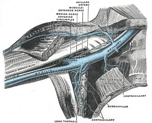
Anterior view of right upper limb and thorax - axillary vein and the distal part of the basilic vein and cephalic vein.

Figure 1: Anterior view of right upper limb and thorax - axillary vein and the distal part of the basilic vein and cephalic vein.
The deltoid muscle covers the shoulder joint, originating from the clavicle, acromion, and spine of the scapula. The medial epicondyle is larger and gives rise to powerful grasping muscles of the anterior forearm. The lateral epicondyle is smaller and gives rise to weaker posterior forearm muscles. Compartment syndrome in the anterior compartment is indicated by swelling.
The shoulder joint is a multiaxial ball-and-socket joint providing flexion, extension, abduction, adduction, circumduction, and rotation. The glenoid labrum extends around the glenoid cavity margin. Intrinsic ligaments, including the coracohumeral and glenohumeral ligaments, provide some support. However, primary support comes from the rotator cuff muscles. [2]
The distal humerus features the medial epicondyle, lateral epicondyle, and lateral supracondylar ridge. These are attachment points for forearm muscles. [3]
| Muscle | Origin | Insertion | Action | Nerve Supply |
|---|---|---|---|---|
| Supraspinatus | Supraspinous fossa | Greater tubercle | Abduction | Suprascapular |
| Infraspinatus | Infraspinous fossa | Greater tubercle | Lateral rotation | Suprascapular |
| Teres minor | Lateral border | Greater tubercle | Lateral rotation | Axillary |
| Subscapularis | Subscapular fossa | Lesser tubercle | Medial rotation | Upper and lower subscapular |
Carpal tunnel syndrome results from nerve compression, causing pain, numbness, and weakness in the hand. [3]
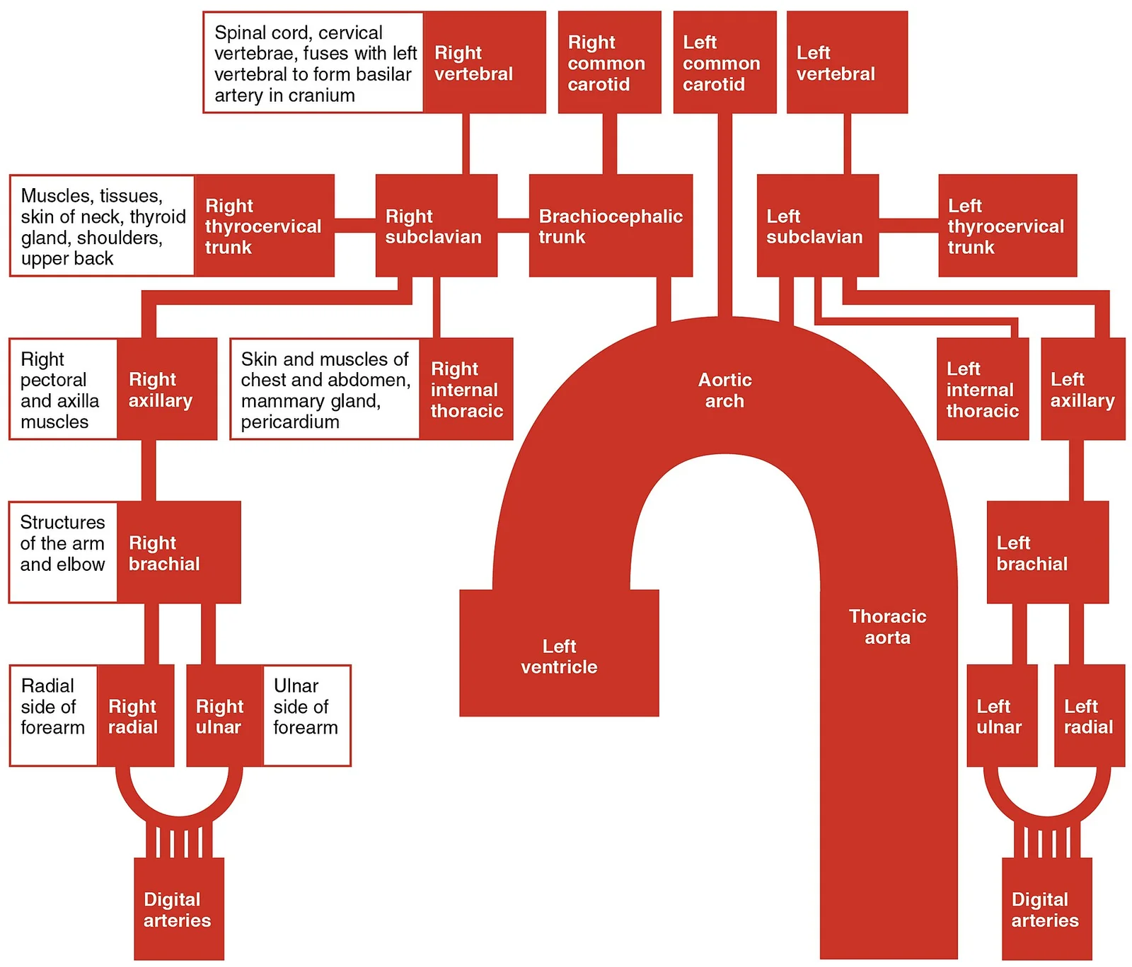
The arterial system is largely symmetrical, with paired vessels running parallel on both sides of the body, although singular midline vessels like the aorta exist[1]. The supply to the upper limb originates from the aortic arch, which gives rise to three major branches: the brachiocephalic artery, the left common carotid artery, and the left subclavian artery[1]. The left subclavian artery, named for its position under the clavicle, is an elastic artery that provides the primary inflow to the left upper limb[1]. On the right side, the brachiocephalic artery bifurcates into the right common carotid and the right subclavian artery, mirroring the left-sided supply[1].
Figure 22128 Thoracic Upper Limb Arteries Chart
Long description
The flow chart summarizes the distribution of the major arteries from the heart into the upper limb.

Figure 2: The flow chart summarizes the distribution of the major arteries from the heart into the upper limb.
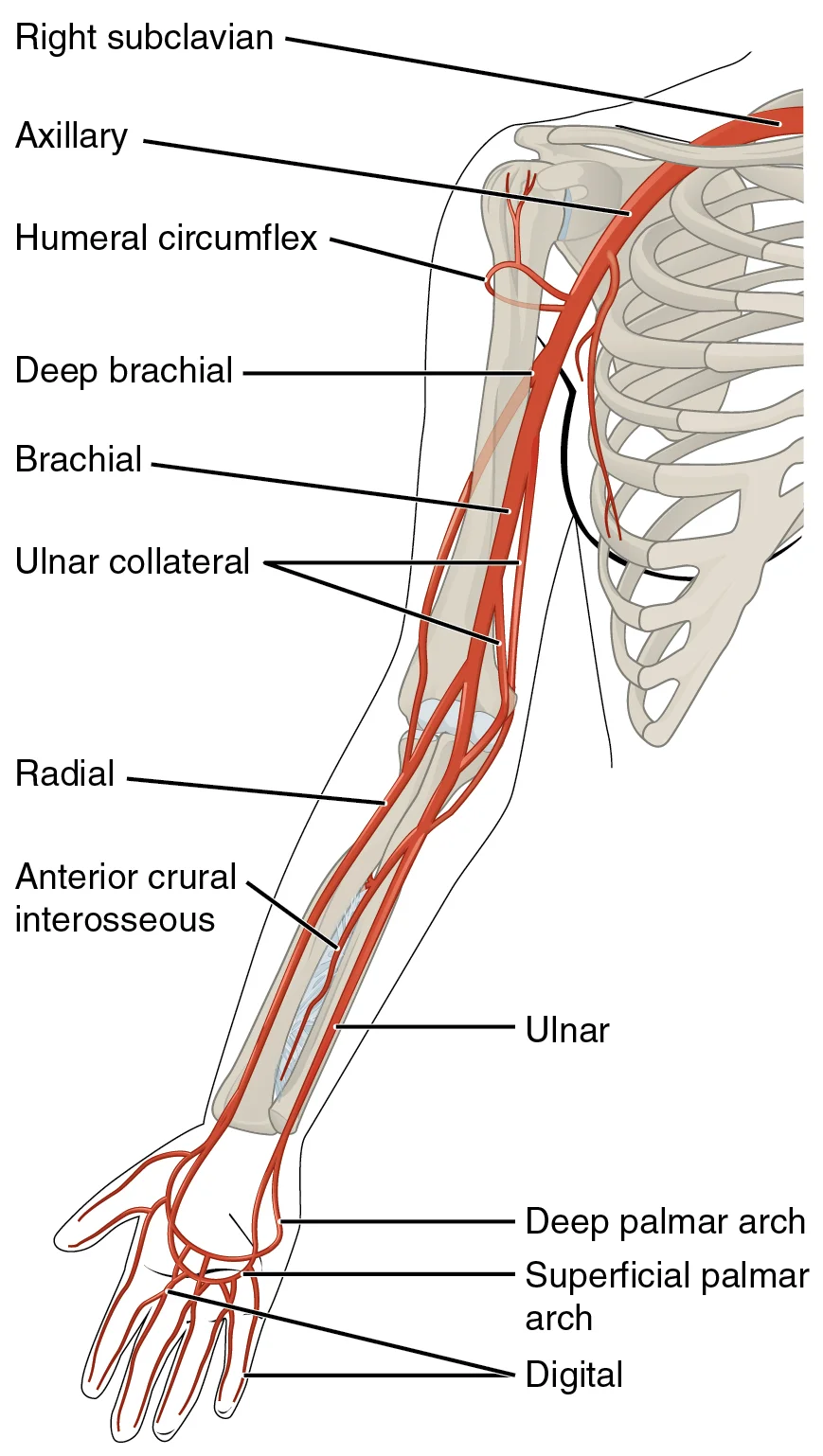
The subclavian artery begins at the aortic arch (left) or brachiocephalic trunk (right) and courses laterally beneath the clavicle[1]. Its course is divided into three parts by the anterior scalene muscle. The first part gives off the vertebral artery, internal thoracic artery, and thyrocervical trunk. The second part gives off the costocervical trunk. The subclavian artery continues as the axillary artery once it passes the lateral border of the first rib.
The axillary artery is the direct continuation of the subclavian artery, extending from the lateral border of the first rib to the inferior border of the teres major muscle[1]. It is divided into three parts by the pectoralis minor muscle. The first part gives off the superior thoracic artery. The second part gives off the thoracoacromial artery and the lateral thoracic artery. The third part gives off the subscapular artery, anterior circumflex humeral artery, and posterior circumflex humeral artery.
The brachial artery begins at the inferior border of the teres major muscle and runs down the medial aspect of the arm[1]. It is the main vessel supplying the anterior compartment of the arm. The brachial artery gives off the deep brachial artery, which runs in the radial groove of the humerus. It terminates at the cubital fossa by dividing into the radial and ulnar arteries.
Figure 32127 Thoracic Upper Limb Arteries
Long description
The arteries that supply blood to the arms and hands are extensions of the subclavian arteries.

Figure 3: The arteries that supply blood to the arms and hands are extensions of the subclavian arteries.
| Structure | Origin | Course | Termination |
|---|---|---|---|
| Subclavian | Aortic arch (L) / Brachiocephalic (R) | Lateral to clavicle | Lateral border of 1st rib |
| Axillary | Lateral border of 1st rib | To inferior border of teres major | Inferior border of teres major |
| Brachial | Inferior border of teres major | Medial arm | Cubital fossa |
The venous system of the upper limb facilitates the return of deoxygenated blood from the distal extremities to the central circulation, ultimately reaching the right atrium via the superior vena cava. This pathway is critical for understanding clinical procedures such as venepuncture and the spread of infection. [1]
Figure 42134 Thoracic Upper Limb Veins
Long description
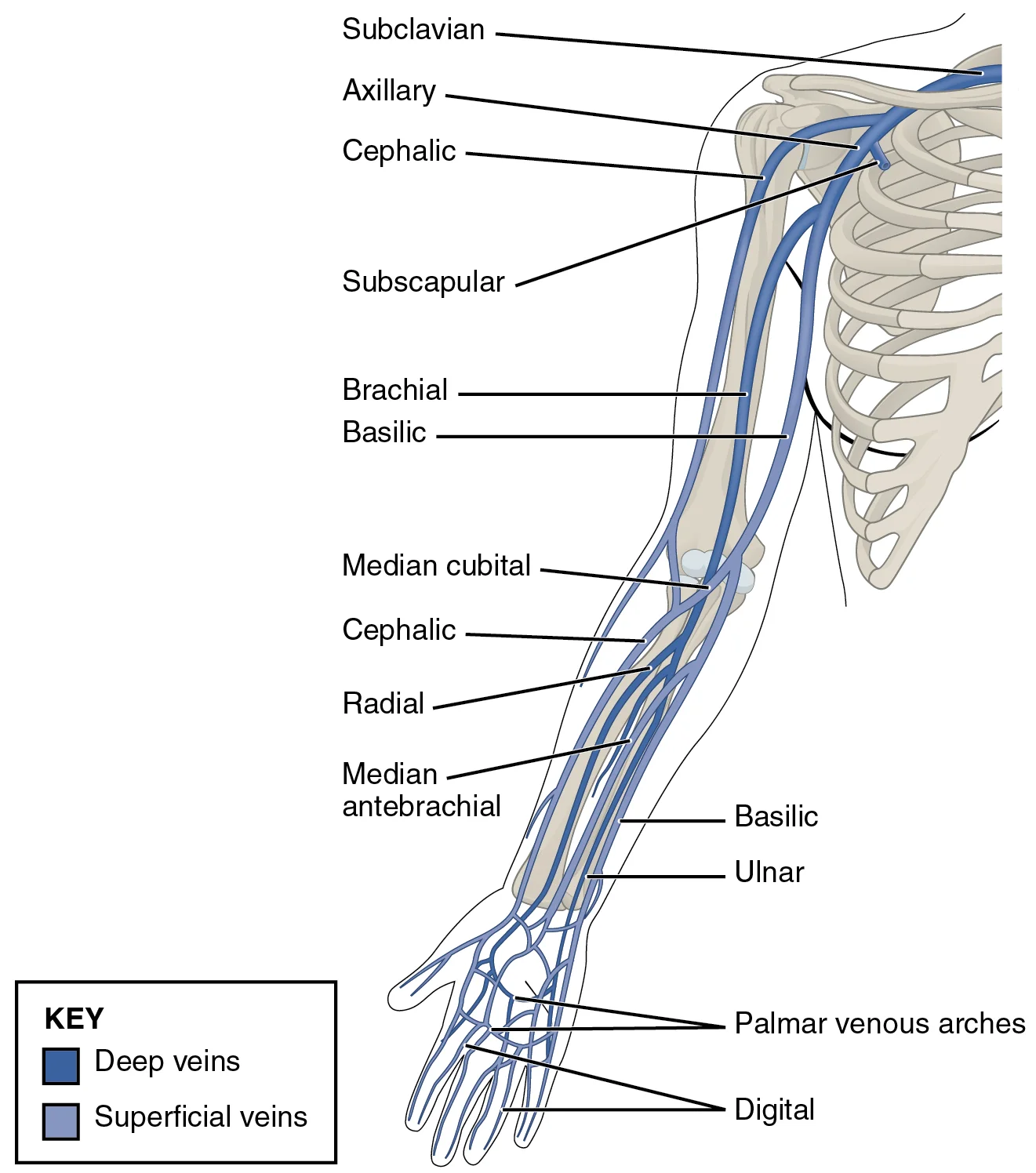
This anterior view shows the veins that drain the upper limb.

Figure 4: This anterior view shows the veins that drain the upper limb.
The process begins in the digits. Digital veins in the fingers converge within the hand to form the palmar venous arches. These arches serve as the initial collecting network before blood moves proximally. [1]
From the palmar arches, blood flows into three primary superficial and deep channels: the radial vein, the ulnar vein, and the median antebrachial vein. [1]
The radial vein and the ulnar vein run parallel to the corresponding bones of the forearm. In the forearm, the median antebrachial vein parallels the ulnar vein but lies more medially. It subsequently joins the basilic vein within the forearm. [1]
In the brachium (upper arm), the radial and ulnar veins unite at the antebrachium to form the brachial vein. This is a deep vein that flows directly into the axillary vein. [1]
The basilic vein travels through the arm in a medial and superficial course before joining the axillary vein. This superficial trajectory is clinically significant for access. [1]
A key anatomical feature in the antecubital region is the median cubital vein. As the basilic vein reaches this area, it gives off the median cubital vein. This vessel crosses at an angle to join the cephalic vein. [1]
The median cubital vein is the most common site for drawing venous blood in humans. Its superficial location and stability make it ideal for venepuncture. [1]
Understanding these relations allows recognition of the median cubital vein as the primary target for blood draws, while the brachial vein represents the deep drainage pathway into the axillary system.
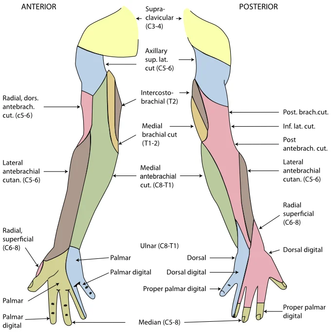
Figure 5Gray812and814
Long description
The cutaneous innervation of the right upper limb. Both areas supplied by the medial brachial cutaneous are colored green. The areas supplied by branches of the radial nerve are colored in pink.

Figure 5: The cutaneous innervation of the right upper limb.
The motor and cutaneous supply of the upper limb is distributed via five major peripheral nerves arising from the brachial plexus. The median nerve provides motor innervation to most flexors in the forearm and thenar muscles. It supplies sensation to the lateral palm and the palmar aspect of the thumb, index, middle, and lateral half of the ring finger. The ulnar nerve innervates the medial forearm flexors and intrinsic hand muscles, including the interossei and medial lumbricals. Cutaneously, it supplies the medial palm and the medial half of the ring finger and the little finger. The radial nerve provides motor supply to all extensors of the arm and forearm. It gives cutaneous innervation to the posterior arm, posterior forearm, and the dorsal aspect of the lateral hand. The musculocutaneous nerve supplies the anterior compartment muscles of the arm and the lateral forearm skin. The axillary nerve innervates the deltoid and teres minor muscles, with cutaneous supply to the lateral shoulder skin.
| Nerve | Key Muscles Supplied | Cutaneous Area |
|---|---|---|
| Median | Flexor digitorum superficialis, Flexor pollicis longus, Thenar muscles | Lateral palm, digits 1-3.5 |
| Ulnar | Flexor carpi ulnaris, Medial lumbricals, Interossei | Medial palm, digit 5 and medial digit 4 |
| Radial | Triceps, Brachioradialis, Extensor carpi radialis | Posterior arm/forearm, dorsal lateral hand |
| Musculocutaneous | Biceps brachii, Brachialis, Coracobrachialis | Lateral forearm |
| Axillary | Deltoid, Teres minor | Lateral shoulder (regimental badge area) |
Lesions of these nerves produce distinct clinical deficits. Median nerve injury at the wrist causes loss of thumb opposition and sensory loss in the lateral digits. Ulnar nerve injury at the elbow results in claw hand deformity and sensory loss in the medial digits. Radial nerve injury leads to wrist drop and sensory loss on the dorsal hand. Understanding these patterns allows precise localisation of nerve damage based on motor weakness and sensory loss distribution.
The lymphatic system of the upper limb serves as a critical drainage pathway for interstitial fluid, immune surveillance, and potential routes for metastatic spread. Understanding this network requires tracing the flow from superficial cutaneous vessels to deep nodal groups, culminating in the thoracic duct or right lymphatic duct.
Superficial lymphatic vessels accompany the superficial veins of the upper limb. They are divided into medial and lateral groups based on their anatomical course.
The medial group accompanies the basilic vein. It begins in the hand and forearm, ascending along the medial aspect of the arm. These vessels typically pass through the epitrochlear nodes, located above the medial epicondyle of the humerus. From there, they continue to the axillary nodes. This pathway is clinically significant because infections of the medial hand or forearm often present with tender epitrochlear lymphadenopathy before involving the axilla.
The lateral group accompanies the cephalic vein. It originates in the lateral hand and forearm, ascending along the lateral aspect of the arm. These vessels generally bypass the epitrochlear nodes and drain directly into the apical and pectoral groups of the axillary nodes. This direct route explains why infections of the lateral hand may present with axillary swelling without prior epitrochlear involvement.
Clinical pearlClinical Pearl
Epitrochlear lymphadenopathy is rare in healthy individuals and suggests pathology in the medial hand, forearm, or ulnar side of the arm, such as cat-scratch disease or syphilis.
Deep lymphatic vessels follow the deep arteries of the upper limb, including the radial, ulnar, and interosseous arteries. They drain the deep structures of the hand, forearm, and arm, including muscles, joints, and bones. These vessels accompany the deep veins and typically drain into the axillary nodes, often bypassing the epitrochlear nodes. They provide a parallel drainage system to the superficial vessels, ensuring comprehensive coverage of the limb's tissues.
The axillary lymph nodes are the primary drainage site for the upper limb. They are grouped into five categories: pectoral, subscapular, lateral, central, and apical.
The lateral nodes receive lymph from the upper limb via both superficial and deep vessels. They are located along the lateral wall of the axilla, accompanying the axillary vein. The pectoral nodes drain the anterior chest wall and breast, while the subscapular nodes drain the posterior chest wall and scapular region. The central nodes receive lymph from the pectoral, subscapular, and lateral groups. Finally, the apical nodes receive lymph from all other axillary groups and drain into the subclavian trunk.
| Group | Location | Drainage |
|---|---|---|
| Lateral | Along axillary vein | Upper limb |
| Pectoral | Anterior axillary wall | Anterior chest, breast |
| Subscapular | Posterior axillary wall | Posterior chest, scapula |
| Central | Axillary fat pad | Pectoral, subscapular, lateral |
| Apical | Apex of axilla | All other axillary nodes |
The efferent vessels from the apical nodes form the subclavian trunk. On the left side, the left subclavian trunk joins the thoracic duct, which empties into the left subclavian vein near its junction with the left internal jugular vein. On the right side, the right subclavian trunk joins the right lymphatic duct, which empties into the right subclavian vein. This asymmetry is crucial for understanding the spread of malignancies; for instance, breast cancer may metastasise to the axillary nodes and subsequently enter the systemic circulation via the thoracic duct.
The axillary artery passes through the axilla, lying anterior to the subscapularis muscle. The cephalic vein ascends in the deltopectoral groove, lateral to the pectoralis major. The basilic vein runs medially, piercing the deep fascia to join the brachial veins. [1]
An interphalangeal joint is one of the articulations between adjacent phalanges of the digits. The thumb (pollex) is digit number 1 and has two phalanges, a proximal phalanx, and a distal phalanx bone. Digits 2 (index finger) through 5 (little finger) have three phalanges each, called the proximal, middle, and distal phalanx bones. The fingers and thumb contain 14 bones, each of which is called a phalanx bone. [3]
DefinitionPhalanges
The bones of the digits, named after the ancient Greek phalanx (a rectangular block of soldiers).
The neurovascular structures are protected by the deep fascia. The median nerve passes between the two heads of the pronator teres. The ulnar nerve passes posterior to the medial epicondyle. The radial nerve winds around the spiral groove of the humerus.
| Muscle | Origin | Insertion | Action | Nerve Supply |
|---|---|---|---|---|
| Biceps Brachii | Scapula | Radius | Flexion, Supination | Musculocutaneous |
| Brachialis | Humerus | Ulna | Flexion | Musculocutaneous |
| Coracobrachialis | Scapula | Humerus | Flexion, Adduction | Musculocutaneous |
The brachiocephalic vein is formed by the fusion of the subclavian and internal jugular veins. Each vertebral vein also flows into the brachiocephalic vein close to this fusion. These veins arise from the base of the brain and the cervical region of the spinal cord. The internal thoracic vein, also known as an internal mammary vein, drains the anterior surface of the chest wall and flows into the brachiocephalic vein. [1]
NoteVenous Drainage
The subclavian vein forms when the axillary vein passes through the body wall from the axillary region. [1]
The axillary vein becomes the subclavian vein as it passes through the body wall and enters the thorax. The superior vena cava drains most of the body superior to the diaphragm. The veins draining the cervical vertebrae and the posterior surface of the skull, including some blood from the occipital sinus, flow into the vertebral veins. These parallel the vertebral arteries and travel through the transverse foramina of the cervical vertebrae. The vertebral veins also flow into the brachiocephalic veins. [1]
The internal jugular vein flows parallel to the common carotid artery and is more or less its counterpart. It empties into the brachiocephalic vein. [1]
Exam trapArterial vs Venous Path
Tracing blood flow through arteries follows the current in the direction of blood flow, so that we move from the heart through the large arteries and into the smaller arteries to the capillaries. [1]
Understanding the upper limb requires correlating surface landmarks with deep structures visible on imaging. This section outlines the appearance of key anatomical features across different modalities, ensuring you can identify pathology and normal variants in clinical practice.
Plain X-rays remain the first-line investigation for trauma. They provide a rapid assessment of bone integrity and joint alignment.
The humeral head articulates with the glenoid fossa of the scapula. The acromion process lies superiorly, forming the roof of the subacromial space. The clavicle extends laterally to meet the acromion at the acromioclavicular joint. The coracoid process projects anteriorly and inferiorly from the scapula. These bony landmarks are critical for identifying fractures or dislocations.
CT offers superior bone detail and three-dimensional reconstruction. It is essential for complex fractures involving the joint surface.
In CT images, the cortical bone appears bright white due to high density. The medullary cavity is less dense. CT arthrography can delineate soft tissue structures like the labrum. The glenoid labrum appears as a triangular fibrocartilaginous structure attached to the rim of the glenoid fossa. This structure deepens the socket, enhancing shoulder stability. Displacement of the labrum may indicate a Bankart lesion, commonly associated with anterior shoulder dislocation.
MRI is the gold standard for soft tissue evaluation. It visualises muscles, tendons, ligaments, and cartilage without ionising radiation.
T1-weighted images show anatomy clearly, with fat appearing bright. T2-weighted images highlight fluid and oedema, which appear bright. The rotator cuff muscles—supraspinatus, infraspinatus, teres minor, and subscapularis—surround the humeral head. Supraspinatus tendon passes under the acromion. Tears often occur at the insertion on the greater tubercle of the humerus. MRI can detect partial-thickness tears that may be missed on X-ray. The subscapularis tendon inserts on the lesser tubercle. Integrity of these tendons is crucial for shoulder function.
Ultrasound is dynamic and real-time. It allows assessment of moving structures and guided injections.
The supraspinatus tendon is easily visualised in the longitudinal plane. It appears as a fibrillar, hyperechoic structure. Loss of this fibrillar pattern suggests tendinopathy or tear. The long head of the biceps tendon runs in the bicipital groove. It is enclosed by the transverse humeral ligament. Ultrasound can detect tendon subluxation or tenosynovitis. The deltoid muscle overlies the shoulder joint. Its contraction can be observed dynamically. This modality is particularly useful for evaluating rotator cuff pathology in an ambulatory setting. [3]
The upper limb is a complex functional unit where surface landmarks guide clinical assessment. Understanding the projection of vessels and nerves is essential for safe procedures and accurate diagnosis of injuries.
The brachial artery is the primary pulse point in the antecubital fossa, lying medial to the biceps tendon. It is often auscultated here for blood pressure measurement. The median nerve runs deep to the bicipital aponeurosis, protecting it from superficial trauma but making it vulnerable in deep lacerations. The radial artery is palpable at the wrist, lateral to the flexor carpi radialis tendon, forming the anatomical snuffbox floor. [3]
Injuries to the upper limb often result from falls or direct trauma. A fracture of the surgical neck of the humerus can damage the axillary nerve, leading to deltoid paralysis and loss of sensation over the lateral shoulder. This is a classic clinical presentation known as Erb’s palsy if the upper trunk is affected, though Erb’s palsy specifically refers to C5-C6 root avulsion. A fracture of the mid-shaft of the humerus risks injury to the radial nerve, causing wrist drop due to paralysis of the extensor muscles. This condition is termed radial nerve palsy.
Venepuncture is commonly performed in the median cubital vein, which connects the cephalic and basilic veins. This vein is superficial and stable, making it ideal for blood collection. The basilic vein is larger and lies closer to the brachial artery, requiring careful technique to avoid arterial puncture. Understanding the relationship between these vessels prevents complications such as haematoma formation or nerve injury.
The superficial branch provides sensory innervation to the dorsum of the hand, while the deep branch innervates the extensor muscles.
Clinical pearlClinical Pearl
Always assess radial pulse and capillary refill after forearm fractures to rule out vascular compromise.
| Nerve | Injury Mechanism | Clinical Deficit |
|---|---|---|
| Axillary | Surgical neck fracture | Deltoid paralysis, sensory loss over lateral shoulder |
| Radial | Mid-shaft humerus fracture | Wrist drop, sensory loss on dorsum of hand |
| Median | Supracondylar fracture | Thenar atrophy, loss of thumb opposition |
It branches into the right subclavian artery and the right common carotid artery. The left subclavian and left common carotid arteries arise independently from the aortic arch. [1]
These branches include the lingual, facial, occipital, maxillary, and superficial temporal arteries. [1]
Exam trapCarotid Sinus
Do not confuse this with the aortic sinuses, though both provide similar information.
| Ligament | Location | Primary Function |
|---|---|---|
| Fibular collateral | Lateral | Resists hyperextension or rotation |
| Tibial collateral | Medial | Resists hyperextension or rotation |
| Anterior cruciate | Intracapsular | Resists hyperextension |
| Posterior cruciate | Intracapsular | Prevents anterior sliding of femur |
Students frequently confuse the structural basis of joint stability with mobility. A primary error is assuming that the shoulder joint’s extensive range of motion derives from robust ligamentous support. In reality, the glenohumeral joint possesses the largest range of motion of any joint in the body precisely because of a lack of structural support, which offsets mobility with a loss of stability. To correct this, remember that freedom of movement here is bought at the cost of inherent instability. [2]
Another frequent mistake involves the elbow’s collateral ligaments. Learners often misidentify the strongest component of the ulnar collateral ligament. The strongest part is the anterior portion, which specifically resists hyperextension of the elbow. This distinction is critical because injury to this ligament is common in baseball pitchers due to frequent, forceful extensions of the forearm. [2]
Students also struggle with the specific articulations of the radius. It is incorrect to state that the head of the radius articulates with the trochlea of the humerus. The small depression on the surface of the head articulates with the capitulum of the humerus as part of the elbow joint. The smooth, outer margin of the head articulates with the radial notch of the ulna at the proximal radioulnar joint. [3]
A further pitfall concerns the ankle joint’s classification. Many assume it allows for inversion and eversion. The ankle is a uniaxial hinge joint that allows only for dorsiflexion and plantar flexion of the foot. Side-to-side motion is prevented by the firm articulations of the talus sides with the medial malleolus of the tibia and the lateral malleolus of the fibula. [2]
Finally, students often overlook the functional role of the longitudinal arches. It is a mistake to view the foot as a flat platform. Body weight is then conveyed from the talus to the ground by the anterior and posterior ends of these arches. [1]
Exam trapShoulder Stability
The shoulder joint has the largest range of motion of any joint in the body, but this freedom is due to a lack of structural support, offsetting mobility with a loss of stability. [2]
At the bedside, understanding the elbow’s ligamentous support is vital for assessing trauma. The medial elbow is supported by the ulnar collateral ligament, while the radial collateral ligament supports the lateral side. These ligaments prevent side-to-side movements and resist hyperextension of the elbow. Clinicians must recognize that the ulnar collateral ligament may be injured by frequent, forceful extensions of the forearm, as is seen in baseball pitchers. [2]
In the operating theatre, knowledge of the proximal radioulnar joint is essential. This is a pivot joint that allows for rotation of the radius during supination and pronation of the forearm. The annular ligament encircles the head of the radius to hold it in place at this joint. Surgeons repairing this area must understand that the radial collateral ligament arises from the lateral epicondyle of the humerus and blends into the lateral side of the annular ligament. [2]
On imaging, correct identification of bony landmarks prevents diagnostic errors. The posterior humerus has the olecranon fossa, a larger depression that receives the olecranon process of the ulna when the forearm is fully extended. Radiologists look for this relationship to confirm full extension. Additionally, the radius runs parallel to the ulna on the lateral side of the forearm. The radial tuberosity, an oval-shaped bony protuberance, serves as a muscle attachment point on the medial side of the radius shaft. [3]
In examinations, students must distinguish between the metatarsals. The first metatarsal bone is shorter and thicker than the others. The second metatarsal is the longest. This landmark is crucial for identifying fractures.
The ankle joint’s structure dictates its function. The superior aspect of the talus bone is square-shaped and has three areas of articulation. The top of the talus articulates with the inferior tibia, carrying the body weight between the leg and foot. This uniaxial hinge design ensures that weight bearing is managed through dorsiflexion and plantar flexion only. [2] [1]
| Ligament | Location | Function |
|---|---|---|
| Ulnar collateral | Medial | Prevents side-to-side movement; anterior part resists hyperextension |
| Radial collateral | Lateral | Prevents side-to-side movement; blends with annular ligament |
| Annular | Lateral/Proximal Radius | Encircles head of radius; supports proximal radioulnar joint |
The glenohumeral joint has the largest range of motion but lacks structural support. The ulnar collateral ligament’s strongest part is the anterior portion resisting hyperextension. The proximal radioulnar joint is a pivot joint for pronation and supination. The annular ligament encircles the head of the radius to stabilize it. The ankle is a uniaxial hinge joint allowing only dorsiflexion and plantar flexion. The talus receives body weight and conveys it via the longitudinal arches. The first metatarsal is shorter and thicker; the second is the longest. The olecranon fossa receives the olecranon process during full extension.
Answer each one in your head first, then open the answer.
Which ligament’s strongest part resists hyperextension of the elbow?
Show answerHide answer
The anterior portion of the ulnar collateral ligamentWhat type of joint is the proximal radioulnar joint?
Show answerHide answer
A pivot jointWhich metatarsal is the longest?
Show answerHide answer
The second metatarsalWhat bone receives the body weight at the top of the longitudinal arches?
Show answerHide answer
The talus boneWhere does the radial collateral ligament arise?
Show answerHide answer
From the lateral epicondyle of the humerus
- Betts JG, Young KA, Wise JA, et al. Circulatory Pathways. OpenStax, Anatomy and Physiology 2e (CC BY-NC-SA 4.0). Accessed .
- Betts JG, Young KA, Wise JA, et al. Anatomy of Selected Synovial Joints. OpenStax, Anatomy and Physiology 2e (CC BY-NC-SA 4.0). Accessed .
- Betts JG, Young KA, Wise JA, et al. Bones of the Upper Limb. OpenStax, Anatomy and Physiology 2e (CC BY-NC-SA 4.0). Accessed .
Figure credits
- Figure 1: Henry Vandyke Carter, via Wikimedia Commons, Public domain, Public domain.
- Figure 2: OpenStax College, via Wikimedia Commons, CC BY 3.0, CC BY 3.0. resized.
- Figure 3: OpenStax College, via Wikimedia Commons, CC BY 3.0, CC BY 3.0.
- Figure 4: OpenStax College, via Wikimedia Commons, CC BY 3.0, CC BY 3.0.
- Figure 5: Henry Vandyke Carter, via Wikimedia Commons, Public domain, Public domain.
Educational content for medical students and doctors. It is not a substitute for clinical judgement or local guidelines.