Peripheral nerve injuries of the upper limb
Anatomical overview of upper limb regions, bones, and brachial plexus origins.
The upper limb is anatomically divided into three distinct regions to facilitate functional description. The arm is the region located between the shoulder and elbow joints. The forearm lies between the elbow and wrist joints. The hand is situated distal to the wrist. Each upper limb contains a total of 30 bones. [2]
The skeleton of the upper limb begins with the humerus, which is the single bone of the upper arm. In the forearm, the ulna is positioned medially and the radius laterally. These are the paired bones of the forearm. The base of the hand contains eight bones, each called a carpal bone. The palm of the hand is formed by five bones, each called a metacarpal bone. The fingers and thumb contain a total of 14 bones, each of which is a phalanx bone of the hand. The articulating surfaces of the distal humerus consist of the trochlea medially and the capitulum laterally. [2]
Nerve supply to the upper limb arises from the brachial plexus. This plexus is one of four major nerve plexuses in the body. The other plexuses are located at the cervical, lumbar, and sacral levels. Spinal nerves C4 through T1 reorganize through this plexus to give rise to the nerves of the arms. The name brachial suggests this association with the arm. [3]
DefinitionBrachial Plexus
The brachial plexus is formed by spinal nerves C4 through T1, which reorganize to give rise to the nerves of the arms.
| Nerve | Origin | Course |
|---|---|---|
| Radial | Brachial Plexus | Continues through the arm |
| Axillary | Radial Nerve | Branches to armpit region |
| Ulnar | Brachial Plexus | Parallels radial nerve |
| Median | Brachial Plexus | Parallels radial nerve |
NoteClinical Relevance
Understanding the brachial plexus origins (C4-T1) is essential for localizing lesions causing upper limb weakness or sensory loss.
For the upper limb, the relevant structures are spinal nerves, which emerge from the spinal cord. There are thirty-one pairs of spinal nerves in total, named according to the vertebral level from which they arise. These include eight cervical pairs (C1–C8), twelve thoracic pairs (T1–T12), five lumbar pairs (L1–L5), five sacral pairs (S1–S5), and one coccygeal pai [3]
The upper trunk is formed by the union of C5 and C6. The middle trunk is a direct continuation of C7. The lower trunk is formed by the union of C8 and T1.
The central structure is the axillary artery. The lateral cord lies anterolateral to the artery. The medial cord lies anteromedial to the artery. The posterior cord lies posterior to the artery. This spatial relationship is critical for understanding the course of the terminal branches. The nerves are not straight continuations of the spinal nerves but represent a reorganization of axons. [3]
The upper limb is a complex anatomical region comprising three distinct parts and a total of thirty bones. Understanding the course of peripheral nerves requires first understanding the bony framework that guides them. The upper arm, situated between the shoulder and elbow, contains the humerus. The proximal humerus features the head, which articulates with the scapula at the glenohumeral joint. This joint is flanked by the greater and lesser tubercles, separated by the intertubercular or bicipi [2]
DefinitionBony Landmarks
The humerus provides the structural scaffold for the upper limb, with specific epicondyles and tubercles serving as attachment points for muscles and guides for neurovascular structures.
Peripheral nerves generally follow the path of major arteries. The radial and ulnar arteries run parallel to their respective bones, giving off smaller branches until they reach the wrist or carpal region. At the wrist, these vessels fuse to form the superficial and deep palmar arches, which supply the hand and digits. Nerves such as the median nerve travel alongside these vascular structures. Compression of the median nerve within the carpal tunnel produces carpal tunnel syndrome, which is char [1][2]
Exam trapNerve Composition
Nerves are not purely motor or sensory; they contain mixed fibers. For instance, the radial nerve carries both cutaneous sensation fibers from the arm and motor fibers to move arm muscles. [3]
These plexuses contain fibers that serve sensory functions or fibers that serve motor functions. The motor fibers send action potentials to cause skeletal. [3]
The ulnar artery runs medially.
| Structure | Location | Clinical Relevance |
|---|---|---|
| Surgical Neck | Proximal humerus | Fracture risk; axillary nerve injury |
| Medial Epicondyle | Distal humerus | Ulnar nerve passes posteriorly [2] |
The upper limb is a complex anatomical region comprising three distinct parts and a total of thirty bones. Understanding the course of peripheral nerves requires first understanding the bony framework that guides them. The upper arm, situated between the shoulder and elbow, contains the humerus. The proximal humerus features the head, which articulates with the scapula at the glenohumeral joint. This joint is flanked by the greater and lesser tubercles, separated by the intertubercular or bicipi | Radial Groove | Humeral shaft | Radial nerve course |
Injury to these nerves can result from trauma to the associated bony structures. For example, a fracture of the humeral shaft may damage the radial nerve as it courses through the radial groove. Similarly, dislocation of the shoulder or fracture of the surgical neck may affect the axillary nerve. Understanding the precise course of these nerves relative to the bones is essential for diagnosing the level of injury.
Figure 1Gray812and814
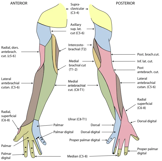
Long description
The cutaneous innervation of the right upper limb. Both areas supplied by the medial brachial cutaneous are colored green. The areas supplied by branches of the radial nerve are colored in pink.

Figure 1: The cutaneous innervation of the right upper limb.
The axillary artery gives off branches that supply blood to the region near the head of the humerus, specifically via the humeral circumflex arteries. [1]
These nerves provide motor innervation to the muscles of the shoulder, arm, forearm, and hand, and sensory innervation to the overlying skin. Understanding the specific branches is critical for localizing lesions.
| Nerve | Motor Supply | Sensory Supply |
|---|---|---|
| Axillary | Deltoid, Teres minor | Skin over lower deltoid |
| Musculocutaneous | Biceps brachii, Brachialis, Coracobrachialis | Lateral forearm |
| Median | Flexors of forearm, Thenar muscles | Lateral palm, digits 1-3 |
| Ulnar | Intrinsic hand muscles, Flexor carpi ulnaris | Medial palm, digit 5 |
| Radial | Extensors of arm and forearm | Posterior arm, forearm, dorsal hand |
Clinical pearlClinical Pearl
Always correlate motor deficits with sensory loss to localize the level of nerve injury.
DefinitionBrachial Plexus
A network of nerves formed by the ventral rami of spinal nerves C5 to T1, supplying the upper limb.
The upper limb is a complex anatomical region where peripheral nerve injuries are classified by their proximity to the spinal cord. Understanding these levels requires a clear view of the brachial plexus and its terminal branches. This figure illustrates the cervical spine and the emergence of nerve roots. The roots exit the intervertebral foramen at their corresponding level. [3]
DefinitionBrachial Plexus
A network of nerves formed by the ventral rami of spinal nerves C5 to T1, which supplies the upper limb.
Injuries are often described by the level of the plexus involved. Root lesions affect the proximal part of the plexus. For example, an injury to the C5 and C6 roots results in Erb’s palsy. This condition presents with the arm hanging by the side, internally rotated, and the forearm extended and pronated. This posture is known as the 'waiter’s tip'. The deficit arises because the muscles responsible for abduction and external rotation of the shoulder are paralysed.
Trunk lesions involve the upper, middle, or lower trunks. An upper trunk lesion affects the suprascapular and axillary nerves. This leads to weakness in shoulder abduction and external rotation. The patient may present with a 'claw hand' deformity due to paralysis of the interossei and lumbricals.
Cord lesions are less common but distinct. A lateral cord lesion affects the musculocutaneous and lateral root of the median nerve. This causes weakness in elbow flexion and supination. A medial cord lesion affects the ulnar nerve and medial root of the median nerve. This leads to sensory loss in the medial forearm and hand. A posterior cord lesion affects the axillary and radial nerves.
Terminal nerve lesions are the most distal. A median nerve lesion at the wrist causes thenar muscle atrophy and loss of opposition. An ulnar nerve lesion at the wrist affects the hypothenar muscles and interossei. A radial nerve lesion at the spiral groove causes wrist drop. The deficit is determined by the specific muscles and sensory areas innervated by the affected nerve.
| Nerve | Level | Key Deficit |
|---|---|---|
| Axillary | Posterior Cord | Weakness in shoulder abduction |
| Radial | Spiral Groove | Wrist drop |
| Median | Wrist | Loss of thumb opposition |
| Ulnar | Wrist | Claw hand deformity |
Clinical pearlClinical Pearl
Always examine the entire limb to distinguish between a peripheral nerve lesion and a plexus lesion.
The clinical examination of peripheral nerve injuries in the upper limb requires a systematic assessment of motor function, sensory distribution, and specific provocative tests. Understanding the anatomical course of each nerve is essential for localising the lesion. The axillary nerve, which supplies the deltoid and teres minor muscles, is vulnerable during shoulder dislocations. Examination involves testing abduction against resistance and assessing sensation over the regimental badge area of the shoulder.
The musculocutaneous nerve innervates the anterior compartment of the arm, including the biceps brachii, brachialis, and coracobrachialis. Injury results in weakness of elbow flexion and forearm supination. Sensory loss occurs on the lateral aspect of the forearm.
Clinical pearlSaturday Night Palsy
This radial nerve palsy results from compression in the spiral groove, typically after prolonged arm compression against a hard surface.
A thorough examination must include testing for Tinel’s sign, where percussion over the nerve elicits tingling in the distal distribution, indicating nerve irritation or regeneration. Phalen’s test is used for carpal tunnel syndrome, requiring the patient to hold their wrists in maximal flexion for 60 seconds to reproduce symptoms.
| Nerve | Motor Deficit | Sensory Deficit | Common Cause |
|---|---|---|---|
| Axillary | Shoulder abduction weakness | Lateral shoulder | Shoulder dislocation |
| Radial | Wrist drop | Dorsal hand | Humeral fracture |
| Median | Thumb opposition weakness | Lateral palm/fingers | Carpal tunnel |
| Ulnar | Claw hand | Medial hand/fingers | Elbow trauma |
When examining the upper limb, always compare strength and sensation bilaterally. Use a tuning fork to assess vibration sense if large fibre involvement is suspected. Remember that nerve injuries can be partial or complete; partial injuries may present with mixed motor and sensory deficits, while complete injuries result in total loss of function below the lesion site. Early recognition and accurate localisation are critical for appropriate management and prognosis.
The brachial artery is the major vessel of the arm and is typically auscultated for blood pressure measurement. It divides into the radial and ulnar arteries in the cubital fossa. The radial artery runs along the lateral aspect of the forearm and is palpable at the wrist.
DefinitionBrachial Pulse
The brachial artery is the primary pulse point for assessing circulation in the upper limb and is used for blood pressure measurement.
Exam trapMedian Nerve Trap
Do not confuse the median nerve with the ulnar nerve. The median nerve does not supply the hypothenar muscles or the medial two lumbricals.
Injury at the elbow causes 'claw hand' deformity of the ring and little fingers.
DefinitionUlnar Nerve Course
The ulnar nerve passes posterior to the medial epicondyle, making it susceptible to injury in fractures of the medial epicondyle.
The radial nerve arises from the posterior cord. It winds around the posterior aspect of the humerus in the radial groove. It supplies the triceps brachii, anconeus, brachioradialis, and extensor muscles of the forearm. Injury to the radial nerve in the radial groove results in 'wrist drop' due to paralysis of the extensor muscles. Sensory loss occurs over the posterior arm and forearm and the dorsal aspect of the lateral hand.
Clinical pearlRadial Nerve Lesion
Wrist drop is the hallmark of radial nerve injury. The patient cannot extend the wrist or fingers.
The musculocutaneous nerve pierces the coracobrachialis muscle and supplies the biceps brachii, brachialis, and coracobrachialis. It terminates as the lateral antebrachial cutaneous nerve. Injury is rare but results in weakness of elbow flexion and supination.
DefinitionMusculocutaneous Nerve
This nerve supplies the anterior compartment of the arm and provides sensory innervation to the lateral forearm.
The axillary nerve arises from the posterior cord and winds around the surgical neck of the humerus. It supplies the deltoid and teres minor muscles. Injury to the axillary nerve results in weakness of shoulder abduction and loss of sensation over the lateral aspect of the shoulder ('regimental badge' area).
Exam trapAxillary Nerve Trap
The axillary nerve is closely associated with the surgical neck of the humerus and the quadrangular space. Fractures here can injure this nerve.
It has roots, trunks, divisions, cords, and branches. The mnemonic 'Randy Travis Drinks Cold Beer' helps remember the order: Roots, Trunks, Divisions, Cords, Branches.
DefinitionBrachial Plexus Mnemonic
Roots (C5-T1), Trunks, Divisions, Cords, Branches. Remember: 'Randy Travis Drinks Cold Beer'.
The Erb-Duchenne palsy involves injury to the upper trunk (C5-C6). It results in 'waiter's tip' posture: arm adducted and medially rotated, elbow extended, forearm pronated, and wrist flexed. This is often caused by excessive separation of the head and shoulder during birth.
Clinical pearlErb-Duchenne Palsy
Upper trunk injury (C5-C6) leads to waiter's tip posture. The patient cannot abduct or externally rotate the arm.
The Klumpke's palsy involves injury to the lower trunk (C8-T1). It results in 'claw hand' deformity due to paralysis of the intrinsic hand muscles. It is often caused by excessive upward traction on the arm, such as grabbing a branch to break a fall.
DefinitionKlumpke's Palsy
Lower trunk injury (C8-T1) leads to claw hand. It may also involve Horner's syndrome if the T1 root is avulsed.
Compression of the median nerve here causes carpal tunnel syndrome, characterized by pain, numbness, and tingling in the median nerve distribution.
Exam trapCarpal Tunnel Content
The carpal tunnel contains the median nerve and nine tendons. The ulnar nerve does NOT pass through the carpal tunnel; it passes through Guyon's canal.
Students frequently confuse the anatomical landmarks of the proximal humerus, leading to errors in identifying fracture sites and nerve relations. A primary error is conflating the anatomical neck with the surgical neck. The anatomical neck is the margin of the smooth area of the head of the humerus, whereas the surgical neck is located at the base of the expanded, proximal end of the humerus, where it joins the narrow shaft. The surgical neck is a common site of arm fractures. Students must rem [2]
Another frequent mistake involves the bony prominences of the proximal humerus. The greater tubercle is located on the lateral side of the proximal humerus, while the lesser tubercle is found on the anterior aspect. Both serve as attachment sites for muscles that act across the shoulder joint. Confusing their positions can lead to incorrect identification of rotator cuff insertions. The intertubercular groove, also known as the bicipital groove, provides passage for a tendon of the biceps brachii muscle. [2]
In the forearm, students often misidentify the orientation of the ulna and radius. The ulna is the medial bone of the forearm, running parallel to the radius, which is the lateral bone. A common error is assuming the ulna is lateral or that the radius is medial. The proximal end of the ulna resembles a crescent wrench with its large, C-shaped trochlear notch, which articulates with the trochlea of the humerus as part of the elbow joint. The posterior and superior portions of the proximal ulna ma [2]
Regarding the distal forearm, the styloid process of the ulna projects from the posterior side of the ulnar head. This serves as an attachment point for a connective tissue structure that unites the distal ends of the ulna and radius. Students should not confuse this with the radial styloid process. The interosseous border of the ulna forms a ridge on the lateral side of the shaft, serving as the line of attachment for the interosseous membrane of the forearm. This membrane unites the ulna and radius bones. [2]
In the hand, students often overlook the functional significance of metacarpal mobility. The first metacarpal bone, at the base of the thumb, is separated from the other metacarpal bones. This allows it a freedom of motion that is independent of the other metacarpal bones, which is very important for thumb mobility. Conversely, the second and third metacarpal bones are firmly anchored in place and are immobile. The fourth and fifth metacarpal bones have limited anterior-posterior mobility, a motion that is greater for the fifth bone. This mobility is important during power gripping with the hand. The anterior movement of these bones, particularly the fifth metacarpal bone, increases the strength of contact for the medial hand during gripping actions. [2]
Finally, students may misinterpret the clinical significance of tongue movement. Loss of function of the tongue can interfere with speech and swallowing[1]. Because the location of the hypoglossal nerve and nucleus is near the cardiovascular center, inspiratory and expiratory areas for respiration, and the vagus nuclei that regulate digestive functions, a tongue that protrudes incorrectly can suggest damage in adjacent structures that have nothing to do with controlling the tongue[1]. This highlights the importance of considering adjacent structures when assessing cranial nerve deficits. [3]
This knowledge is used at the bedside to localise neurological deficits following trauma. When a patient presents with a fracture of the surgical neck of the humerus, clinicians must assess for axillary nerve injury due to its proximity to this landmark. The surgical neck is a common site of arm fractures. Understanding that the greater and lesser tubercles serve as attachment sites for muscles that act across the shoulder joint helps in assessing rotator cuff integrity. [2]
In the operating theatre, precise identification of bony landmarks is crucial for surgical approaches. The intertubercular groove provides passage for a tendon of the biceps brachii muscle. Surgeons must navigate this groove carefully to avoid tendon damage. The deltoid tuberosity is a roughened, V-shaped region located on the lateral side in the middle of the humerus shaft, serving as the site of attachment for the deltoid muscle. This landmark is essential for deltoid muscle reattachment proce [2]
On imaging, radiologists use the ulna and radius relationships to diagnose fractures. The ulna is the medial bone of the forearm, running parallel to the radius, which is the lateral bone. The proximal end of the ulna resembles a crescent wrench with its large, C-shaped trochlear notch, which articulates with the trochlea of the humerus as part of the elbow joint. The posterior and superior portions of the proximal ulna make up the olecranon process, which forms the bony tip of the elbow. Disruption of these articulations indicates significant elbow injury. [2]
In examinations, students are tested on the functional anatomy of the hand. The first metacarpal bone, at the base of the thumb, is separated from the other metacarpal bones. This allows it a freedom of motion that is independent of the other metacarpal bones, which is very important for thumb mobility. Questions often focus on the mobility of the fourth and fifth metacarpal bones, which have limited anterior-posterior mobility, a motion that is greater for the fifth bone. This mobility is important during power gripping with the hand. The anterior movement of these bones, particularly the fifth metacarpal bone, increases the strength of contact for the medial hand during gripping actions.
The PNS is not as contained as the CNS because it is defined as everything that is not the CNS. Some peripheral structures are incorporated into the other organs of the body. In describing the anatomy of the PNS, it is necessary to describe the common structures, the nerves and the ganglia, as they are found in various parts of the body. Many of the neural structures that are incorporated into other organs are features of the digestive system; these structures are known as the enteric nervous system and are a special subset of the PNS. Understanding this distinction is vital for clinical neurology. [3]
DefinitionSurgical Neck
The surgical neck is located at the base of the expanded, proximal end of the humerus, where it joins the narrow shaft of the humerus.
DefinitionUlna
The ulna is the medial bone of the forearm, running parallel to the radius, which is the lateral bone of the forearm.
| Structure | Location | Clinical Significance |
|---|---|---|
| Anatomical Neck | Margin of the smooth area of the head | Boundary of articular surface |
| Surgical Neck | Base of expanded proximal end, joins shaft | Common site of arm fractures |
| Greater Tubercle | Lateral side of proximal humerus | Attachment for shoulder muscles |
| Lesser Tubercle | Anterior aspect of humerus | Attachment for shoulder muscles |
| Intertubercular Groove | Between greater and lesser tubercles | Passage for biceps brachii tendon |
Upper Limb Trauma Assessment
- Patient presents with upper limb traumaIdentify fracture site
- Question: Fracture at surgical neck?
Yes
- Assess axillary nerve functionCheck deltoid sensation and power
No
- Proceed to next landmarkCheck intertubercular groove
- Question: Fracture at intertubercular groove?
Yes
- Assess biceps brachii tendonCheck flexion and supination
No
- Continue assessmentCheck distal forearm
The surgical neck is a common site of arm fractures. The ulna is the medial bone of the forearm, running parallel to the radius. The first metacarpal is separated from other metacarpals for independent motion. The second and third metacarpals are firmly anchored and immobile. The fourth and fifth metacarpals have limited anterior-posterior mobility. The PNS is defined as everything that is not the CNS. The tongue's proximity to vital centres means dysfunction suggests adjacent damage[1]. The interosseous membrane unites the ulna and radius bones.
Answer each one in your head first, then open the answer.
Which neck of the humerus is a common site of arm fractures?
Show answerHide answer
The surgical neckWhich bone is the medial bone of the forearm?
Show answerHide answer
The ulnaWhich metacarpal bones are firmly anchored and immobile?
Show answerHide answer
The second and third metacarpal bonesWhat is the PNS defined as?
Show answerHide answer
Everything that is not the CNSWhich process forms the bony tip of the elbow?
Show answerHide answer
The olecranon process
- Betts JG, Young KA, Wise JA, et al. Circulatory Pathways. OpenStax, Anatomy and Physiology 2e (CC BY-NC-SA 4.0). Accessed .
- Betts JG, Young KA, Wise JA, et al. Bones of the Upper Limb. OpenStax, Anatomy and Physiology 2e (CC BY-NC-SA 4.0). Accessed .
- Betts JG, Young KA, Wise JA, et al. The Peripheral Nervous System. OpenStax, Anatomy and Physiology 2e (CC BY-NC-SA 4.0). Accessed .
Figure credits
Educational content for medical students and doctors. It is not a substitute for clinical judgement or local guidelines.