Cutaneous innervation of the upper limb
Innervation of the upper limb via the brachial plexus and associated reflexes.
The upper limb is innervated by the brachial plexus, a complex network formed by the anterior rami of spinal nerves C4 through T1. This plexus reorganizes to give rise to the major nerves of the arm, ensuring both motor control and sensory perception. Understanding the dermatomal distribution is essential for localising neurological lesions. Referred pain often follows these dermatomal patterns, allowing clinicians to infer the level of spinal cord or nerve root involvement based on the area of perceived pain. [2]
Dermatomes represent the area of skin supplied by a single spinal nerve. Myotomes refer to the group of muscles primarily innervated by a single spinal nerve level. Testing muscle strength against resistance helps identify the specific myotome affected. For instance, shoulder abduction tests C5, elbow flexion tests C5 and C6, and finger extension tests C7.
Reflexes provide a rapid assessment of the integrity of the reflex arc, involving sensory input, spinal integration, and motor output. The muscle is quickly stretched, resulting in activation of the muscle spindle that sends a signal into the spinal cord through the dorsal root. The fiber synapses directly on the ventral horn motor neuron that activates the muscle, causing contraction. The reflexes are physiologically useful for stability. If a muscle is stretched, it reflexively contracts to return the muscle to compensate for the change in length. In the context of the neurological exam, reflexes indicate that the LMN is functioning properly. [1]
Clinical pearlReflex Arc
The reflex arc involves a sensory neuron, an interneuron (or direct synapse), and a motor neuron. It bypasses the brain for speed, ensuring immediate protective or postural responses.
The musculocutaneous nerve arises from the lateral cord of the brachial plexus (C5-C7). It pierces the coracobrachialis muscle and descends between the biceps brachii and brachialis. It provides motor innervation to these anterior compartment muscles and sensory innervation to the lateral forearm via the lateral antebrachial cutaneous nerve.
The ulnar nerve arises from the medial cord (C8-T1). It travels posterior to the medial epicondyle of the humerus in the cubital tunnel. It supplies the flexor carpi ulnaris and the medial half of the flexor digitorum profundus in the forearm.
| Feature | Description |
|---|---|
| Motor | Claw hand deformity (hyperextension at MCP, flexion at IP joints of 4th/5th digits) |
| Sensory | Numbness in medial 1.5 digits and corresponding palm/hand |
| Reflex | No specific deep tendon reflex, but intrinsic hand muscle weakness |
A lesion here may spare sensation but cause significant intrinsic hand muscle weakness, leading to difficulties with fine motor tasks and grip strength.
DefinitionLower Motor Neuron Lesion
A lesion on the LMN would result in paralysis, or at least partial loss of voluntary muscle control, which is known as paresis. The paralysis observed in LMN diseases is referred to as flaccid paralysis, referring to a complete or partial loss of muscle tone, in contrast to the loss of control in UMN lesions in which tone is retained and spasticity is exhibited. Other signs of an LMN lesion are fibrillation, fasciculation, and compromised or lost reflexes resulting from the denervation of the muscle fibers. [1]
The nerves are classified as cranial nerves or spinal nerves on the basis of their connection to the brain or spinal cord. [2]
DefinitionNerve Plexus
A network of intersecting nerve fibers where axons from different spinal nerves reorganize to form systemic nerves with distinct courses and territories.
The upper limb is innervated by the brachial plexus, a plexus formed by spinal nerves C4 through T1. This structure represents the reorganization of axons from the spinal nerves to follow specific peripheral courses. [2]
The roots (C5–T1) emerge from the vertebral column. They unite to form three trunks: superior, middle, and inferior. Each trunk divides into anterior and posterior divisions. The posterior divisions unite to form the posterior cord. The anterior divisions of the superior and middle trunks join to form the lateral cord, while the anterior division of the inferior trunk continues as the medial cord. The terminal branches arise from these cords. The axillary nerve arises from the posterior cord. The musculocutaneous nerve arises from the lateral cord. The radial nerve arises from the posterior cord. The median nerve arises from both the lateral and medial cords. The ulnar nerve arises from the medial cord.
NoteSensory Testing
Clinical examination of the upper limb involves testing the cutaneous territories of these terminal nerves to localize lesions within the plexus or at specific nerve sites.
The axillary nerve supplies the skin over the lower deltoid. The musculocutaneous nerve supplies the skin of the lateral forearm. The median nerve supplies the skin of the palmar aspect of the lateral three and a half digits. The ulnar nerve supplies the skin of the palmar and dorsal aspects of the medial one and a half digits.
The lateral cord lies lateral to the artery. The medial cord lies medial to the artery. The posterior cord lies posterior to the artery. The cutaneous nerves are clearly distinguished from the motor branches supplying the underlying musculature.
| Nerve | Origin (Cord) | Primary Cutaneous Territory |
|---|---|---|
| Axillary | Posterior | Lower deltoid region |
| Musculocutaneous | Lateral | Lateral forearm |
| Radial | Posterior | Posterior arm/forearm, dorsal lateral hand |
| Median | Lateral & Medial | Palmar lateral 3.5 digits |
| Ulnar | Medial | Palmar & dorsal medial 1.5 digits |
Understanding this formation allows for precise localisation of neurological deficits. Lesions at the root level affect multiple nerves, whereas lesions at the cord or peripheral level affect specific cutaneous distributions. These neurons transmit action potentials from the cutaneous surface into the central nervous system via the dorsal root. [2]
The cutaneous innervation of the upper limb is derived from the brachial plexus, a complex network of nerve fibres formed by the anterior rami of spinal nerves C5 to T1. Understanding this distribution requires tracing the course of these nerves from their origin in the spinal cord to their termination in the skin of the arm, forearm, and hand. The brachial plexus is organized into roots, trunks, divisions, cords, and terminal branches. The roots emerge from the intervertebral foramina and unite to form the superior, middle, and inferior trunks. These trunks divide into anterior and posterior divisions within the axilla, which then recombine to form the lateral, posterior, and medial cords. The cords give rise to the major peripheral nerves that supply the limb.
Figure 1Gray811
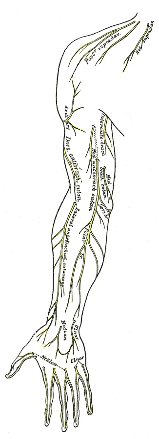
Long description
Cutaneous nerves of right upper extremity. Anterior view.

Figure 1: Cutaneous nerves of right upper extremity (anterior view).
Figure 2Gray814
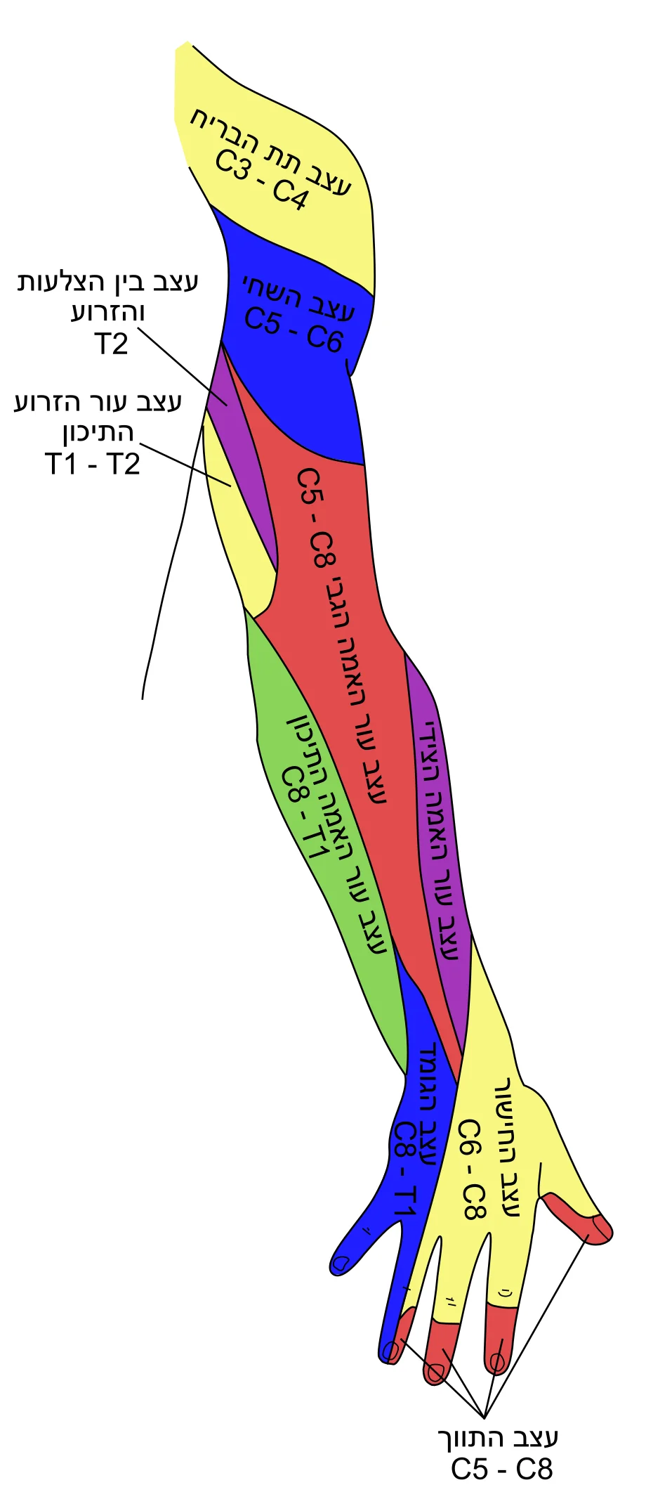
Long description
Diagram of segmental distribution of the cutaneous nerves of the right upper extremity. Posterior view.

Figure 2: Diagram of segmental distribution of the cutaneous nerves of the right upper extremity (posterior view).
Figure 3Gray813
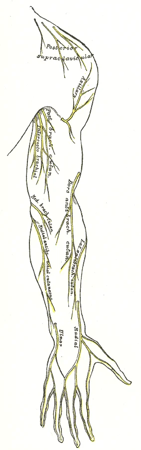
Long description
Cutaneous nerves of right upper extremity. Posterior view.

Figure 3: Cutaneous nerves of right upper extremity (posterior view).
Figure 4Gray812
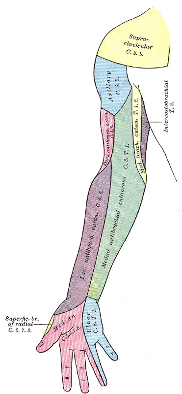
Long description
Diagram of segmental distribution of the cutaneous nerves of the right upper extremity. Anterior view.

Figure 4: Diagram of segmental distribution of the cutaneous nerves of the right upper extremity (anterior view).
The medial cord is positioned medial to the axillary artery, contributing the medial root of the median nerve and the ulnar nerve. The posterior cord lies posterior to the axillary artery, giving rise to the radial nerve and the axillary nerve. For instance, the musculocutaneous nerve pierces the coracobrachialis muscle and runs between the biceps brachii and brachialis muscles, terminating as the lateral antebrachial cutaneous nerve. The axillary nerve winds around the surgical neck of the humerus, supplying the skin over the deltoid muscle. The radial nerve descends in the radial groove of the humerus, giving off posterior cutaneous branches to the arm and forearm before dividing into superficial and deep branches in the forearm. The ulnar nerve travels medially in the arm, passing behind the medial epicondyle of the humerus to enter the forearm, where it supplies the medial aspect of the hand. The median nerve runs down the anterior compartment of the forearm, giving off the palmar cutaneous branch before entering the hand via the carpal tunnel.
The skin of the upper limb is supplied by specific dermatomes, which correspond to the spinal nerve roots. These dermatomal patterns are crucial for localizing nerve lesions.
The brachial plexus also gives rise to several important branches that supply the shoulder and upper arm. The long thoracic nerve, originating from the roots C5-C7, innervates the serratus anterior muscle, which is essential for scapular movement. The thoracodorsal nerve, from the posterior cord, supplies the latissimus dorsi muscle. These nerves are closely related to the bones and muscles of the shoulder girdle, and their course is influenced by the movements of the shoulder joint.
In the forearm, the cutaneous innervation is provided by the medial antebrachial cutaneous nerve, the lateral antebrachial cutaneous nerve, and the posterior antebrachial cutaneous nerve. The medial antebrachial cutaneous nerve arises from the medial cord and supplies the medial aspect of the forearm. The lateral antebrachial cutaneous nerve is the terminal branch of the musculocutaneous nerve and supplies the lateral aspect of the forearm. The posterior antebrachial cutaneous nerve arises from the radial nerve and supplies the posterior aspect of the forearm. These nerves run alongside the major arteries and veins of the forearm, providing sensory innervation to the skin.
In the hand, the cutaneous innervation is more complex, with overlapping distributions from the median, ulnar, and radial nerves. The median nerve supplies the palmar aspect of the thumb, index, middle, and radial half of the ring finger. The ulnar nerve supplies the palmar aspect of the little finger and the ulnar half of the ring finger. The radial nerve supplies the dorsal aspect of the thumb, index, and middle fingers, as well as the dorsal aspect of the radial side of the hand. This overlapping innervation ensures that loss of one nerve does not result in complete sensory loss in a specific area, although it may lead to reduced sensation.
The clinical significance of this innervation pattern is evident in nerve injuries. For example, a fracture of the surgical neck of the humerus may damage the axillary nerve, leading to sensory loss over the deltoid muscle and weakness in shoulder abduction. A fracture of the mid-shaft of the humerus may damage the radial nerve, resulting in wrist drop and sensory loss over the dorsal aspect of the hand. Understanding the course and relations of these nerves is essential for diagnosing and managing such injuries.
Figure 5Gray812and814
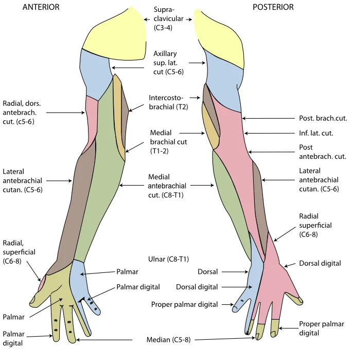
Long description
The cutaneous innervation of the right upper limb. Both areas supplied by the medial brachial cutaneous are colored green. The areas supplied by branches of the radial nerve are colored in pink.

Figure 5: The cutaneous innervation of the right upper limb.
The cutaneous innervation of the upper limb is derived from the brachial plexus, a complex network of nerves formed by the ventral rami of spinal nerves. Understanding this distribution requires tracing the pathway from the spinal roots through the plexus to the terminal cutaneous branches.
Figure 62127 Thoracic Upper Limb Arteries
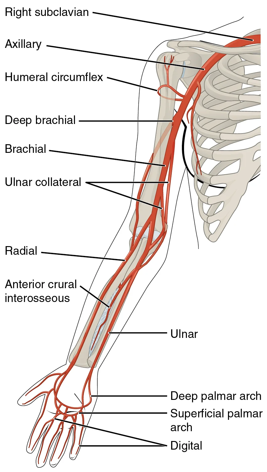
Long description
The arteries that supply blood to the arms and hands are extensions of the subclavian arteries.

Figure 6: The arteries that supply blood to the arms and hands are extensions of the subclavian arteries.
Moving distally, the musculocutaneous nerve pierces the coracobrachialis muscle and terminates as the lateral antebrachial cutaneous nerve, which supplies the lateral aspect of the forearm. The median nerve, running down the anterior arm and forearm, gives off the palmar cutaneous branch proximal to the wrist, supplying the central palm, before continuing into the hand to innervate the lateral palm and the palmar aspects of the lateral three and a half digits. The ulnar nerve, arising from the medial cord, travels down the medial arm and forearm, passing posterior to the medial epicondyle of the humerus. It supplies the medial one and a half digits and the corresponding part of the hand. Finally, the radial nerve, the largest branch, supplies the posterior arm and forearm, and the dorsal aspect of the lateral hand, sparing the fingertips.
| Nerve | Origin | Sensory Supply | Motor Supply |
|---|---|---|---|
| Axillary | Posterior Cord | Skin over lower deltoid (regimental badge) | Deltoid, Teres minor |
| Musculocutaneous | Lateral Cord | Lateral forearm (via lateral antebrachial cutaneous) | Coracobrachialis, Biceps brachii, Brachialis |
| Median | Lateral and Medial Cords | Lateral palm, palmar aspect of lateral 3.5 digits | Flexors of forearm, thenar muscles |
| Ulnar | Medial Cord | Medial 1.5 digits, medial palm | Intrinsic hand muscles (hypothenar, interossei) |
| Radial | Posterior Cord | Posterior arm, posterior forearm, dorsal lateral hand | Extensors of arm, forearm, and hand |
Clinical assessment of these nerves involves testing both sensory and motor functions. Sensory functions are associated with the dorsal regions of the spinal cord, whereas motor function is associated with the ventral side[1]. Localizing damage to the spinal cord is related to assessments of the peripheral projections mapped to dermatomes[1]. For example, loss of sensation in the lateral forearm suggests musculocutaneous nerve injury, while weakness in wrist extension points to radial nerve involvement. The reflexes that are tested in the neurological exam are classified into two groups[1]. A deep tendon reflex is commonly known as a stretch reflex, and is elicited by a strong tap to a tendon, such as in the knee-jerk reflex[1]. A superficial reflex is elicited through gentle stimulation of the skin and causes contraction of the associated muscles[1]. [2]
When examining the upper limb, it is essential to distinguish between peripheral nerve lesions and root lesions. A ganglion is a group of neuron cell bodies in the periphery. Ganglia can be categorized, for the most part, as either sensory ganglia or autonomic ganglia, referring to their primary functions. The most common type of sensory ganglion is a dorsal (posterior) root ganglion. These ganglia are the cell bodies of neurons with axons that are sensory endings in the periphery, such as in the skin, and that extend into the CNS through the dorsal nerve root. Damage to these ganglia or the roots themselves will result in a dermatomal pattern of sensory loss, distinct from the peripheral nerve distribution. [2]
Clinical pearlDermatome vs Nerve
Dermatomes overlap significantly; a single nerve lesion causes more extensive sensory loss than a single root lesion. Always map the deficit to the specific nerve territory, not just the dermatome.
The corticospinal tract represents the neurons that send output from the primary motor cortex[1]. These fibers travel through the deep white matter of the cerebrum, then through the midbrain and pons, into the medulla where most of them decussate, and finally through the spinal cord white matter in the lateral (crossed fibers) or anterior (uncrossed fibers) columns[1]. These fibers synapse on motor neurons in the ventral horn[1]. The ventral horn motor neurons then project to skeletal muscle and cause contraction[1]. These two cells are termed the upper motor neuron (UMN) and the lower motor neuron (LMN)[1]. Voluntary movements require these two cells to be active[1].
In the context of the upper limb, the brachial plexus serves as the conduit for both these motor commands and sensory feedback. This integration allows for precise coordination of movement and sensation. This test, known as the pronator drift, is a sensitive indicator of upper motor neuron pathology affecting the corticospinal tract.
Exam trapSensory Testing
Do not rely solely on light touch. Use pinprick for spinothalamic tract function and vibration for dorsal column function.
Reflexes combine the spinal sensory and motor components with a sensory input that directly generates a motor response[1]. Understanding the anatomy of the brachial plexus and its cutaneous branches is fundamental to interpreting these clinical findings accurately.
Each nerve is colour-coded to match its corresponding dermatome on the skin surface.
Lesions of the upper limb nerves produce specific sensory deficits depending on the level of injury. A lesion at the level of the cervical nerve roots affects the corresponding dermatome. For example, a C6 root lesion causes sensory loss over the lateral forearm and thumb. A C7 root lesion affects the middle finger.
Peripheral nerve lesions produce deficits in the distribution of that specific nerve. A median nerve lesion at the wrist causes sensory loss over the palmar aspect of the lateral three and a half digits. An ulnar nerve lesion at the elbow causes sensory loss over the medial one and a half digits and the corresponding palm.
| Nerve | Site of Lesion | Sensory Deficit |
|---|---|---|
| Radial | Spiral groove | Posterior arm, forearm, dorsal hand |
| Median | Wrist | Palmar lateral 3.5 digits |
| Ulnar | Elbow | Palmar medial 1.5 digits |
| Axillary | Surgical neck of humerus | Lateral shoulder (regimental badge area) |
| Musculocutaneous | Coracobrachialis | Lateral forearm |
The sensitivity to fine touch, which can give indications of the texture and detailed shape of objects, is highest in the fingertips. To assess the limit of this sensitivity, two-point discrimination is measured by simultaneously touching the skin in two locations, such as could be accomplished with a pair of forceps. Failure to recognize two points may be an indication of a dorsal column pathway deficit. [1]
The median nerve supplies the anterior forearm flexors and lateral palm sensation. The ulnar nerve supplies the medial hand sensation and intrinsic hand muscles. The axillary nerve supplies the deltoid and teres minor, with sensation over the lateral shoulder. The musculocutaneous nerve supplies the anterior arm muscles and lateral forearm sensation.
The sensory examination assesses somatic senses, which are those consciously perceived by the patient. This process begins by identifying the specific regions known as dermatomes, which connect to the cortical area where somatosensation is processed in the postcentral gyrus. To evaluate these sensory fields, a clinician applies a simple stimulus, such as the light touch of the soft end of a cotton-tipped applicator, at various locations on the skin. The spinal nerves contain sensory fibers with dendritic endings in the skin and connect with the skin in a topographically organized manner, which is illustrated as dermatomes. [1]
In addition to testing perception at different positions on the skin, it is necessary to test sensory perception within the dermatome from distal to proximal locations in the appendages, or lateral to medial locations in the trunk. When testing the eighth cervical nerve, the patient would be asked if the touch of the cotton to the fingers or the medial forearm was perceptible, and whether there were any differences in the sensations. This systematic approach ensures that specific nerve pathways are functioning correctly and helps localize any potential lesions. [1]
The sense of position and motion, known as proprioception, is tested by moving the fingers or toes and asking the patient if they sense the movement. If the distal locations are not perceived, the test is repeated at increasingly proximal joints. This method allows the examiner to determine the extent of any sensory deficit and identify the specific level of the nervous system that may be affected. [1]
A final subtest of sensory perception that concentrates on the sense of proprioception is known as the Romberg test. The patient is asked to stand straight with feet together. Once the patient has achieved their balance in that position, they are asked to close their eyes. Without visual feedback that the body is in a vertical orientation relative to the surrounding environment, the patient must rely on the proprioceptive stimuli of joint and muscle position, as well as information from the inner ear, to maintain balance. This test can indicate deficits in dorsal column pathway proprioception, as well as problems with proprioceptive projections to the cerebellum through the spinocerebellar tract. [1]
Bundles of axons in the peripheral nervous system are referred to as nerves. These structures in the periphery are different than the central counterpart, called a tract. Nerves are composed of more than just nervous tissue; they have connective tissues invested in their structure, as well as blood vessels supplying the tissues with nourishment. The outer surface of a nerve is a surrounding layer of fibrous connective tissue called the epineurium. Within the nerve, axons are further bundled into fascicles, which are each surrounded by their own layer of fibrous connective tissue called perineurium. Finally, individual axons are surrounded by loose connective tissue called the endoneurium. [2]
DefinitionDermatome
A region of skin innervated by a single spinal nerve.
Clinical pearlProprioception
The sense of position and motion, tested by moving fingers or toes.
The cutaneous innervation of the upper limb is derived from the brachial plexus, formed by the ventral rami of C5 to T1. Understanding this distribution is critical for localizing neurological lesions.
The brachial plexus originates from the ventral rami of spinal nerves C5 through T1. These roots combine to form three trunks: the superior trunk (C5–C6), the middle trunk (C7), and the inferior trunk (C8–T1). This organization allows for the coordinated motor and sensory supply to the shoulder and arm.
The musculocutaneous nerve arises from the lateral cord (C5–C7). It pierces the coracobrachialis muscle and terminates as the lateral antebrachial cutaneous nerve. This nerve supplies the skin of the lateral aspect of the forearm. A lesion here results in sensory loss over the lateral forearm and weakness in elbow flexion.
The axillary nerve arises from the posterior cord (C5–C6). It winds around the surgical neck of the humerus. It provides cutaneous innervation to the skin over the lower part of the deltoid muscle, known as the regimental badge area. Damage to this nerve, often from anterior shoulder dislocation, causes sensory loss in this specific patch and deltoid weakness.
It descends in the radial groove of the humerus. Its terminal branch, the posterior antebrachial cutaneous nerve, supplies the skin on the posterior aspect of the forearm.
A classic trap is confusing the median nerve's sensory territory with the radial nerve's dorsal supply; the median nerve does not supply the dorsal hand except for the distal phalanges.
The ulnar nerve arises from the medial cord (C8–T1). It passes behind the medial epicondyle of the humerus. It provides cutaneous innervation to the palmar and dorsal aspects of the medial one and a half digits (little finger and ulnar half of ring finger). It also supplies the skin of the medial palm and the corresponding dorsal hand area.
Lesions of the brachial plexus can be upper (Erb’s palsy, C5–C6) or lower (Klumpke’s palsy, C8–T1). Erb’s palsy results in a 'waiter’s tip' posture and sensory loss over the lateral arm. Klumpke’s palsy affects the hand intrinsics and results in a claw hand, with sensory loss in the medial forearm and hand.
Testing sensation involves mapping these specific dermatomes. For example, loss of sensation in the 'regimental badge' area localizes the lesion to the axillary nerve. Loss of sensation in the lateral forearm suggests musculocutaneous nerve involvement.
Clinical pearlDermatome Mapping
Always test the fingertips and the lateral forearm to distinguish between median, ulnar, and musculocutaneous nerve lesions.
Exam trapCommon Trap
Do not confuse the radial nerve's dorsal hand supply with the median nerve's. The median nerve only supplies the dorsal tips of the lateral three and a half digits.
| Nerve | Primary Cutaneous Territory |
|---|---|
| Musculocutaneous | Lateral forearm |
| Axillary | Lateral shoulder (regimental badge) |
| Radial | Posterior forearm, dorsal lateral hand |
| Median | Palmar lateral 3.5 digits, dorsal tips |
| Ulnar | Palmar/dorsal medial 1.5 digits, medial palm |
Students frequently confuse the structural definitions of the peripheral nervous system with those of the central nervous system, leading to errors in identifying cutaneous innervation patterns. The peripheral nervous system is defined broadly as everything that is not the central nervous system. This definition implies that many neural structures are incorporated into other organs, such as the digestive system, rather than existing as isolated nerves. When studying cutaneous innervation, it is crucial to remember that the peripheral structures are not as contained as the central structures. A common error is treating peripheral nerves as simple linear cables; in reality, they form complex networks. For instance, terminal ganglia below the head and neck are often incorporated into the wall of the target organ as a plexus. While this concept is most famously applied to the enteric nervous system, the principle of plexus formation applies to somatic nerves as well. A plexus, in a general sense, is a network of fibers or vessels. Students must distinguish between the main trunk of a nerve and its terminal branches. Just as a large artery referred to as a trunk gives rise to several smaller arteries, a major peripheral nerve trunk gives rise to several smaller cutaneous branches. Confusing the trunk with its branches leads to incorrect localization of sensory loss. [2]
Another significant pitfall involves the lateralization of innervation. Students often assume that sensory deficits are always ipsilateral to the lesion in the peripheral nerve, which is correct, but they may confuse this with central pathways. Furthermore, when describing the anatomy of the PNS, it is necessary to describe the common structures, the nerves and the ganglia, as they are found in various parts of the body. This means that the same nerve may have different names or relations depend
A third pitfall is the misinterpretation of vascular and neural relationships. The pulmonary trunk curves posteriorly and rapidly bifurcates (divides) into two branches, a left and a right pulmonary artery. This bifurcation pattern is analogous to the division of major nerves like the brachial plexus. However, students must be careful not to confuse vascular bifurcations with neural divisions. The single vessel exiting the right ventricle is the pulmonary trunk. To prevent confusion between thes
Finally, students often fail to appreciate the complexity of the networks. The right and left anterior cerebral arteries join together to form an anastomosis called the anterior communicating artery. The initial segments of the anterior cerebral arteries and the anterior communicating artery form the anterior portion of the arterial circle. This anastomotic nature ensures redundancy. Peripheral nerves also have overlapping innervation zones. The basilar artery is an anastomosis that begins at the junction of the two vertebral arteries and sends branches to the cerebellum and brain stem. It flows into the posterior cerebral arteries. This redundancy is a key feature of both vascular and neural systems. Summarizes the veins of the thoracic region that flow into the superior vena cava. Understanding the drainage patterns helps in understanding the sensory pathways. The veins of the thoracic region that flow into the superior vena cava mirror the arterial supply. This symmetry is a useful mnemonic for remembering innervation patterns.
Knowledge of cutaneous innervation is fundamental at the bedside for localizing neurological lesions. When a patient presents with sensory loss, the clinician must map the deficit to a specific dermatome or nerve distribution. The PNS is not as contained as the CNS because it is defined as everything that is not the CNS. This broad definition means that lesions can occur anywhere along the pathway from the spinal root to the skin. In the operating theatre, surgeons must identify nerves to avoid iatrogenic injury. For example, during shoulder surgery, the surgeon must be aware of the brachial plexus branches. As you study this section, imagine you are on a “Voyage of Discovery” similar to Lewis and Clark’s expedition in 1804–1806, which followed rivers and streams through unfamiliar territory, seeking a water route from the Atlantic to the Pacific Ocean. This approach is effective for mastering major circulatory patterns. Similarly, tracing nerve pathways from the spine to the skin is essential for surgical planning. Another approach that works well for many students is to create simple line drawings similar to the ones provided, labeling each of the major vessels. Drawing nerve pathways helps in visualizing their course and relations.
On imaging, radiologists use cutaneous innervation patterns to interpret nerve compression syndromes. The external iliac artery exits the body cavity and enters the femoral region of the lower leg. As it passes through the body wall, it is renamed the femoral artery. This renaming at anatomical boundaries is a key concept for identifying nerve entrapment sites. For instance, the femoral nerve passes under the inguinal ligament, analogous to the artery. The femoral artery also gives rise to the genicular artery, which provides blood to the region of the knee. This branching pattern is mirrored by the saphenous nerve, a branch of the femoral nerve, which supplies the medial leg. In examinations, students are often tested on the specific branches of major nerves. The popliteal artery branches into the anterior and posterior tibial arteries. The tibial and common peroneal nerves branch from the sciatic nerve in the popliteal fossa. Understanding these branching points is crucial for answering questions on nerve lesions. [3]
The pulmonary trunk curves posteriorly and rapidly bifurcates (divides) into two branches, a left and a right pulmonary artery. This bifurcation is a key landmark in thoracic surgery, similar to the bifurcation of the brachial plexus into cords. To prevent confusion between these vessels, it is important to refer to the vessel exiting the heart as the pulmonary trunk, rather than also calling it a pulmonary artery. Precision in naming is tested in exams. The internal carotid arteries along with the vertebral arteries are the two primary suppliers of blood to the human brain. Given the central role and vital importance of the brain to life, it is critical that blood supply to this organ remains uninterrupted. This principle of critical supply applies to nerves; interruption of nerve supply can result in temporary loss of function. Loss of blood flow for longer periods, typically between 3 and 4 minutes, will likely produce irreversible brain damage or a stroke. Prolonged nerve compression can lead to similar irreversible damage. [3]
In clinical practice, the concept of plexus formation is vital. Terminal ganglia below the head and neck are often incorporated into the wall of the target organ as a plexus. A plexus, in a general sense, is a network of fibers or vessels. The brachial plexus is a network of nerve fibers. The right and left anterior cerebral arteries join together to form an anastomosis called the anterior communicating artery. The initial segments of the anterior cerebral arteries and the anterior communicating artery form the anterior portion of the arterial circle. This anastomotic nature ensures redundancy, which is also seen in nerve innervation. The basilar artery is an anastomosis that begins at the junction of the two vertebral arteries and sends branches to the cerebellum and brain stem. It flows into the posterior cerebral arteries. This redundancy is a key feature of both vascular and neural systems. Summarizes the veins of the thoracic region that flow into the superior vena cava. Understanding the drainage patterns helps in understanding the sensory pathways. The veins of the thoracic region that flow into the superior vena cava mirror the arterial supply. This symmetry is a useful mnemonic for remembering innervation patterns.
DefinitionPlexus
A network of intersecting nerve fibers, blood vessels, or other structures.
| Nerve | Origin | Cutaneous Territory | Clinical Significance |
|---|---|---|---|
| Musculocutaneous | Lateral cord | Lateral forearm | Loss of sensation on lateral forearm |
| Radial | Posterior cord | Posterior arm, forearm, hand | Wrist drop, sensory loss on dorsal hand |
| Median | Medial and lateral cords | Palmar aspect of lateral 3.5 digits | Carpal tunnel syndrome |
| Ulnar | Medial cord | Palmar aspect of medial 1.5 digits | Claw hand, sensory loss on medial hand |
| Axillary | Posterior cord | Lateral shoulder | "Regimental badge" area sensory loss |
Localizing Upper Limb Sensory Deficits
- Identify Sensory LossMap the area of numbness or paresthesia
- Question: Is it on the lateral forearm?
Yes
- Check Musculocutaneous Nerve
No
- Proceed to next decision
The peripheral nervous system is defined as everything that is not the central nervous system. Terminal ganglia below the head and neck are often incorporated into the wall of the target organ as a plexus. A plexus, in a general sense, is a network of fibers or vessels. The external iliac artery exits the body cavity and enters the femoral region of the lower leg. As it passes through the body wall, it is renamed the femoral artery. The popliteal artery branches into the anterior and posterior tibial arteries. The internal carotid arteries along with the vertebral arteries are the two primary suppliers of blood to the human brain. Loss of blood flow for longer periods, typically between 3 and 4 minutes, will likely produce irreversible brain damage or a stroke.
Answer each one in your head first, then open the answer.
How is the peripheral nervous system defined?
Show answerHide answer
Everything that is not the central nervous systemWhat is a plexus in the context of nervous tissue?
Show answerHide answer
A network of fibers or vesselsWhere does the external iliac artery enter?
Show answerHide answer
The femoral region of the lower legWhat happens to the external iliac artery as it passes through the body wall?
Show answerHide answer
It is renamed the femoral arteryWhat are the two primary suppliers of blood to the human brain?
Show answerHide answer
The internal carotid arteries and the vertebral arteries
- Betts JG, Young KA, Wise JA, et al. The Sensory and Motor Exams. OpenStax, Anatomy and Physiology 2e (CC BY-NC-SA 4.0). Accessed .
- Betts JG, Young KA, Wise JA, et al. The Peripheral Nervous System. OpenStax, Anatomy and Physiology 2e (CC BY-NC-SA 4.0). Accessed .
- Betts JG, Young KA, Wise JA, et al. Circulatory Pathways. OpenStax, Anatomy and Physiology 2e (CC BY-NC-SA 4.0). Accessed .
Figure credits
- Figure 1: Henry Vandyke Carter, via Wikimedia Commons, Public domain, Public domain.
- Figure 2: Henry Vandyke Carter, via Wikimedia Commons, Public domain, Public domain.
- Figure 3: Henry Vandyke Carter, via Wikimedia Commons, Public domain, Public domain.
- Figure 4: Henry Vandyke Carter, via Wikimedia Commons, Public domain, Public domain.
- Figure 5: Henry Vandyke Carter, via Wikimedia Commons, Public domain, Public domain.
- Figure 6: OpenStax College, via Wikimedia Commons, CC BY 3.0, CC BY 3.0.
Educational content for medical students and doctors. It is not a substitute for clinical judgement or local guidelines.