Upper limb: lymphatic drainage
Anatomical pathways of lymphatic drainage in the upper limb and associated nodal groups.
The lymphatic system operates as a parallel circulatory network, distinct from the cardiovascular system in that it lacks a central pump. Instead, lymph is propelled through vessels by skeletal muscle contraction and respiratory movements. This system begins with blind-ending lymphatic capillaries, also known as terminal lymphatics, which permeate almost every soft connective tissue in the body. Interstitial fluid enters these capillaries to become lymph, which then travels through progressively larger vessels. [1]
DefinitionLymphatic Capillaries
Blind-ending vessels where interstitial fluid enters the lymphatic system to become lymph fluid, found in almost every tissue except the CNS and avascular structures.
The structure of lymphatic vessels mirrors that of veins, featuring a three-tunic structure and one-way semi-lunar valves. These valves are spaced closely together, causing bulges that give the vessels a characteristic beaded appearance. Lymph flows from capillaries into larger vessels, eventually merging into lymphatic trunks. The drainage of the upper limb follows specific anatomical routes. Superficial lymphatics of the subcutaneous skin generally follow the course of veins, while deep lymphatic vessels of the viscera follow arterial paths. [1] [3]
DefinitionLymph Nodes
Small, bean-shaped organs located throughout the lymphatic system that act as filters for pathogens and staging areas for immune responses. [1]
Lymph nodes are encapsulated organs separated into compartments by trabeculae. Lymph enters via afferent vessels into the subcapsular sinus, passes through the cortex containing lymphoid follicles, and exits via efferent vessels from the medulla. Dendritic cells and macrophages within the node internalize and kill pathogens, while T cells and B cells mediate adaptive immune responses. [1]
The upper limb is anatomically divided into the arm (shoulder to elbow), forearm (elbow to wrist), and hand (distal to wrist). Lymphatic drainage from the hand and forearm primarily targets the epitrochlear nodes, located medial to the humerus above the elbow. From there, lymph drains into the axillary nodes. The axillary nodes are grouped into five clusters: pectoral, subscapular, humeral, central, and apical. The apical nodes drain into the subclavian trunk.
Exam trapDrainage Asymmetry
The right lymphatic duct drains only the upper right quadrant, whereas the thoracic duct drains the rest of the body, including the left upper limb. [1]
The right upper limb drains into the right lymphatic duct, which empties into the junction of the right jugular and subclavian veins. The left upper limb drains into the thoracic duct, which originates from the cisterna chyli and empties into the left subclavian vein. Disruption of these pathways, such as through surgical removal of axillary nodes, can lead to lymphoedema. This condition results from the accumulation of protein-rich fluid in the interstitial spaces due to impaired lymphatic return. [1]
The lymphatic system serves as a vital drainage network, removing excess interstitial fluid and returning it to the bloodstream to maintain fluid balance[1]. In humans, capillary filtration releases approximately 20 liters of plasma into the interstitial space daily[1]. While 17 liters are reabsorbed directly by blood vessels, the remaining 3 liters are collected by the lymphatic system[1]. This fluid, now termed lymph, travels through a series of vessels, trunks, and ducts[1].
DefinitionLymph
Interstitial fluid that has entered the lymphatic system is referred to as lymph.
The system also transports dietary lipids and immune cells[1]. Lymphatic capillaries are blind-ended tubes formed by a single layer of endothelial cells[1]. These cells overlap to form initial flaps that act as one-way valves[1]. When interstitial pressure is low, the flaps close to prevent backflow[1]. As pressure increases, collagen filaments anchor the capillaries to surrounding structures and pull the flaps open, allowing fluid entry[1].
NoteMechanism
Collagen filaments pull on endothelial cell flaps when interstitial pressure increases, facilitating fluid entry.
In the upper limb, lymphatic drainage follows a predictable pattern. Superficial lymphatic vessels accompany the superficial veins, primarily the cephalic and basilic veins[1]. These vessels drain the skin and subcutaneous tissues of the hand and forearm. Deep lymphatic vessels accompany the deep arteries and drain the muscles, joints, and bones[1].
Figure 1H. Gray, Anatomy Descriptive and Surgical.
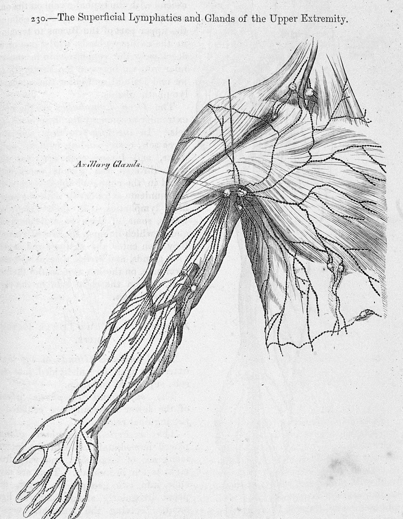
Long description
Illustration 230. The superficial lymphatics and glands of the upper extremity. General Collections Keywords: h v carter, md - illustrations; superficial lymphatics/ glands; Anatomy; Henry Gray; H.V. Carter

Figure 1: Illustration 230.
At the elbow, the vessels converge towards the cubital lymph nodes. From here, the main trunks ascend through the arm, passing deep to the biceps brachii. They terminate in the axillary lymph nodes, which are located in the axilla. The axillary nodes then drain into the subclavian trunk.
The axillary lymph nodes are grouped into five sets: pectoral, subscapular, humeral, central, and apical[1]. The pectoral nodes receive drainage from the anterior chest wall and breast[1]. The subscapular nodes drain the posterior chest wall and back[1]. The humeral nodes drain the upper limb[1]. The central nodes receive lymph from the previous three groups[1]. The apical nodes receive lymph from the central nodes and the upper part of the breast[1].
Clinical pearlClinical Relevance
The apical axillary nodes are the final common pathway for upper limb lymphatic drainage before entering the venous system.
The efferent vessels from the apical nodes form the subclavian trunk[1]. On the right side, the right subclavian trunk typically joins the right jugular trunk to form the right lymphatic duct[1]. The right lymphatic duct drains into the junction of the right internal jugular and right subclavian veins[1]. On the left side, the left subclavian trunk usually joins the left jugular trunk to form the thoracic duct[1]. The thoracic duct drains into the junction of the left internal jugular and left subclavian veins[1].
DefinitionThoracic Duct
The thoracic duct is the larger of the two main lymphatic trunks, draining the left upper limb and the rest of the body below the diaphragm.
This termination ensures that lymph returns to the venous circulation at the subclavian veins, completing the cycle of fluid balance[1]. The immune cells transported within this system can then access the bloodstream to mount responses against pathogens[1].
The lymphatic drainage of the upper limb follows a predictable pathway, moving from superficial cutaneous vessels to deep vessels accompanying the major arteries, ultimately converging on the axillary lymph nodes. Understanding this hierarchy is essential for clinical assessment of metastatic spread and lymphoedema.
All these pathways converge towards the axillary region, where they terminate in the axillary lymph nodes.
DefinitionSuperficial Drainage
Superf lymphatic vessels follow the course of the cephalic and basilic veins, draining the skin and subcutaneous tissues of the hand and forearm.
The superficial lymphatic vessels are numerous and form an extensive network. They begin in the digital pulp and the palm, draining into the dorsal lymphatic plexus of the hand. From here, vessels travel along the radial (lateral) border, accompanying the cephalic vein, to reach the deltopectoral groove. Here, they drain into the infraclavicular (pectoral) group of axillary nodes. Other superficial vessels travel along the ulnar (medial) border, accompanying the basilic vein. These vessels pierce the deep fascia in the mid-arm and drain directly into the central or apical groups of the axillary nodes.
Figure 2Gray608
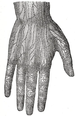
Long description
Lymphatic vessels of the dorsal surface of the hand. (Sappey.)

Figure 2: Lymphatic vessels of the dorsal surface of the hand (Sappey.).
Exam trapEpitrochlear Nodes
Enlargement of the epitrochlear nodes is not a normal finding and suggests pathology in the ulnar aspect of the forearm or hand, such as infection or malignancy.
The deep lymphatic vessels accompany the major arteries of the upper limb: the radial, ulnar, and interosseous arteries. These vessels originate from the deep structures of the hand, including the muscles, joints, and bones. They travel proximally within the neurovascular bundles. The deep vessels generally bypass the epitrochlear nodes and drain directly into the axillary lymph nodes, specifically the central and apical groups. This deep pathway is crucial for draining the intrinsic muscles of the hand and the forearm flexors and extensors.
| Structure | Primary Drainage Pathway | Terminal Nodes |
|---|---|---|
| Digital Pulp | Superficial plexus | Infraclavicular (Pectoral) |
| Medial Forearm Skin | Superficial (Basilic vein) | Central / Apical |
| Lateral Forearm Skin | Superficial (Cephalic vein) | Infraclavicular (Pectoral) |
| Deep Hand Muscles | Deep (Radial/Ulnar arteries) | Central / Apical |
| Medial Elbow Region | Superficial | Epitrochlear |
These groups are the pectoral (anterior), subscapular (posterior), lateral, central, and apical (apical) nodes. The pectoral nodes receive drainage from the anterior chest wall, breast, and upper abdominal wall. The subscapular nodes drain the posterior chest wall and back. The central nodes receive lymph from the previous three groups and drain into the apical nodes. The apical nodes are the final common pathway for the upper limb, draining into the subclavian trunk. [1]
Clinical pearlClinical Relevance
The apical nodes are the last station for upper limb lymph; obstruction here causes severe lymphoedema of the entire arm.
In summary, the lymphatic drainage of the upper limb is a dual system of superficial and deep vessels. The superficial system follows the superficial veins, while the deep system follows the major arteries. Both systems ultimately converge on the axillary lymph nodes, with the lateral nodes being the primary recipients for the limb itself. This organized drainage pattern allows for precise clinical localization of pathology.
The lymphatic drainage of the upper limb follows a predictable superficial and deep pattern, ultimately converging on the axillary lymph nodes. Understanding this pathway is essential for identifying the spread of pathology, such as malignancy or infection, from the extremity to the central thoracic structures.
The axillary nodes are arranged in groups: pectoral (anterior), subscapular (posterior), lateral, central, and apical. The apical nodes are located at the apex of the axilla, near the clavicle. From the apical nodes, efferent vessels form the subclavian lymphatic trunk. On the right side, this trunk typically drains into the right lymphatic duct, which empties into the junction of the right internal jugular and right subclavian veins. On the left side, the trunk drains into the thoracic duct, which empties into the left venous angle. This anatomical arrangement ensures that lymph from the entire upper limb, including the hand, forearm, arm, and lateral chest wall, is filtered through the axillary nodes before entering the systemic circulation. [1]
DefinitionLymphedema
When the lymphatic system is damaged in some way, such as by being blocked by cancer cells or destroyed by injury, protein-rich interstitial fluid accumulates (sometimes “backs up” from the lymph vessels) in the tissue spaces. This inappropriate accumulation of fluid referred to as lymphedema may lead to serious medical consequences. [1]
The clinical significance of this drainage pattern is profound. Because the axillary nodes are the primary filter for the upper limb, any obstruction or infiltration of these nodes can result in significant swelling of the arm. This is commonly seen following mastectomy or axillary lymph node dissection for breast cancer. The accumulation of fluid is not merely water; it is protein-rich, leading to chronic inflammation and fibrosis if not managed.
| Structure | Primary Drainage Site | Clinical Relevance |
|---|---|---|
| Superficial vessels (medial arm/forearm) | Axillary nodes (lateral & central groups) | Common site for spread of skin cancers |
| Deep vessels (accompanying arteries) | Axillary nodes (apical group) | Spread from deep muscle or bone pathology |
| Lateral chest wall | Axillary nodes (pectoral group) | Spread from breast tissue pathology |
| Apical nodes | Subclavian lymphatic trunk | Entry point into systemic venous circulation |
The convergence of lymphatic vessels at the axilla mirrors the arterial supply, which also undergoes name changes at anatomical landmarks. For example, the left subclavian artery becomes the axillary artery as it passes through the body wall and into the axillary region, and then becomes the brachial artery as it flows from the axillary region into the upper arm (or brachium)[1]. Similarly, lymphatic vessels change their grouping and nomenclature as they progress from the peripheral tissues to the central thoracic duct or right lymphatic duct. This parallel organization highlights the integrated nature of the circulatory and lymphatic systems in maintaining fluid homeostasis.
NoteClinical Pearl
Always consider the lymphatic drainage when assessing swelling in the upper limb. Swelling may indicate not just local injury but also obstruction at the axillary level, potentially signaling underlying malignancy or infection.
The detailed mapping of these territories allows clinicians to predict the spread of disease. For instance, carcinoma of the breast often metastasizes to the pectoral lymph nodes first, which are part of the axillary group. The superficial vessels, running with the basilic vein, are particularly important as they are often the first to be involved in cutaneous malignancies of the upper limb.
The lymphatic system of the upper limb relies on a network of channels that ensure drainage even if one pathway is obstructed. This redundancy is achieved through extensive anastomoses, which are connections between vessels that allow blood or lymph to bypass blockages. Understanding these pathways is crucial for clinical practice, particularly in cases of lymphoedema or after surgical intervention.
The deep lymphatic vessels accompany the major arteries, such as the brachial artery.
The superficial lymphatic vessels are more numerous and follow the venous drainage patterns. They begin in the digital lymphatics of the fingers and palm. From the fingers, they drain into the palmar lymphatic plexus. This plexus connects with the dorsal lymphatic vessels. The dorsal vessels drain the back of the hand and fingers. They converge to form two main trunks: the radial and ulnar trunks. The radial trunk follows the cephalic vein, while the ulnar trunk follows the basilic vein. These trunks anastomose in the forearm, creating a collateral pathway. This anastomosis ensures that lymph can flow from the ulnar side to the radial side, or vice versa, if one path is compromised.
DefinitionAnastomosis
A connection between two vessels, allowing flow to bypass an obstruction.
The deep lymphatic vessels are fewer in number but follow a more direct route. They accompany the brachial arteries and veins. These vessels drain the muscles and bones of the arm. They converge at the elbow, where they anastomose with the superficial vessels. This connection is vital for draining the elbow joint and surrounding structures. The deep vessels then pass through the axilla, joining the superficial vessels at the axillary lymph nodes.
The axillary lymph nodes receive lymph from both the superficial and deep systems. They are arranged in groups: pectoral, subscapular, lateral, central, and apical. The pectoral nodes drain the anterior chest wall and breast. The subscapular nodes drain the posterior chest wall and back. The lateral nodes drain the upper limb. The central nodes receive lymph from the other groups. The apical nodes drain the central nodes and the upper part of the breast. Lymph from the apical nodes passes into the subclavian trunk.
Clinical pearlClinical Relevance
Damage to the axillary lymph nodes can lead to lymphoedema of the upper limb.
The subclavian trunk then joins the jugular trunk to form the right lymphatic duct on the right side, or the thoracic duct on the left side. These ducts empty into the venous system at the junction of the internal jugular and subclavian veins. This final connection ensures that lymph returns to the bloodstream.
The anastomoses between the superficial and deep systems are particularly important. They allow for collateral drainage in case of obstruction. For example, if the axillary vein is thrombosed, lymph can still drain via the deep vessels. Similarly, if the superficial vessels are damaged, the deep vessels can compensate. This redundancy is a key feature of the lymphatic system.
| Pathway | Origin | Termination | Key Anastomosis |
|---|---|---|---|
| Superficial | Digital lymphatics | Axillary nodes | Radial-Ulnar |
| Deep | Brachial arteries | Axillary nodes | Elbow |
| Axillary | Superficial/Deep | Subclavian trunk | Central-Apical |
Understanding these pathways is essential for diagnosing and treating lymphatic disorders. Surgeons must be aware of these connections to avoid unnecessary damage. Clinicians can use this knowledge to assess the extent of lymphatic obstruction. The anastomoses provide a safety net, ensuring that lymph can always find a route back to the heart.
The lymphatic drainage of the upper limb is not a rigid, unchanging highway but a dynamic network subject to significant individual variation. While standard anatomical textbooks describe a predictable pattern of superficial and deep drainage, clinical practice and dissection reveal that these pathways can diverge considerably from the norm. Understanding these variations is critical for accurate diagnosis, surgical planning, and interpreting imaging studies, as deviations can alter the spread of pathology such as infection or malignancy.
The superficial lymphatic vessels generally follow the venous drainage patterns of the upper limb. However, the specific routes taken by these vessels can vary. For instance, while most superficial lymphatics from the hand and forearm drain into the axillary nodes, some may bypass these and drain directly into the supraclavicular nodes. This variation is particularly relevant in the context of breast cancer metastasis, where atypical drainage patterns can lead to unexpected nodal involvement.
The initial lymphatic capillaries in the skin of the upper limb form a dense plexus. From this plexus, collecting vessels emerge and travel towards the axilla. In some individuals, these vessels may take a more medial or lateral course than typically described, potentially influencing the clinical presentation of lymphoedema or the spread of cutaneous melanoma.
Figure 3Gray607
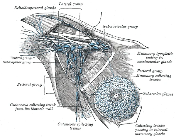
Long description
Lymphatics of the mamma, and the axillary glands (semidiagrammatic). (Poirier and Charpy.)

Figure 3: Lymphatics of the mamma, and the axillary glands (semidiagrammatic) (Poirier and Charpy.).
Deep lymphatic vessels accompany the deep veins of the upper limb. These vessels are generally more consistent in their course than their superficial counterparts but still exhibit notable variations. The deep lymphatics from the hand and forearm typically drain into the brachial nodes, which are located along the brachial vein. From there, the lymph flows into the axillary nodes.
Figure 4Gray609
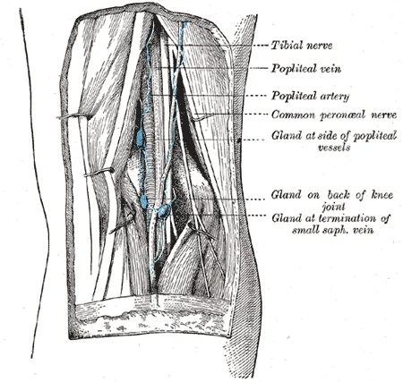
Long description
Lymph glands of popliteal fossa. (Poirier and Charpy.)

Figure 4: Lymph glands of popliteal fossa (Poirier and Charpy.).
However, variations in the number and position of the brachial nodes can occur. Some individuals may have an increased number of nodes, while others may have fewer, with lymph draining directly into the axillary group. This variation can affect the staging of cancers arising in the deep structures of the upper limb. [1]
The axillary lymph nodes are divided into several groups: pectoral, subscapular, humeral, central, and apical. The drainage patterns into these groups can vary. For example, while most lymph from the lateral aspect of the breast drains into the pectoral nodes, some lymph may drain directly into the central or apical nodes. This variation is crucial for surgeons performing axillary lymph node dissection, as missing an atypical node can lead to incomplete staging or treatment.
Furthermore, the connections between the axillary nodes and the supraclavicular nodes can be more prominent in some individuals. This can facilitate the spread of cancer from the upper limb or breast to the supraclavicular region, a finding that has significant prognostic implications.
Recognizing these variations is essential for medical students and clinicians alike. Misinterpretation of lymphatic drainage patterns can lead to diagnostic errors or suboptimal surgical outcomes. For example, in the case of lymphoedema, understanding the specific drainage pathways can help in designing effective compression therapy or surgical interventions. Similarly, in oncology, accurate knowledge of lymphatic variations ensures that staging is precise and that treatment plans are tailored to the individual patient's anatomy.
As noted in the literature, individual variations in circulation patterns are not uncommon. This statement underscores the importance of a flexible approach to anatomical knowledge, one that acknowledges the diversity of human anatomy rather than relying solely on idealized models. By integrating an understanding of these variations into their clinical reasoning, medical students can better navigate the complexities of upper limb lymphatic pathology. [2]
The clinical assessment of the upper limb relies heavily on understanding the relationship between bony landmarks, vascular access points, and potential sites of injury.
Above this lateral epicondyle lies the roughened lateral supracondylar ridge. These bony features are not merely structural; they are key landmarks for identifying the course of neurovascular bundles and lymphatic channels that traverse the cubital fossa and the medial aspect of the arm.
In the context of the upper limb, the distal humerus serves as a critical anchor. The medial epicondyle, being the origin for the anterior forearm muscles, is a site where swelling or trauma can impact the nearby ulnar nerve and associated lymphatic vessels. Similarly, the lateral epicondyle and supracondylar ridge are attachment sites for posterior forearm muscles. Injury to these areas can lead to inflammation that may obstruct local lymphatic flow.
Access points for venous blood, such as the median cubital vein, are located superficially in the cubital fossa, anterior to the distal humerus. Ligation of vessels in this region is rarely performed due to the risk of compromising blood supply to the hand, but understanding the bony landmarks like the medial and lateral epicondyles helps in locating these vessels safely. The roughened lateral supracondylar ridge provides a stable surface for muscle attachment, and any fracture in this region can disrupt the surrounding soft tissue planes, potentially affecting lymphatic drainage. [2]
Clinical pearlLandmark Recognition
The medial epicondyle is larger and more robust than the lateral epicondyle because it anchors the powerful anterior forearm muscles.
| Structure | Location | Clinical Significance |
|---|---|---|
| Medial Epicondyle | Medial side of distal humerus | Attachment for anterior forearm muscles; landmark for ulnar nerve |
| Lateral Epicondyle | Lateral side of distal humerus | Attachment for posterior forearm muscles |
| Lateral Supracondylar Ridge | Above lateral epicondyle | Roughened ridge for muscle attachment |
The lymphatic drainage of the upper limb follows a predictable superficial and deep pattern, converging towards the axilla. Superficial vessels from the hand and forearm generally accompany the cephalic and basilic veins. Deep vessels follow the major arteries, such as the radial, ulnar, and brachial arteries. All lymph from the upper limb ultimately drains into the axillary lymph nodes. These nodes are arranged in groups based on their location relative to the axillary vein and surrounding structures.
The axillary lymph nodes are the primary drainage site for the upper limb. They are divided into five groups: pectoral, subscapular, humeral, central, and apical. The pectoral nodes receive lymph from the anterior chest wall and the breast. The subscapular nodes drain the posterior chest wall and back. The humeral nodes receive lymph from the upper limb. The central nodes receive efferent vessels from the other four groups. The apical nodes receive lymph from the central nodes and the upper part of the breast. Efferent vessels from the apical nodes form the subclavian lymphatic trunk.
Clinical pearlDrainage Pathway
Superficial lymphatics from the lateral side of the hand and forearm accompany the cephalic vein. Deep lymphatics from the medial side accompany the basilic vein and brachial artery.
Breast cancer frequently metastasises to the axillary lymph nodes. The pectoral nodes are particularly important in breast cancer staging. Infection of the upper limb can cause lymphadenopathy in the axillary nodes. The apical nodes are a critical checkpoint for metastatic spread from the upper limb and breast.
| Group | Location | Drainage Area |
|---|---|---|
| Pectoral | Anterior axillary wall | Anterior chest wall, breast |
| Subscapular | Posterior axillary wall | Posterior chest wall, back |
| Humeral | Lateral axillary wall | Upper limb |
| Central | Axillary fat | Efferents from other groups |
| Apical | Apex of axilla | Efferents from central nodes, upper breast |
Exam trapCommon Mistake
Do not confuse the drainage of the thumb with the little finger. The thumb drains laterally, while the little finger drains medially. Both ultimately reach the axillary nodes, but via different initial pathways.
Superficial lymphatic vessels are numerous and follow a course parallel to the superficial veins. They have thin walls and numerous valves to prevent backflow. Deep lymphatic vessels accompany the deep veins and arteries. They are fewer in number than superficial vessels. Both superficial and deep vessels converge towards the axillary nodes. The vessels from the medial side of the forearm and hand often cross the cubital fossa to reach the epitrochlear nodes before proceeding to the axillary nodes.
DefinitionEpitrochlear Nodes
Small lymph nodes located above the medial epicondyle of the humerus. They drain the medial side of the forearm and hand.
Carcinoma of the hand or forearm may first present with enlarged epitrochlear nodes. This is a rare but important clinical sign. Metastasis to the axillary nodes is common in breast cancer. The pattern of spread can help determine the primary site of the tumour. Surgical removal of axillary nodes is a key part of breast cancer treatment.
Students frequently confuse the lymphatic drainage of the upper limb with the venous drainage, assuming they follow identical superficial pathways. While the deep lymphatics generally follow the deep veins, the superficial lymphatics of the upper limb do not strictly parallel the superficial veins. A common error is assuming that all lymph from the hand drains directly into the axillary nodes. In reality, lymph from the lateral aspect of the hand and forearm drains into the epitrochlear nodes before reaching the axillary group, whereas the medial aspect may drain directly to the axillary nodes or via the epitrochlear nodes. Misidentifying this pathway can lead to incorrect localisation of pathology, such as understanding why an infection in the index finger might present with swelling in the epitrochlear region rather than the axilla. [1]
Another significant pitfall involves the terminology of lymph node groups. Students often use "axillary nodes" as a single homogeneous group. It is crucial to distinguish between the pectoral, subscapular, lateral, central, and apical groups. The lateral group receives lymph from the upper limb, while the pectoral group receives lymph from the anterior chest wall and breast. Confusing these can lead to errors in clinical examination, particularly when assessing for metastatic spread from breast cancer, which typically involves the pectoral nodes first, not the lateral nodes which serve the arm.
A third mistake is neglecting the role of the epitrochlear nodes in systemic disease. These nodes are not always palpable in healthy individuals, but their enlargement is a specific sign of infection or inflammation in the hand and forearm, particularly involving the ulnar side. Assuming they are insignificant ignores their role as a filter for the medial aspect of the upper limb.
Fourth, students often overlook the anatomical relations of the axillary vein during lymph node dissection. The axillary vein lies medial to the axillary artery. In surgical contexts, such as axillary lymph node clearance, damaging the vein can cause severe haemorrhage or thrombosis. Understanding that the lymph nodes are arranged around the axillary vein and artery is critical for safe surgical navigation.
Fifth, there is confusion regarding the drainage of the breast. While the breast is not part of the upper limb, its lymphatic drainage is intimately linked to the axillary nodes. Students may incorrectly assume the breast drains only to the axilla, ignoring the significant drainage to the internal thoracic nodes, which is relevant for breast cancer staging. However, focusing strictly on the upper limb, the error lies in failing to recognise that the lateral quadrants of the breast drain to the pectoral nodes, which are part of the axillary chain.
Sixth, students often misidentify the boundaries of the axilla. The apex of the axilla is the thoracic outlet, where structures pass between the neck and the arm. Confusing the boundaries can lead to errors in understanding how infections or tumours might spread from the neck to the arm or vice versa. The lateral boundary is the humerus, specifically the surgical neck, which is a common site for fractures that can disrupt local structures.
Seventh, there is a tendency to ignore the deep lymphatic pathways. Deep lymphatics follow the major arteries, such as the brachial and radial arteries. Assuming all drainage is superficial misses potential sites of metastasis or infection that travel with these deep vessels.
Eighth, students may confuse the lymphatic drainage with the nerve supply. For example, the ulnar nerve supplies the medial hand, and its sensory distribution is often confused with the lymphatic drainage area. While the ulnar nerve supplies the medial one and a half digits, the lymphatic drainage of the medial hand is complex and involves both direct axillary drainage and epitrochlear drainage.
In the operating theatre, knowledge of upper limb lymphatic drainage is critical for axillary lymph node dissection, particularly in breast cancer surgery or melanoma staging. Surgeons must identify and preserve the axillary vein while removing nodes from the pectoral, subscapular, and lateral groups. Understanding the anatomical relations, such as the position of the long thoracic nerve and the thoracodorsal nerve relative to the lymph nodes, prevents iatrogenic injury. The surgical neck of the humerus is a landmark for the proximal extent of the axillary contents, and fractures here can disrupt lymphatic flow.
On imaging, radiologists use contrast-enhanced ultrasound or MRI to visualise enlarged lymph nodes in the axilla and epitrochlear regions. Enlargement of epitrochlear nodes is a specific sign of infection in the hand or forearm, such as cat-scratch disease or sporotrichosis. Recognising this pattern helps in diagnosing the primary source of infection. In breast cancer staging, sentinel lymph node biopsy targets the first draining node, often in the pectoral group, to determine if cancer has spread.
At the bedside, clinicians palpate the epitrochlear nodes in patients with hand infections or systemic conditions like lymphoma. Palpation of the axillary nodes is routine in breast examination and general physical assessment. Identifying tender, enlarged nodes in the epitrochlear region can guide further investigation into hand or forearm pathology, such as cellulitis or lymphangitis.
In examinations, students are often tested on the pathway of lymph from specific digits. For instance, knowing that the index finger drains to the lateral axillary nodes, while the little finger may drain to the epitrochlear nodes, is a classic question. Understanding the distinction between superficial and deep drainage is also frequently tested.
The knowledge is also applied in understanding lymphoedema. After axillary node dissection, patients are at risk of lymphoedema in the arm. Understanding the collateral drainage pathways helps in managing this complication. Compression garments and physiotherapy are used to promote lymphatic flow, relying on the understanding of the remaining functional pathways.
Furthermore, in trauma, understanding the lymphatic anatomy helps in assessing the risk of infection spread. A deep laceration to the forearm may introduce bacteria into the deep lymphatic channels, leading to rapid spread to the axillary nodes. Recognising this risk prompts early antibiotic treatment and monitoring of the nodes.
The anatomical relations of the lymph nodes to the axillary vessels are also crucial in interventional radiology. Procedures such as lymphangiography or node biopsy require precise knowledge of the anatomy to avoid vascular injury. The axillary vein, which lies medial to the artery, is a key landmark.
Finally, in dermatology, the pattern of lymphatic spread can help in diagnosing skin cancers. Melanoma on the arm may metastasise to the axillary nodes, and understanding the drainage pattern helps in planning sentinel node biopsy. The lateral aspect of the arm drains to the lateral axillary nodes, while the medial aspect may involve the subscapular or central nodes.
Lymphatic Drainage Pathway
- StartIdentify the region of the upper limb
- Question: Is it the lateral aspect?
Yes
- Drains to Lateral Axillary Nodes
No
- Question: Is it the medial aspect?
Yes
- Question: Is there hand involvement?
Yes
- Drains to Epitrochlear Nodes
No
- Drains to Central Axillary Nodes
No
- Drains to Pectoral or Subscapular Nodes
Lymph from the lateral upper limb drains to the lateral axillary nodes. Lymph from the medial upper limb may drain to the epitrochlear nodes. The epitrochlear nodes are located medial to the humerus. Axillary lymph nodes are grouped into pectoral, subscapular, lateral, central, and apical groups. The axillary vein lies medial to the axillary artery. Enlargement of epitrochlear nodes suggests hand or forearm pathology. Sentinel lymph node biopsy is used in breast cancer staging. Lymphoedema is a risk after axillary node dissection.
Answer each one in your head first, then open the answer.
Which nodes receive lymph from the lateral aspect of the upper limb?
Show answerHide answer
Lateral axillary nodesWhere are the epitrochlear nodes located?
Show answerHide answer
Medial to the humerus, above the elbowWhich group of axillary nodes receives lymph from the anterior chest wall?
Show answerHide answer
Pectoral nodesWhat is a common sign of hand infection in the lymphatic system?
Show answerHide answer
Enlargement of epitrochlear nodesWhich vessel lies medial to the axillary artery?
Show answerHide answer
Axillary vein
- Betts JG, Young KA, Wise JA, et al. Anatomy of the Lymphatic and Immune Systems. OpenStax, Anatomy and Physiology 2e (CC BY-NC-SA 4.0). Accessed .
- Betts JG, Young KA, Wise JA, et al. Circulatory Pathways. OpenStax, Anatomy and Physiology 2e (CC BY-NC-SA 4.0). Accessed .
- Betts JG, Young KA, Wise JA, et al. Bones of the Upper Limb. OpenStax, Anatomy and Physiology 2e (CC BY-NC-SA 4.0). Accessed .
Figure credits
- Figure 1: https://wellcomeimages.org/indexplus/obf_images/78/4c/f23a478c1ac6a86138f43d6a58cb.jpg Gallery: https://wellcomeimages.org/indexplus/image/L0028363.html Wellcome Collection gallery (2018-03-29): https://wellcomecollection.org/works/dky8hn3w CC-BY-4.0, via Wikimedia Commons, CC BY 4.0, CC BY 4.0.
- Figure 2: Henry Vandyke Carter, via Wikimedia Commons, Public domain, Public domain.
- Figure 3: Henry Vandyke Carter, via Wikimedia Commons, Public domain, Public domain.
- Figure 4: Henry Vandyke Carter, via Wikimedia Commons, Public domain, Public domain.
Educational content for medical students and doctors. It is not a substitute for clinical judgement or local guidelines.