Upper limb: venous drainage
Anatomical overview of superficial and deep venous drainage of the upper limb.
The venous drainage of the upper limb is organised into two distinct systems: superficial veins and deep veins. This dual arrangement serves both circulatory and thermoregulatory functions.
The superficial venous system lies within the subcutaneous tissue and does not typically accompany arteries directly. These veins play a significant role in thermoregulation. When ambient temperatures are warm, blood flow is diverted to these superficial channels to facilitate heat dissipation into the environment. Conversely, in cold conditions, vasoconstriction of these veins shunts blood deeper into the body to conserve heat. [1]
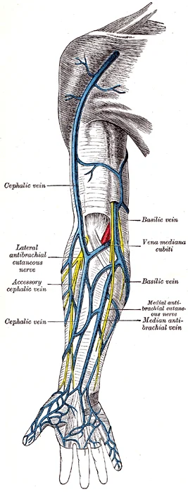
The primary superficial veins are the cephalic, basilic, and median cubital veins. The basilic vein originates in the forearm, running medially. It parallels the ulnar vein and is situated more medially than the median antebrachial vein. As the basilic vein ascends through the arm, it travels superficially along the medial aspect before piercing the deep fascia to join the axillary vein. [1]
Figure 1Gray574
Long description
The superficial veins of the upper extremity.

Figure 1: The superficial veins of the upper extremity.
The cephalic vein runs along the lateral aspect of the arm. The connection between the medial and lateral superficial systems is provided by the median cubital vein. Due to its superficial location and stability, the median cubital vein is the most common site for venepuncture and drawing blood samples in humans. [1]
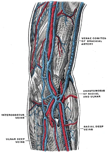
Deep veins accompany the major arteries of the upper limb. They generally correspond to the complementary arteries, forming venae comitantes. These vessels are responsible for the majority of venous return from the limb to the central circulation. [1]
Figure 2Gray575
Long description
The deep veins of the upper extremity. (Bourgery.)

Figure 2: The deep veins of the upper extremity (Bourgery.).
Clinical access to the venous system is most frequently achieved via the superficial veins. The median cubital vein is preferred due to its anatomical position in the antecubital fossa, where it bridges the basilic and cephalic systems. Its superficial course makes it easily accessible for cannulation.
While venous thromboembolism is more commonly discussed in the lower limbs, the principles of venous stasis and vessel wall integrity apply to the upper limb as well. Thrombus formation is influenced by the anatomical features of the veins.
DefinitionSuperficial vs Deep Veins
Superficial veins lack direct arterial counterparts and aid thermoregulation, while deep veins accompany arteries and handle primary blood return. [1]
| Vein | Course | Clinical Significance |
|---|---|---|
| Basilic | Medial forearm to axilla | Joins deep system in axilla |
| Cephalic | Lateral arm to deltopectoral groove | Drains into axillary/subclavian vein |
| Median Cubital | Connects basilic to cephalic | Most common site for venepuncture |
NoteThermoregulation
Superficial veins dilate in warmth to dissipate heat and constrict in cold to retain core body heat.
The venous drainage of the upper limb follows a hierarchical pattern, moving from superficial to deep systems before converging into the axillary vein. This arrangement ensures efficient return of blood against gravity, aided by skeletal muscle pumps and one-way valves.
The superficial system lies within the subcutaneous tissue and is clinically significant for venous access and assessing collateral circulation. It comprises two main trunks: the cephalic and basilic veins.
The cephalic vein originates from the dorsal venous network of the hand. It ascends along the lateral aspect of the forearm and arm, passing between the deltoid and pectoralis major muscles.
The basilic vein arises from the medial side of the dorsal venous network. It travels up the medial forearm and arm, piercing the deep fascia in the mid-arm. It then joins the brachial veins to form the axillary vein.
Deep veins accompany the arteries of the upper limb. They are paired in the forearm and arm, known as venae comitantes. These veins have thin walls and numerous valves to prevent backflow. They converge to form the axillary vein.
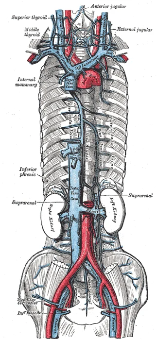
Figure 3Gray577
Long description
The venæ cavæ and azygos veins, with their tributaries.

Figure 3: The venæ cavæ and azygos veins, with their tributaries.
The axillary vein begins at the lower border of the teres major muscle, formed by the union of the basilic vein and the brachial veins. It runs alongside the axillary artery. At the lateral border of the first rib, it becomes the subclavian vein. The subclavian vein joins the internal jugular vein to form the brachiocephalic vein.
DefinitionVenous Drainage
The process by which deoxygenated blood returns from the tissues of the upper limb to the heart via the superior vena cava.
The valves in the deep veins are crucial. They ensure unidirectional flow. Without them, venous hypertension can occur, leading to oedema and skin changes. The superficial veins also contain valves, but they are less numerous. Communication between superficial and deep systems occurs via perforating veins. These perforators allow blood to flow from superficial to deep veins, facilitating drainage.
| Structure | Origin | Termination |
|---|---|---|
| Cephalic Vein | Dorsal venous network | Axillary vein |
| Basilic Vein | Dorsal venous network | Axillary vein |
| Axillary Vein | Union of basilic and brachial veins | Subclavian vein |
| Subclavian Vein | Lateral border of first rib | Brachiocephalic vein |
Understanding this anatomy is vital for procedures such as central line insertion. The subclavian vein is a common site for catheter placement. Knowledge of its course prevents complications such as pneumothorax. Similarly, understanding the superficial veins aids in venipuncture and cannulation. The cephalic vein is often used for peripheral intravenous access due to its accessibility and size.
Exam trapValve Failure
Valve incompetence in the deep veins can lead to chronic venous insufficiency, causing swelling and skin ulcers.
The lymphatic system runs parallel to the venous system, draining interstitial fluid. However, this topic focuses solely on venous drainage. The efficiency of venous return depends on the integrity of the valves and the patency of the veins. Obstruction or thrombosis can disrupt this flow, leading to significant clinical issues.
The venous drainage of the upper limb follows a hierarchical pattern, moving from superficial to deep systems before converging into the axillary vein. This arrangement ensures efficient return of blood against gravity, relying on valves and muscular pumps.
DefinitionSuperficial Veins
The superficial venous system lies within the subcutaneous tissue and does not generally accompany arteries, except in specific regions. It is clinically significant for venous access and mapping.
The cephalic vein originates from the dorsal venous network of the hand. It ascends along the lateral aspect of the forearm and arm, lying superficial to the biceps brachii muscle. It pierces the clavipectoral fascia to join the axillary vein.
The basilic vein arises from the medial side of the dorsal venous network. It travels up the medial aspect of the arm, deep to the biceps brachii. In the mid-arm, it pierces the deep fascia to join the brachial veins, forming the axillary vein.
This vein connects the cephalic and basilic veins in the cubital fossa. It is a common site for venipuncture due to its superficial location and stability.
| Structure | Origin | Course | Termination |
|---|---|---|---|
| Cephalic Vein | Dorsal venous network | Lateral forearm/arm | Axillary vein |
| Basilic Vein | Dorsal venous network | Medial forearm/arm | Axillary vein |
| Median Cubital Vein | Cephalic vein | Cubital fossa | Basilic vein |
The basilic vein is on the medial side. The median cubital vein arches between them, forming a 'Y' shape. The brachial veins are deep to the biceps, accompanying the brachial artery. The axillary vein is formed by the union of the basilic and brachial veins.
The deep venous system consists of paired veins that accompany the arteries of the upper limb. These veins are named after their accompanying arteries and are connected by numerous valves.
The brachial veins are the deep veins of the arm. They accompany the brachial artery on both sides. They begin in the cubital fossa and ascend to the lower border of the teres major muscle, where they join the basilic vein to form the axillary vein.
The axillary vein is the continuation of the brachial and basilic veins. It runs alongside the axillary artery. It terminates at the lateral border of the first rib, becoming the subclavian vein.
The subclavian vein continues from the axillary vein. It joins the internal jugular vein to form the brachiocephalic vein. The right and left brachiocephalic veins unite to form the superior vena cava.
NoteClinical Pearl
The superior vena cava receives systemic venous return from the head, neck, chest, shoulders, and upper limbs. This is a key anatomical landmark for central venous access. [1]
The venous drainage of the upper limb is organized into superficial and deep systems that correspond to the arterial supply. The superficial system collects blood from the skin and subcutaneous tissues, while the deep system drains the muscles, bones, and joints. These systems communicate via perforating veins and ultimately converge to form the major axial veins of the arm.
The superficial veins include the cephalic, basilic, and median antebrachial veins. The cephalic vein drains the lateral aspect of the upper limb. It begins in the dorsal venous network of the hand and ascends along the radial side of the forearm. In the arm, it runs in the deltopectoral groove. The median antebrachial vein drains the anterior aspect of the forearm. It often anastomoses with the basilic vein.
The deep veins include the radial, ulnar, and brachial veins. The radial and ulnar veins accompany their respective arteries in the forearm. They drain the deep muscles of the forearm. The brachial veins accompany the brachial artery in the arm. They drain the deep muscles of the arm. The brachial veins join to form the axillary vein.
DefinitionSuperficial vs Deep
Superficial veins drain the skin and subcutaneous tissue, while deep veins accompany arteries and drain muscles and bones.
The venous drainage follows a hierarchical pattern. Small tributaries join to form larger veins. The superficial veins communicate with the deep veins via perforators. The deep veins converge to form the axillary vein. The axillary vein becomes the subclavian vein. The subclavian vein joins the internal jugular vein to form the brachiocephalic vein.
| Vein | Origin | Course | Termination |
|---|---|---|---|
| Cephalic | Dorsal venous network | Lateral forearm and arm | Axillary vein |
| Basilic | Dorsal venous network | Medial forearm and arm | Brachial veins |
| Median antebrachial | Anterior forearm | Anterior forearm | Basilic vein |
The deep veins are named according to the arteries they accompany. They are paired in the forearm and arm. They have valves to prevent backflow. The valves are more numerous in the distal limbs. The deep veins drain the majority of the blood from the upper limb.
NoteClinical Relevance
Understanding the territories is essential for interpreting venous ultrasound and planning surgical access.
The venous system of the upper limb is efficient. It relies on muscle pumps to return blood to the heart. The valves ensure unidirectional flow. Obstruction of any major vein can lead to swelling and pain. Collateral circulation can develop if a major vein is blocked.
The venous architecture of the upper limb relies on a dual system of superficial and deep vessels that communicate extensively. This network ensures that if one pathway is compromised, blood can still return to the central circulation via alternative routes. Understanding these connections is vital for interpreting clinical signs of obstruction or congenital anomalies.
The superficial veins lie within the subcutaneous tissue and are not accompanied by arteries. They begin in the dorsal venous arch of the hand, which receives blood from the dorsal digital veins. From this arch, two major trunks ascend: the cephalic vein and the basilic vein. The cephalic vein runs along the radial (lateral) aspect of the forearm and arm, eventually piercing the clavipectoral fascia to drain into the axillary vein. The basilic vein travels along the ulnar (medial) aspect, piercing the deep fascia in the mid-arm to join the brachial veins. [1]
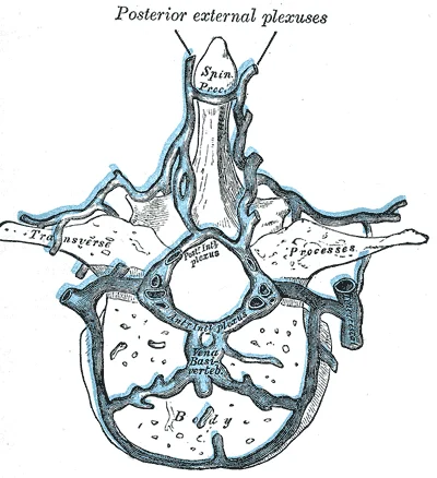
Figure 4Gray578
Long description
Transverse section of a thoracic vertebra, showing the vertebral venous plexuses.

Figure 4: Transverse section of a thoracic vertebra, showing the vertebral venous plexuses.
Figure 5Gray573
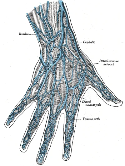
Long description
The veins on the dorsum of the hand. (Bourgery.)

Figure 5: The veins on the dorsum of the hand (Bourgery.).
DefinitionSuperficial Veins
Veins located in the subcutaneous tissue that do not accompany arteries directly, such as the cephalic and basilic veins.
The deep veins accompany the major arteries of the upper limb. In the forearm, the radial and ulnar veins run alongside their respective arteries. These merge to form the brachial veins in the arm. The brachial veins then unite with the basilic vein to form the axillary vein, which becomes the subclavian vein at the lateral border of the first rib. These deep veins are equipped with numerous valves to prevent backflow, especially important during muscle contraction.
Anastomoses are direct connections between veins that allow blood to bypass obstructions. In the upper limb, significant anastomoses occur between the superficial and deep systems. For instance, the median cubital vein connects the cephalic and basilic veins in the antecubital fossa, providing a crucial collateral route. Additionally, perforating veins link the superficial and deep systems at various levels, allowing bidirectional flow depending on pressure gradients.
| Structure | Connection | Clinical Significance |
|---|---|---|
| Median cubital vein | Cephalic to Basilic | Common site for venipuncture; collateral flow |
| Perforating veins | Superficial to Deep | Allows bidirectional flow based on pressure |
| Dorsal venous arch | Digital veins to Cephalic/Basilic | Initial drainage from hand digits |
Figure 6Gray576
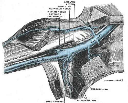
Long description
Anterior view of right upper limb and thorax - axillary vein and the distal part of the basilic vein and cephalic vein.

Figure 6: Anterior view of right upper limb and thorax - axillary vein and the distal part of the basilic vein and cephalic vein.
NoteClinical Pearl
The extensive anastomotic network means that unilateral venous obstruction may not cause immediate severe swelling, as collateral pathways can compensate.
The venous drainage of the upper limb is not a rigid, singular pathway but a complex network subject to significant anatomical variation. Understanding these variations is critical for interpreting imaging and performing interventions, as standard textbook diagrams often present an idealised version of reality. The primary distinction lies between the superficial and deep systems, and the variability often centres on the connections between them, the termination points, and the presence of accessory channels.
The superficial venous system comprises the cephalic and basilic veins, along with the median antebrachial vein. While the cephalic vein typically drains into the axillary vein, variations in its termination are common. It may drain directly into the subclavian vein or join the axillary vein at a higher level. The basilic vein usually pierces the deep fascia in the mid-arm to join the brachial veins, forming the axillary vein. However, the site of this piercing can vary from the mid-humerus to the lower axilla. The median antebrachial vein, which arises from the palmar venous plexus, shows considerable variation. It may ascend in the midline of the forearm to join the basilic vein, or it may bifurcate, sending branches to both the cephalic and basilic systems. In some individuals, a prominent median cubital vein connects the cephalic and basilic veins at the elbow, serving as a common site for venipuncture.
Deep venous drainage follows the arteries, with paired venae comitantes accompanying most major arteries. Variations here often involve the number and size of these companion veins. For instance, the brachial veins may be single or double, and their confluence with the basilic vein to form the axillary vein can occur at varying levels. The axillary vein itself is defined as the continuation of the brachial veins, but its proximal termination can vary. It typically becomes the subclavian vein at the lateral border of the first rib, but in some cases, this transition occurs more medially or laterally.
The subclavian vein continues as the brachiocephalic vein, which joins its counterpart to form the superior vena cava. Variations in the formation of the brachiocephalic veins are rare but significant. An aberrant right subclavian artery, for example, can alter the anatomical relationships and potentially compress the adjacent venous structures. Additionally, the presence of accessory veins, such as the internal jugular vein joining the subclavian vein at different angles, can influence venous flow dynamics.
NoteClinical Relevance
Variations in venous anatomy can complicate central line insertion and affect the interpretation of venography. Awareness of these variations is essential for safe practice.
| Structure | Typical Anatomy | Common Variation |
|---|---|---|
| Cephalic Termination | Axillary Vein | Subclavian Vein |
| Basilic Piercing | Mid-arm | Lower axilla |
| Median Antebrachial | Joins Basilic | Bifurcates to both |
| Brachial Veins | Paired | Single |
| Axillary/Subclavian Border | Lateral 1st Rib | Variable |
Understanding these variations requires a systematic approach. One must trace the flow from the distal extremities proximally, noting where channels join or diverge. This detailed mapping allows for accurate diagnosis and tailored treatment plans, ensuring that interventions are based on the patient's unique anatomy rather than a generic template. [2]
The clinical management of upper limb venous pathology requires precise anatomical knowledge to guide interventions, imaging, and surgical decisions. A fundamental principle in assessing venous competence is the reliance on duplex ultrasound, which necessitates proper blood flow for accurate evaluation. To fully assess vein patency, direction of flow, and valve behaviour, provocation maneuvers are essential to induce movement within the veins. This dynamic assessment is critical because static images may miss subtle incompetence or thrombosis. [2]
When considering endovascular interventions, such as stenting for venous obstructions, the selection of the device is paramount. An ideal venous stent must possess flexibility and rigidity proportional to the anatomy of the involved segment, particularly at stress points. However, stent design involves trade-offs; thinner struts are associated with fewer post-stenting obstructions and better deliverability but offer less radio-opacity, complicating follow-up evaluations. [2]
It is crucial to remember that the degree of stenosis does not always correlate with clinical severity. Even a 20% obstruction can cause significant impairment of quality of life, venous hypertension, and severe symptoms. Therefore, stenting decisions should be based on the patient's symptoms and quality of life impairment rather than the degree of stenosis alone. This principle applies to both upper and lower limbs, although most data focuses on the lower limb. [2]
Understanding these landmarks is vital for safe access and identifying pathological changes such as thrombosis or malformations.
Clinical pearlClinical Pearl
Always consider provocation maneuvers during duplex ultrasound to fully evaluate venous valve competence and flow direction.
| Structure | Origin | Termination | Clinical Note |
|---|---|---|---|
| Cephalic Vein | Dorsal venous network | Axillary vein | Common site for AV fistula creation |
| Basilic Vein | Dorsal venous network | Axillary vein | Pierces deep fascia in mid-arm |
| Median Cubital Vein | Cephalic to Basilic | N/A | Preferred site for venipuncture |
Exam trapCommon Trap
Do not rely solely on the degree of stenosis to decide on stenting; symptoms and quality of life are more important indicators.
The venous drainage of the upper limb follows a hierarchical pattern, mirroring the arterial supply but with significant anastomotic potential. Understanding the transition of vessel names at anatomical landmarks is fundamental.
The superficial venous system consists of the cephalic and basilic veins, which drain the skin and subcutaneous tissue. These vessels are clinically vital for venous access and surgical flaps.
- Cephalic Vein: Originates from the dorsal venous network of the hand. It ascends along the lateral aspect of the forearm and arm, passing between the deltoid and pectoralis major muscles. It terminates by draining into the axillary vein.
- Basilic Vein: Also originates from the dorsal venous network. It ascends along the medial aspect of the arm. In the mid-arm, it pierces the deep fascia to join the brachial veins, forming the axillary vein.
Clinical pearlAnastomosis
Anastomoses are especially common in veins, helping maintain flow if one vessel is blocked. [1]
The deep venous system accompanies the arteries and is divided into regional segments.
| Segment | Accompanying Artery | Termination |
|---|---|---|
| Digital/Palmar | Digital arteries | Median/Ulnar veins |
| Forearm | Radial/Ulnar arteries | Brachial veins |
| Arm | Brachial artery | Axillary vein |
| Axilla | Axillary artery | Subclavian vein |
- Radial and Ulnar Veins: These paired veins accompany their respective arteries in the forearm. They unite to form the brachial vein.
- Brachial Veins: Two brachial veins accompany the brachial artery. They merge with the basilic vein to form the axillary vein.
- Axillary Vein: This vein begins at the lower border of the teres major muscle. It is formed by the union of the brachial and basilic veins. It continues as the subclavian vein at the lateral border of the first rib.
DefinitionNaming Convention
An artery or vein can change names as it passes an anatomical landmark. [1]
The subclavian vein continues from the axillary vein. It joins the internal jugular vein to form the brachiocephalic vein.
- Brachiocephalic Veins: The left and right brachiocephalic veins unite to form the superior vena cava.
- Superior Vena Cava: Formed by the union of the brachiocephalic veins, it drains into the right atrium.
Exam trapLaterality
The left subclavian artery becomes the axillary artery as it passes through the body wall. Note that venous naming follows similar landmark transitions. [1]
Venous access is frequently performed in the antecubital fossa, utilising the median cubital vein which connects the cephalic and basilic veins. Thrombosis in these deep veins can lead to swelling and pain, requiring imaging for diagnosis.
NoteImaging
Computed tomography venogram is an imaging modality capable of predicting stent length with a high degree of accuracy. [2]
Students frequently confuse the systemic venous return with arterial supply, assuming a strict one-to-one correspondence that ignores the complexity of the venous system. While systemic arteries distribute oxygenated blood from the left ventricle, systemic veins return deoxygenated blood to the right atrium. A critical distinction is that while many veins share names with their accompanying arteries, the venous circulation exhibits significantly greater anatomical variability than the arterial system. Relying solely on arterial naming conventions can lead to errors when identifying veins in clinical practice or dissection, as the "complementary" pattern is not universal. [1]
Another frequent error involves the termination and drainage pathways of the upper limb. Students often assume that all upper limb veins drain directly into the superior vena cava without acknowledging the intermediate steps through the axillary and subclavian veins. The deep veins of the upper limb generally accompany the arteries, but the superficial veins, such as the cephalic and basilic veins, follow distinct courses that do not always mirror arterial branches. For instance, the basilic vein pierces the deep fascia in the mid-arm to join the brachial veins, forming the axillary vein, whereas the cephalic vein drains into the axillary vein laterally. Misunderstanding these junctions can lead to incorrect localisation of pathology, such as thrombosis or varicosities. [1]
A third pitfall is the confusion between the pulmonary and systemic circulations, particularly regarding vessel nomenclature. It is essential to refer to the vessel exiting the right ventricle as the pulmonary trunk, rather than calling it a pulmonary artery, to avoid confusion with the paired pulmonary arteries that branch from it. Similarly, while the aorta is the largest artery in the body, arising from the left ventricle, its structural divisions (ascending, arch, descending) are distinct from the venous return pathways. Confusing the aortic arch with venous structures or misidentifying the level of bifurcation can lead to significant errors in interpreting imaging or surgical anatomy.
Finally, students often overlook the clinical significance of venous valves and flow direction. Unlike arteries, which have high pressure and unidirectional flow driven by the heart, veins rely on skeletal muscle pumps and valves to prevent backflow. Failure to appreciate this can result in misunderstanding conditions like venous insufficiency. The variability in venous patterns means that anatomical variations are common, and assuming a standard textbook diagram applies to every patient can be dangerous in surgical contexts.
Knowledge of upper limb venous drainage is fundamental at the bedside for assessing circulatory status and performing procedures. Clinicians use superficial veins, such as the cephalic vein, for venipuncture and intravenous access because they are easily accessible and relatively superficial. Understanding the course of these veins helps avoid complications such as infiltration or nerve injury. Furthermore, recognizing the pattern of venous return aids in the diagnosis of conditions like deep vein thrombosis (DVT) or superior vena cava syndrome, where obstruction of venous flow leads to swelling and collateral circulation.
In the operating theatre, precise knowledge of venous anatomy is crucial for surgeries involving the upper limb, neck, and thorax. Surgeons must identify and ligate or preserve specific veins to control bleeding and ensure adequate drainage. For example, during mastectomy or axillary lymph node dissection, knowledge of the axillary vein and its tributaries is essential to avoid iatrogenic injury. Additionally, in vascular surgery, understanding the variability of venous patterns is vital for procedures such as arteriovenous fistula creation for dialysis, where the cephalic vein is often used. The choice of stent and its extension into the inferior vena cava (IVC) also requires precise anatomical knowledge to prevent complications like contralateral jailing, with extensions typically ranging from 0.5 to 2 cm[1].
On imaging, radiologists rely on standard venous pathways to interpret scans such as CT venography or MRI. The complementary pattern of veins and arteries helps in identifying normal anatomy versus pathological changes. However, radiologists must also be aware of anatomical variations, as these can mimic pathology or complicate interventions. For instance, identifying the pulmonary trunk and its bifurcation into left and right pulmonary arteries is critical in assessing pulmonary embolism, where distinguishing the trunk from the arteries prevents misdiagnosis.
In examinations, students are tested on their ability to describe the origin, course, relations, branches, and termination of major veins. Questions often focus on the transformation of the basilic vein into the axillary vein or the drainage of the cephalic vein. Understanding the "Voyage of Discovery" approach, imagining the flow of blood from the periphery to the heart, helps in memorising these pathways. Creating simple line drawings and labeling major vessels is an effective study method for mastering these patterns.
- Systemic veins return deoxygenated blood to the right atrium.
- Venous circulation has greater variability than arterial circulation. [1]
- The pulmonary trunk bifurcates into left and right pulmonary arteries. [1]
- The aorta bifurcates at the level of the fourth lumbar vertebra. [1]
- Stent extension into the IVC typically ranges from 0.5 to 2 cm[1]. [2]
- Brain blood flow is approximately 20 percent of total cardiac output. [1]
- Irreversible brain damage may occur after 3 to 4 minutes of ischemia. [1]
- Simple line drawings aid in mastering major circulatory patterns. [1]
Answer each one in your head first, then open the answer.
What is the primary function of systemic veins?
Show answerHide answer
Return deoxygenated blood to the right atriumAt what vertebral level does the aorta bifurcate?
Show answerHide answer
Fourth lumbar vertebraWhat is the typical range for stent extension into the IVC?
Show answerHide answer
0.5 to 2 cmHow long can blood flow interruption cause irreversible brain damage?
Show answerHide answer
3 to 4 minutes
- Betts JG, Young KA, Wise JA, et al. Circulatory Pathways. OpenStax, Anatomy and Physiology 2e (CC BY-NC-SA 4.0). Accessed .
- Salimi J, Chinisaz F, Yazdi SAM. A comprehensive study on venous endovascular management and stenting in deep veins occlusion and stenosis: A review study. Surgery open science 2024;19:131-140. doi:10.1016/j.sopen.2024.04.001. Accessed .
Figure credits
- Figure 1: Henry Vandyke Carter, via Wikimedia Commons, Public domain, Public domain.
- Figure 2: Henry Vandyke Carter, via Wikimedia Commons, Public domain, Public domain.
- Figure 3: Henry Vandyke Carter, via Wikimedia Commons, Public domain, Public domain.
- Figure 4: Henry Vandyke Carter, via Wikimedia Commons, Public domain, Public domain.
- Figure 5: Henry Vandyke Carter, via Wikimedia Commons, Public domain, Public domain.
- Figure 6: Henry Vandyke Carter, via Wikimedia Commons, Public domain, Public domain.
Educational content for medical students and doctors. It is not a substitute for clinical judgement or local guidelines.