Upper limb: arterial supply
Arterial supply, venous drainage, and lymphatic pathways of the upper limb.
Figure 1Gray522
Long description
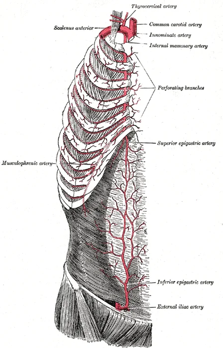
The internal mammary artery and its branches.

Figure 1: The internal mammary artery and its branches.
Figure 2Gray521
Long description
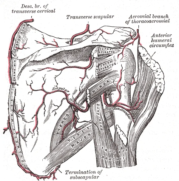
The scapular and circumflex arteries. Labelled illustration from Wikimedia Commons, used under its open licence.

Figure 2: The scapular and circumflex arteries.
Figure 32127 Thoracic Upper Limb Arteries
Long description
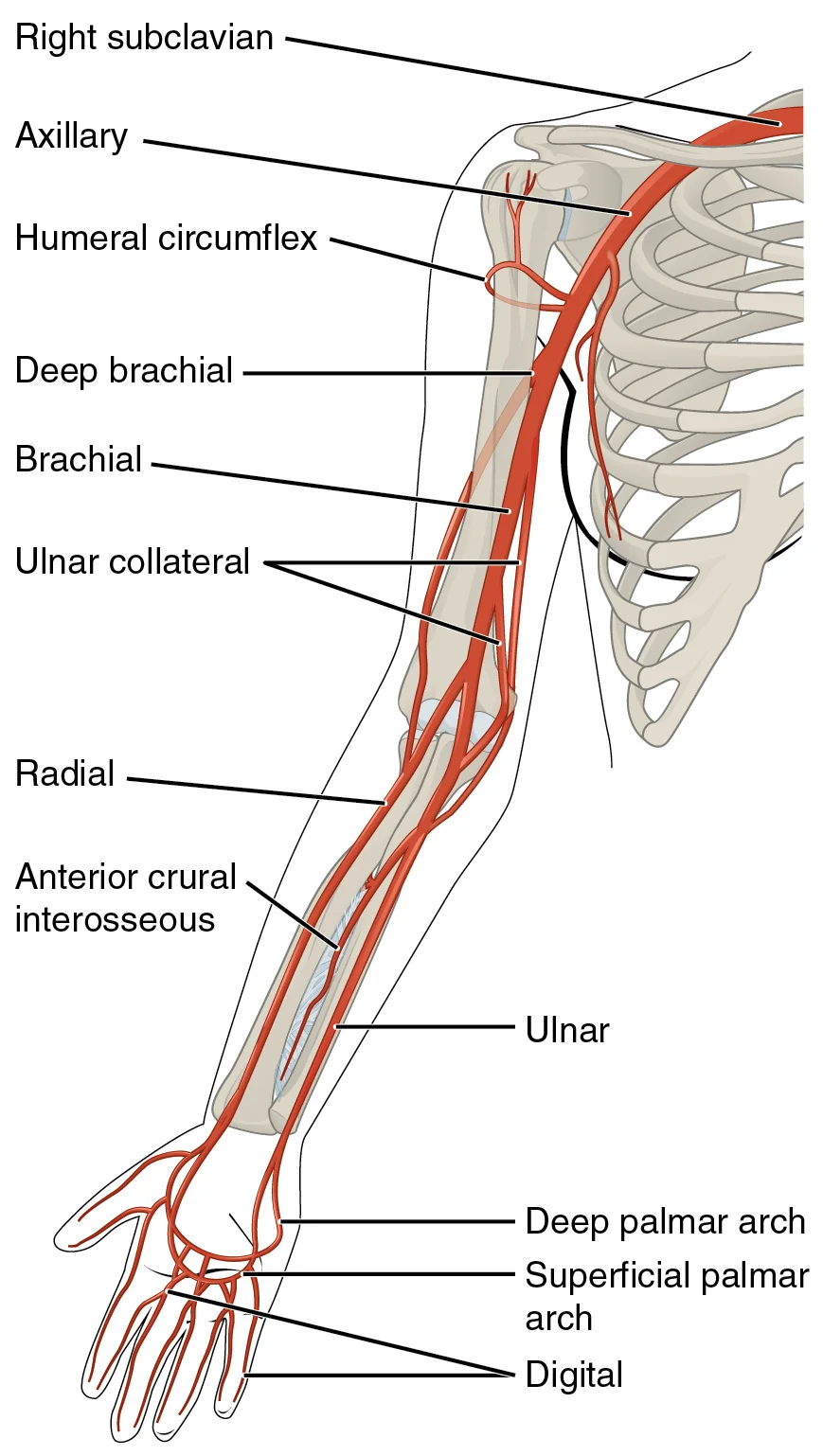
The arteries that supply blood to the arms and hands are extensions of the subclavian arteries.

Figure 3: The arteries that supply blood to the arms and hands are extensions of the subclavian arteries.
Figure 4Brachial a
Long description
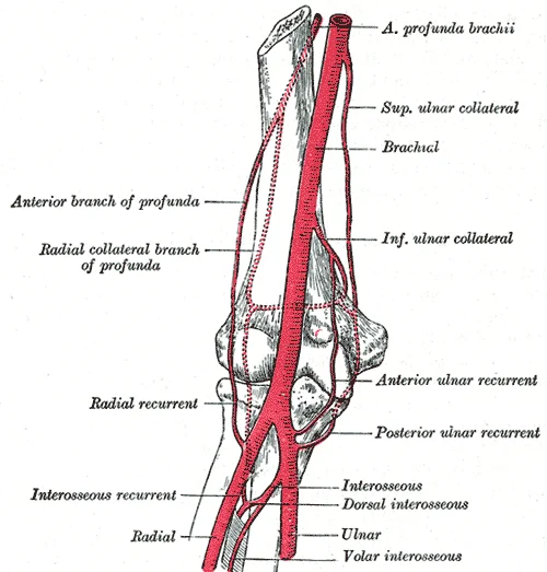
brachial artery and its branches. Labelled illustration from Wikimedia Commons, used under its open licence.

Figure 4: Brachial artery and its branches.
Although it does branch and supply blood to the region near the head of the humerus (via the humeral circumflex arteries), the majority of the vessel continues into the upper arm, or brachium, and becomes the brachial artery. [1]
The brachial artery supplies blood to much of the brachial region. It divides at the elbow into several smaller branches, including the deep brachial arteries, which provide blood to the posterior surface of the arm. Additionally, the ulnar collateral arteries supply blood to the region of the elbow. As the brachial artery approaches the coronoid fossa, it bifurcates into the radial and ulnar arteries. [1]
The radial artery and ulnar artery parallel their namesake bones. They give off smaller branches until they reach the wrist, or carpal region. At this level, they fuse to form the superficial and deep palmar arches. These arches supply blood to the hand, as well as the digital arteries that supply blood to the digits. [1]
In both the neck and limb regions, there are often both superficial and deeper levels of veins. The deeper veins generally correspond to the complementary arteries. The superficial veins do not normally have direct arterial counterparts. In addition to returning blood, they also make contributions to the maintenance of body temperature. When the ambient temperature is warm, more blood is diverted to the superficial veins where heat can be more easily dissipated to the environment. In colder weather, there is more constriction of the superficial veins and blood is diverted deeper where the body can retain more of the heat. [1]
The cephalic and basilic veins are the major superficial veins of the upper limb. They drain into the deep venous system via perforating veins. The cephalic vein typically drains into the axillary vein, while the basilic vein joins the brachial veins to form the axillary vein. [1]
Lymph from the upper limb drains into the axillary lymph nodes. These nodes are grouped into pectoral, subscapular, and humeral groups. Efferent vessels from these nodes drain into the subclavian lymphatic trunk.
Hydrostatic pressure is the force exerted by a fluid due to gravitational pull. One form of hydrostatic pressure is blood pressure, the force exerted by blood upon the walls of the blood vessels or the chambers of the heart. Blood pressure may be measured in capillaries and veins, as well as the vessels of the pulmonary circulation. However, the term blood pressure without any specific descriptors typically refers to systemic arterial blood pressure. In clinical practice, this pressure is measured in mm Hg and is usually obtained using the brachial artery of the arm. [2]
When systemic arterial blood pressure is measured, it is recorded as a ratio of two numbers (e.g., 120/80 is a normal adult blood pressure). The systolic pressure is the higher value (typically around 120 mm Hg) and reflects the arterial pressure resulting from the ejection of blood during ventricular contraction, or systole. The diastolic pressure is the lower value (usually about 80 mm Hg) and represents the arterial pressure of blood during ventricular relaxation, or diastole. [2]
DefinitionPulse Pressure
Pulse pressure is the difference between systolic and diastolic measures. [2]
DefinitionMean Arterial Pressure
Mean arterial pressure is the “average” pressure of blood in the arterial system, driving blood into the tissues. [2]
NoteVenous Return
Since approximately 64 percent of the total blood volume resides in systemic veins, any action that increases the flow of blood through the veins will increase venous return to the heart. [2]
The arterial supply to the upper limb originates from the subclavian artery, which transitions into the axillary artery. This vessel then becomes the brachial artery as it flows from the axillary region into the upper arm. The brachial artery travels down the anterior compartment of the arm, lying medial to the humerus and deep to the biceps brachii. It terminates by dividing into the radial and ulnar arteries as the brachial artery approaches the coronoid fossa, ju [1]
The axillary artery is divided into three parts by the pectoralis minor muscle. The first part lies medial to the muscle, the second part posterior to it, and the third part lateral to it. Each part gives off specific branches. The first part typically gives off the superior thoracic artery. The second part gives off the thoracoacromial artery and the lateral thoracic artery. The third part gives off the subscapular artery, the anterior circumflex humeral artery, and the posterior circumflex hum [1]
The brachial artery is the main blood supply to the arm. It gives off the deep artery of the arm (profunda brachii) and the superior ulnar collateral artery. The profunda brachii artery accompanies the radial nerve in the radial groove of the humerus, supplying the triceps brachii muscle and the humerus itself. The brachial artery also gives off the inferior ulnar collateral artery, which anastomoses with the recurrent ulnar artery.
At the cubital fossa, the brachial artery bifurcates into the radial and ulnar arteries. The radial artery runs down the lateral aspect of the forearm, deep to the brachioradialis muscle. It crosses the anatomical snuffbox and contributes to the deep palmar arch. The ulnar artery runs down the medial aspect of the forearm, deep to the flexor muscles. It passes into the hand via Guyon’s canal and contributes to the superficial palmar arch. These two arteries form extensive anastomoses in the hand [1]
DefinitionAxillary Artery Parts
The axillary artery is divided into three parts by the pectoralis minor muscle: first part (medial), second part (posterior), and third part (lateral).
| Part | Branches |
|---|---|
| First | Superior thoracic artery |
| Second | Thoracoacromial artery, Lateral thoracic artery |
| Third | Subscapular artery, Anterior circumflex humeral artery, Posterior circumflex humeral artery |
The radial and ulnar arteries continue into the hand, forming the superficial and deep palmar arches. The superficial palmar arch is formed mainly by the ulnar artery and gives rise to the common palmar digital arteries. The deep palmar arch is formed mainly by the radial artery and gives rise to the palmar metacarpal arteries. These arches ensure collateral circulation to the hand, which is crucial for maintaining blood flow if one of the main arteries is compromised.
Clinical pearlPalmar Arches
The superficial palmar arch is primarily ulnar, while the deep palmar arch is primarily radial. This arrangement provides redundant blood supply to the hand.
The arterial supply to the upper limb originates from the aortic arch, which gives rise to three major branches. These are the brachiocephalic artery, the left common carotid artery, and the left subclavian artery. Each of these vessels is classified as an elastic artery due to their proximity to the heart. The left subclavian artery, literally meaning “under the clavicle,” is the direct source of the upper limb circulation on the left side. On the right side, the brachiocephalic artery branches into the right subclavian artery and the right common carotid artery. [1]
The subclavian artery passes laterally beneath the clavicle to become the axillary artery. The brachial artery supplies blood to much of the brachial region. At the cubital fossa, it bifurcates into the radial and ulnar arteries. The radial artery runs along the lateral aspect of the forearm, while the ulnar artery runs medially.
The axillary artery is divided into three parts by the pectoralis minor muscle. The first part lies medial to the muscle and gives off the superior thoracic artery. The second part lies posterior to the muscle and gives off the thoracoacromial artery and the lateral thoracic artery. The third part lies lateral to the muscle and gives off the subscapular artery, the anterior circumflex humeral artery, and the posterior circumflex humeral artery. The subscapular artery is the largest branch and further divides into the thoracodorsal artery and the circumflex scapular artery.
The brachial artery is the main vessel of the arm. It runs along the medial side of the humerus in the anterior compartment. Its major branches include the deep brachial artery, which accompanies the radial nerve in the radial groove of the humerus. The brachial artery terminates at the level of the neck of the radius, where it divides into the radial and ulnar arteries. The radial artery is smaller and runs along the lateral aspect of the forearm. The ulnar artery is larger and runs along the medial aspect. Both arteries contribute to the formation of the palmar arches, which supply the hand. [1]
| Artery | Origin | Key Branches | Termination |
|---|---|---|---|
| Subclavian | Aortic arch (left) / Brachiocephalic (right) | Vertebral, internal thoracic, thyrocervical trunk | Becomes axillary artery at lateral border of first rib |
| Axillary | Lateral border of first rib | Superior thoracic, thoracoacromial, lateral thoracic, subscapular, circumflex humeral | Becomes brachial artery at inferior border of teres major [1] |
| Brachial | Inferior border of teres major | Deep brachial, superior ulnar collateral, inferior ulnar collateral | Bifurcates into radial and ulnar arteries at cubital fossa |
| Radial | Cubital fossa | Radial recurrent, palmar carpal, dorsal carpal, princeps pollicis | Deep palmar arch |
| Ulnar | Cubital fossa | Common interosseous, anterior ulnar recurrent, posterior ulnar recurrent | Superficial palmar arch |
The radial and ulnar arteries anastomose extensively in the hand. The superficial palmar arch is formed primarily by the ulnar artery and is completed by the superficial palmar branch of the radial artery. The deep palmar arch is formed primarily by the radial artery and is completed by the deep palmar branch of the ulnar artery. These arches give rise to the common palmar digital arteries and the proper palmar digital arteries, which supply the fingers. [1]
Clinical pearlClinical Pearl
The pulse of the brachial artery is commonly used to measure blood pressure, while the radial pulse is used for heart rate assessment.
Before continuing as the axillary artery, the subclavian artery gives rise to three major branches: the internal thoracic artery, the vertebral artery, and the thyrocervical artery. [1]
The internal thoracic artery, also known as the mammary artery, descends along the inner surface of the anterior chest wall. It supplies blood to the thymus, the pericardium of the heart, and the anterior chest wall structures. This vessel is clinically significant for its use in coronary artery bypass grafting due to its reliable flow and resistance to atherosclerosis. [1]
The vertebral artery ascends through the vertebral foramina of the cervical vertebrae. It then passes through the foramen magnum into the cranial cavity to supply blood to the brain and spinal cord. The paired vertebral arteries join together to form the large basilar artery at the base of the medulla oblongata. This junction represents a critical anastomosis within the cerebral circulation. [1]
The thyrocervical artery provides blood to the thyroid gland, the cervical region of the neck, and the upper back and shoulder. It serves as a key collateral pathway between the subclavian system and the cervical vasculature. [1]
Figure 5Gray520
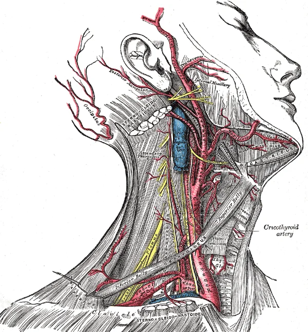
Long description
Superficial dissection of the right side of the neck, showing the carotid and subclavian arteries.

Figure 5: Superficial dissection of the right side of the neck, showing the carotid and subclavian arteries.
Figure 6Gray1235
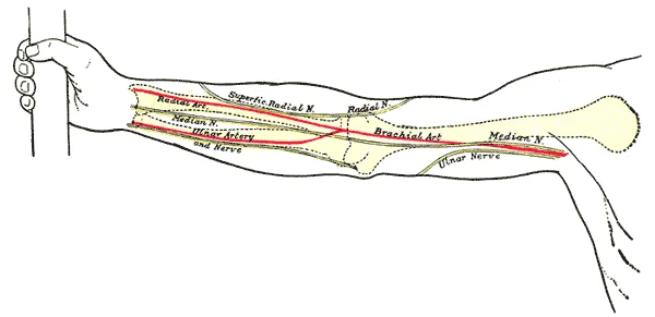
Long description
Front of right upper extremity, showing surface markings for bones, arteries, and nerves.

Figure 6: Front of right upper extremity, showing surface markings for bones, arteries, and nerves.
DefinitionAnastomosis
An anastomosis is a connection between two blood vessels, such as the joining of the vertebral arteries to form the basilar artery.
The continuity of blood flow from the subclavian artery ensures that the upper limb receives adequate perfusion. Any obstruction in the subclavian artery can compromise blood supply to the arm and potentially affect collateral flow to the brain via the vertebral arteries. Understanding these territories is essential for interpreting clinical signs of vascular compromise, such as claudication or neurological deficits.
| Branch | Primary Territories Supplied |
|---|---|
| Internal thoracic artery | Thymus, pericardium, anterior chest wall |
| Vertebral artery | Brain, spinal cord |
| Thyrocervical artery | Thyroid, cervical neck, upper back, shoulder |
The subclavian artery thus serves as a central hub, linking the systemic circulation of the upper limb with the vital structures of the head, neck, and thorax.
Arteries change names as they pass anatomical landmarks, much like a street changing name at an intersection. The left subclavian artery becomes the axillary artery as it passes through the body wall and into the axillary region, and then becomes the brachial artery as it flows from the axillary region into the upper arm. On the right side, the brachiocephalic artery branches into the right subclavian artery and the right common carotid artery. The left subclavian and left common carotid arteries ar [1]
DefinitionAnastomosis
A connection between two blood vessels that allows blood to bypass an obstruction or maintain flow if one vessel is blocked.
Anastomoses are especially common in veins, where they help maintain blood flow even when one vessel is blocked or narrowed. However, there are some important anastomoses in the arteries supplying the brain. In the upper limb, collateral circulation is vital for maintaining perfusion during compression or occlusion. [1]
The subclavian artery gives rise to the thyrocervical trunk, which contributes to the scapular anastomosis. The axillary artery, a continuation of the subclavian, gives off the superior and inferior thoracic arteries, the thoracoacromial artery, and the lateral thoracic artery. The subscapular artery, the largest branch of the axillary artery, divides into the circumflex scapular and thoracodorsal arteries, which anastomose with branches of the suprascapular and dorsal scapular arteries around the scapula. This scapular anastomosis provides a collateral route between the subclavian and axillary arteries.
At the elbow, the brachial artery divides into the radial and ulnar arteries. The anterior and posterior circumflex humeral arteries anastomose around the surgical neck of the humerus. The radial and ulnar arteries form extensive anastomoses around the elbow joint. The superior ulnar collateral artery and the radial recurrent artery anastomose. The inferior ulnar collateral artery and the anterior and posterior interosseous recurrent arteries also participate in this network. The interosseous artery, a branch of the ulnar artery, runs between the radius and ulna and anastomoses with branches of the radial artery. These anastomoses ensure blood supply to the forearm and hand even if the brachial artery is occluded.
Figure 7Gray529
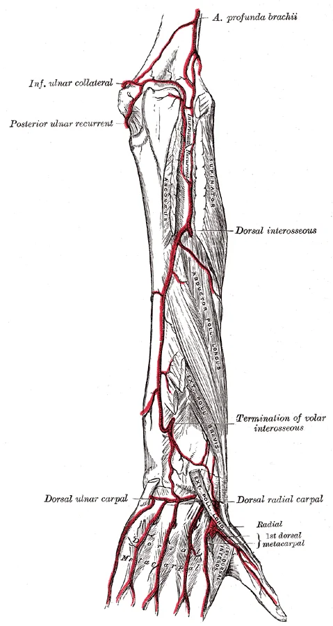
Long description
Arteries of the back of the forearm and hand.

Figure 7: Arteries of the back of the forearm and hand.
Figure 8Gray526
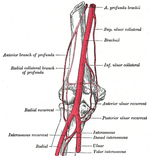
Long description
Diagram of the anastomosis around the elbow-joint.

Figure 8: Diagram of the anastomosis around the elbow-joint.
Pulse can be palpated manually by placing the tips of the fingers across an artery that runs close to the body surface and pressing lightly. Common sites include the radial artery in the wrist and the brachial artery in the upper arm. The radial artery is a major branch of the brachial artery and runs along the lateral aspect of the forearm. The ulnar artery runs along the medial aspect of the forearm. Both arteries contribute to the superficial and deep palmar arches in the hand, ensuring digit [1]
| Structure | Origin | Key Anastomosis |
|---|---|---|
| Subclavian Artery | Aortic Arch (Left) / Brachiocephalic (Right) | Scapular anastomosis |
| Axillary Artery | Continuation of Subclavian | Scapular anastomosis |
| Brachial Artery | Continuation of Axillary | Elbow anastomosis |
| Radial Artery | Brachial Artery | Palmar arches |
| Ulnar Artery | Brachial Artery | Palmar arches |
Clinical pearlClinical Pearl
Palpation of the brachial pulse is useful in cardiac arrest, while the radial pulse is common for routine assessment.
The arterial supply to the upper limb is generally consistent, yet individual variations in circulation patterns are often present. Understanding these anatomical deviations is crucial for clinical procedures such as cannulation or surgical intervention. The primary arterial trunk, the subclavian artery, transitions into the axillary artery at the lateral border of the first rib. This vessel then becomes the brachial artery as it continues distally. [1]
The axillary artery is divided into three parts by the pectoralis minor muscle. The first part lies medial to the muscle, the second part posterior to it, and the third part lateral to it. Variations in the branching pattern of the axillary artery are frequent. For instance, the thoracoacromial artery may arise from the second part rather than the first. Additionally, the lateral thoracic artery may originate directly from the axillary artery proper or from the thoracoacromial trunk. These variations do not typically compromise blood flow to the upper limb due to extensive collateral circulation, but they can complicate surgical dissection.
The brachial artery is the major blood supply to the arm. It runs down the medial side of the humerus. A common variation is the high origin of the radial artery. Instead of branching from the brachial artery in the cubital fossa, the radial artery may arise from the axillary artery or the upper part of the brachial artery. This high origin can lead to a superficial course of the radial artery in the forearm, making it easily accessible for arterial blood gas sampling but also more susceptible to injury. Another variation involves the ulnar artery, which may arise from the brachial artery at a higher level than usual.
Figure 9Gray525
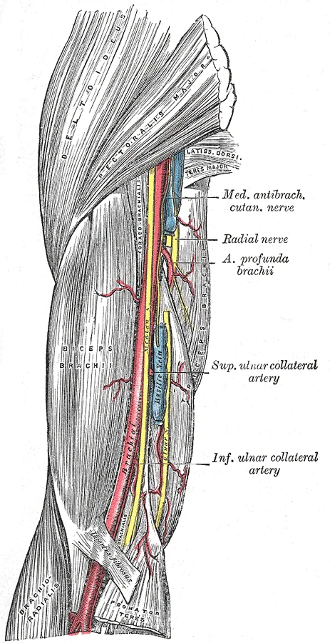
Long description
The brachial artery. Labelled illustration from Wikimedia Commons, used under its open licence.

Figure 9: The brachial artery.
The radial and ulnar arteries form the dual blood supply to the hand. They anastomose to form the deep and superficial palmar arches. Variations in these arches are significant. A superficial palmar arch may be formed primarily by the ulnar artery with minimal contribution from the radial artery. Conversely, a deep palmar arch may be incomplete or absent. In some cases, the median artery, a persistent embryonic vessel, may contribute significantly to the blood supply of the hand. This artery usually regresses but can remain patent and supply the median nerve or the palmar arches. [1]
Figure 10Gray528
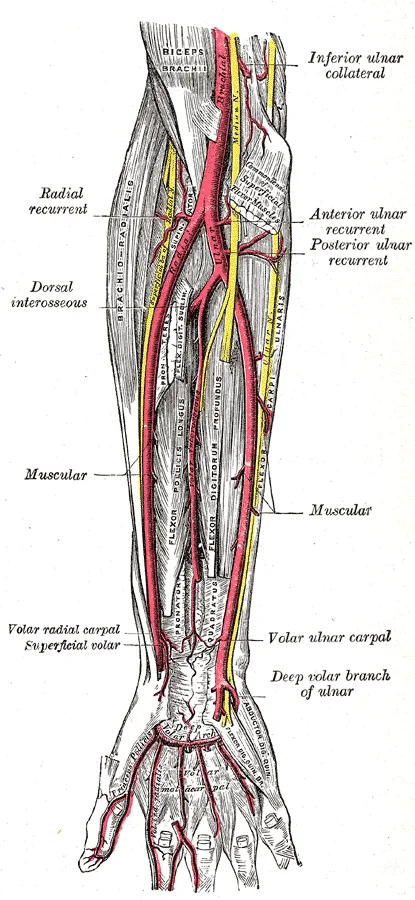
Long description
Ulnar and radial arteries. Deep view. Labelled illustration from Wikimedia Commons, used under its open licence.

Figure 10: Ulnar and radial arteries (deep view).
Figure 11Gray527
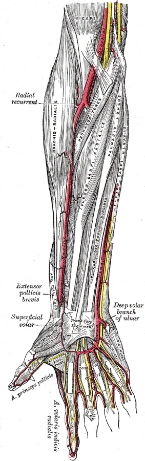
Long description
The radial and ulnar arteries. Labelled illustration from Wikimedia Commons, used under its open licence.

Figure 11: The radial and ulnar arteries.
The upper limb has rich collateral networks that can maintain perfusion even if the main arterial trunk is obstructed. This network can provide alternative routes for blood flow if the subclavian or axillary artery is compromised. Similarly, the elbow anastomosis connects the brachial artery to the radial and ulnar arteries via the profunda brachii, radial recurrent, and ulnar recurrent arteries. These collaterals are vital for preserving limb viability in cases of arterial injury or compression.
The arterial supply of the upper limb is clinically significant due to its role in perfusion and its accessibility for diagnostic and therapeutic interventions. Understanding the course of the major arteries allows for the identification of pulse points, sites of potential aneurysm, and critical access points for catheterisation or ligation in cases of severe haemorrhage.
Clinical pearlPulse Points
The radial artery is the most accessible pulse point for clinical assessment of heart rate and peripheral perfusion.
DefinitionAneurysm
A localized dilation of an artery, often due to weakness in the vessel wall, which can lead to rupture or thrombosis.
| Artery | Location | Clinical Significance |
|---|---|---|
| Brachial | Medial to biceps tendon in antecubital fossa | Blood pressure measurement |
| Radial | Lateral to flexor carpi radialis at wrist | Pulse assessment, arterial blood gas |
| Ulnar | Medial to flexor carpi ulnaris at wrist | Allen's test for collateral flow |
Arterial injury often results from penetrating trauma or iatrogenic causes. The axillary artery is vulnerable in anterior shoulder dislocations. The brachial artery is at risk in supracondylar fractures of the humerus. Thrombosis can lead to acute limb ischaemia, characterised by the six Ps: pain, pallor, pulselessness, paresthesia, paralysis, and poikilothermia. Early recognition and intervention are critical to prevent tissue necrosis and limb loss.
Exam trapCollateral Circulation
Do not assume ligation of a major artery leads to immediate ischaemia; collateral networks often preserve distal perfusion.
The anatomy of the arterial supply is essential for interpreting imaging studies such as angiography and Doppler ultrasound. Lesions may appear as stenoses, occlusions, or aneurysmal dilations. Understanding the spatial relationships between arteries, nerves, and bones aids in surgical planning and trauma management.
The arterial supply of the upper limb follows a predictable proximal-to-distal progression, mirroring the venous drainage but with distinct branching patterns. Understanding the origin and course of these vessels is critical for interpreting clinical signs such as pulse deficits or ischaemic changes.
The subclavian artery becomes the axillary artery at the lateral border of the first rib. This transition marks the entry into the upper limb proper. The axillary artery is divided into three parts by the pectoralis minor muscle, a key anatomical landmark for identifying branches.
The first part, proximal to the pectoralis minor, gives rise to the superior thoracic artery. The third part, distal to the pectoralis minor, gives off branches supplying the shoulder girdle and scapular region. [1]
The axillary artery continues as the brachial artery as it flows from the axillary region into the upper arm. [1]
The brachial artery supplies blood to much of the brachial region. [1]
In the cubital fossa, the brachial artery bifurcates into the radial and ulnar arteries. The radial artery is the preferred site for palpating the pulse in the wrist due to its superficial location against the radius. [1]
The radial artery descends along the lateral aspect of the forearm. It gives off the radial recurrent artery, which anastomoses with the radial collateral artery of the brachial artery. This anastomosis is crucial for maintaining blood flow to the elbow joint during flexion and extension.
The superficial palmar arch is formed primarily by the ulnar artery and completed by the superficial palmar branch of the radial artery. It lies deep to the palmar aponeurosis and gives rise to the common palmar digital arteries.
The deep palmar arch is formed primarily by the radial artery and completed by the deep palmar branch of the ulnar artery. It lies on the interosseous muscles and gives rise to the palmar metacarpal arteries. These arches ensure collateral circulation to the hand, allowing for continued perfusion even if one of the main arteries is compromised.
Clinical pearlPulse Points
Palpate the radial artery at the wrist for routine pulse checks. The brachial artery is used for blood pressure measurement in the antecubital fossa.
Exam trapMedian Nerve Relation
Misidentifying this can lead to accidental nerve injury during procedures.
DefinitionAnastomosis
A connection between two blood vessels that allows blood to bypass a blocked or narrowed vessel, ensuring continued perfusion to the tissue.
| Part | Branch | Supply Region |
|---|---|---|
| First | Superior thoracic | Upper medial chest wall |
| Second | Thoracoacromial | Shoulder and pectoral region |
| Second | Lateral thoracic | Lateral chest wall |
| Third | Subscapular | Scapular region and shoulder |
| Third | Anterior circumflex humeral | Shoulder joint |
| Third | Posterior circumflex humeral | Shoulder joint and deltoid |
Students often struggle with the arterial supply of the upper limb because they attempt to memorise isolated vessel names rather than understanding the continuous pathway of blood flow. A primary error is confusing the anatomical landmarks that define the transition from the thorax to the axilla. The subcl artery becomes the axillary artery specifically at the lateral border of the first rib, not at the clavicle or the sternoclavicular joint. Many learners incorrectly assume the brachial artery begins at the shoulder; in reality, it is the continuation of the axillary artery, terminating at the cubital fossa where it bifurcates.
Another frequent mistake involves the branching pattern of the axillary artery. Students often forget that the branches are defined by their relationship to the pectoralis minor muscle. The first part gives off one branch, the second part gives off two, and the third part gives off three. This mnemonic is crucial because the branches supply different regions: the first part supplies the neck and shoulder, the second supplies the thoracic wall and shoulder, and the third supplies the shoulder joint and scapular region. Failing to remember this spatial relationship leads to errors in identifying which vessels supply specific muscles.
A third pitfall is the confusion between the radial and ulnar arteries regarding their termination and anastomoses. The radial artery winds around the lateral aspect of the wrist, while the ulnar artery passes through the carpal tunnel. Both contribute to the deep and superficial palmar arches, but the ulnar artery is the primary contributor to the superficial palmar arch. Students often misattribute the formation of these arches, leading to incorrect predictions about blood supply to the finger [1]
The fourth common error is neglecting the clinical significance of the pulse points. The brachial artery is palpable in the cubital fossa, medial to the biceps tendon. The radial artery is palpable at the wrist, lateral to the flexor carpi radialis tendon. The ulnar artery is less superficial and harder to palpate. Misidentifying these points can lead to errors in blood pressure measurement and assessment of peripheral perfusion.
The fifth pitfall is the misunderstanding of collateral circulation. The anastomoses around the shoulder and elbow are vital for maintaining blood flow if a major artery is occluded. The scapular anastomosis connects the subclavian and axillary arteries, while the elbow anastomosis connects the brachial, radial, and ulnar arteries. Students often overlook these connections, failing to appreciate how they protect against ischaemia.
The sixth error is the confusion between the arterial supply and the venous drainage. While arteries follow a relatively consistent pattern, veins often accompany arteries but also have superficial counterparts. The cephalic and basilic veins are superficial and do not always follow the deep arterial paths exactly. Confusing these can lead to errors in understanding venous return and potential sites for cannulation.
The seventh pitfall is the assumption that the arterial supply is uniform across all individuals. Anatomical variations are common, particularly in the branching of the axillary and brachial arteries. Students should be aware that while the standard pattern is taught, variations exist and can impact surgical approaches and diagnostic imaging.
The eighth error is the failure to integrate the arterial supply with the innervation of the upper limb. The nerves and arteries often travel together in neurovascular bundles. Understanding the relationship between the brachial plexus and the axillary artery is essential for understanding the clinical presentation of injuries. For example, an injury to the axillary artery may also damage the axillary nerve, leading to specific motor and sensory deficits.
Exam trapAxillary Branches
The branches of the axillary artery are determined by the pectoralis minor muscle, not the clavicle or humerus.
DefinitionSubclavian to Axillary
The subclavian artery becomes the axillary artery at the lateral border of the first rib.
Knowledge of the upper limb arterial supply is fundamental in the operating theatre, particularly during orthopaedic and plastic surgery procedures. Surgeons must identify and preserve key vessels to prevent ischaemia of the limb. For instance, during a shoulder replacement, the circumflex humeral arteries must be carefully managed to maintain blood supply to the humeral head. Similarly, in forearm fracture fixation, the radial and ulnar arteries must be protected to ensure adequate perfusion of the hand.
In the emergency department, understanding arterial anatomy is critical for assessing trauma. A penetrating injury to the neck or axilla may compromise the subclavian or axillary arteries, leading to life-threatening haemorrhage. Rapid identification of the injured vessel is essential for surgical repair. Additionally, in cases of compartment syndrome, knowledge of the arterial supply helps in determining the extent of ischaemia and the urgency of fasciotomy.
On imaging, radiologists use their knowledge of arterial anatomy to interpret angiograms and CT scans. They look for stenosis, aneurysms, or occlusions in the major arteries of the upper limb. For example, a subclavian steal syndrome involves reversed flow in the vertebral artery due to stenosis of the subclavian artery proximal to its origin. Recognising this pattern on imaging is crucial for diagnosis and management.
In examinations, students are often tested on the branching patterns and clinical correlations of the upper limb arteries. Questions may ask for the origin of a specific branch, the territory supplied by a particular artery, or the clinical significance of a pulse point. Understanding the first principles of arterial flow and anatomical relationships is essential for answering these questions correctly.
At the bedside, clinicians use arterial pulses to assess peripheral perfusion. The radial pulse is commonly used to estimate heart rate and rhythm. The brachial pulse is used in blood pressure measurement. The absence of a pulse in a specific artery may indicate occlusion or compression, prompting further investigation.
Assessment of Upper Limb Ischaemia
- Patient presents with limb painAssess for pallor, pulselessness, paresthesia
- Question: Pulse present?
No
- Suspect acute occlusionImmediate vascular surgery consult
Yes
- Check capillary refillIf >2 seconds, suspect microvascular issue
NoteClinical Relevance
Arterial pulses are key bedside indicators of perfusion and vascular integrity.
| Artery | Origin | Termination | Key Branches |
|---|---|---|---|
| Subclavian | Aortic Arch | Lateral border of 1st rib | Vertebral, Internal Thoracic |
| Axillary | Lateral border of 1st rib | Inferior border of teres major | Thoracoacromial, Lateral Thoracic |
| Brachial | Inferior border of teres major | Cubital fossa | Deep Brachial, Ulnar, Radial |
| Radial | Cubital fossa | Deep palmar arch | Superficial Palmar Branch |
| Ulnar | Cubital fossa | Superficial palmar arch | Anterior/Posterior Interosseous |
The subclavian artery becomes the axillary artery at the lateral border of the first rib. The axillary artery is divided into three parts by the pectoralis minor muscle. The brachial artery bifurcates into the radial and ulnar arteries. [1] The radial artery winds around the lateral wrist, while the ulnar artery passes through the carpal tunnel. Both radial and ulnar arteries contribute to the palmar arches. [1] Collateral circulation around the shoulder and elbow protects against ischaemia during occlusion. [1] Pulse points at the radial and brachial arteries are useful for clinical assessment. [1] Anatomical variations in arterial branching are often seen and must be considered in surgery. [1]
Answer each one in your head first, then open the answer.
At which anatomical landmark does the subclavian artery become the axillary artery?
Show answerHide answer
Lateral border of the first ribWhich muscle divides the axillary artery into three parts?
Show answerHide answer
Pectoralis minorWhat is the termination of the brachial artery?
Show answerHide answer
Cubital fossaWhich artery primarily supplies the superficial palmar arch?
Show answerHide answer
Ulnar arteryWhere is the radial pulse best palpated?
Show answerHide answer
Lateral to the flexor carpi radialis tendon at the wrist
- Betts JG, Young KA, Wise JA, et al. Circulatory Pathways. OpenStax, Anatomy and Physiology 2e (CC BY-NC-SA 4.0). Accessed .
- Betts JG, Young KA, Wise JA, et al. Blood Flow, Blood Pressure, and Resistance. OpenStax, Anatomy and Physiology 2e (CC BY-NC-SA 4.0). Accessed .
Figure credits
- Figure 1: Henry Vandyke Carter, via Wikimedia Commons, Public domain, Public domain.
- Figure 2: Henry Vandyke Carter, via Wikimedia Commons, Public domain, Public domain.
- Figure 3: OpenStax College, via Wikimedia Commons, CC BY 3.0, CC BY 3.0.
- Figure 4: Henry Vandyke Carter, via Wikimedia Commons, Public domain, Public domain.
- Figure 5: Henry Vandyke Carter, via Wikimedia Commons, Public domain, Public domain.
- Figure 6: Henry Vandyke Carter, via Wikimedia Commons, Public domain, Public domain.
- Figure 7: Henry Vandyke Carter, via Wikimedia Commons, Public domain, Public domain.
- Figure 8: Henry Gray, via Wikimedia Commons, Public domain, Public domain.
- Figure 9: Henry Vandyke Carter, via Wikimedia Commons, Public domain, Public domain.
- Figure 10: Henry Vandyke Carter, via Wikimedia Commons, Public domain, Public domain.
- Figure 11: Henry Vandyke Carter, via Wikimedia Commons, Public domain, Public domain.
Educational content for medical students and doctors. It is not a substitute for clinical judgement or local guidelines.