Hand anatomy
Comprehensive anatomical overview of the hand covering boundaries, carpal tunnel, compartments, and deformities.
The hand is the distal segment of the upper limb, bound proximally by the wrist crease and containing complex osteofascial compartments. The superficial dorsal lamina is located less than 1 mm from the skin and contains no accompanying structures. The anatomical snuffbox is a triangular depression on the radial dorsum bounded by the extensor pollicis brevis and abductor pollicis longus tendons laterally, and the extensor pollicis longus tendon medially. The dorsum of the hand contains the extensor tendons and superficial veins. The carpal tunnel forms a channel through which tendons and the median nerve pass through, bounded by wrist bones and the transverse carpal ligaments. [2]
DefinitionCarpal tunnel boundaries
The floor is formed by the carpal bones and the roof by the transverse carpal ligament, enclosing the median nerve and nine flexor tendons.
Normal carpal tunnel pressure varies between 2 mmHg and 10 mmHg. Wrist extension increases this pressure to more than 10 times its initial level, while wrist flexion causes an eight times increase. [3]
Distortions of the hand include the ulnar claw hand (intrinsic minus hand affecting ulnar-innervated digits), the hand of benediction (inability to flex index and middle fingers due to high median nerve lesion), and the median claw. Hand infections can spread rapidly through fascial spaces and tendon sheaths.
Carpal Tunnel Pressure Dynamics
- Baseline pressureNormal pressure is 2 mmHg to 10 mmHg
- Question: Wrist movement?
Extension
- Pressure increases to more than 10 times initial level
Flexion
- Pressure increases by eight times
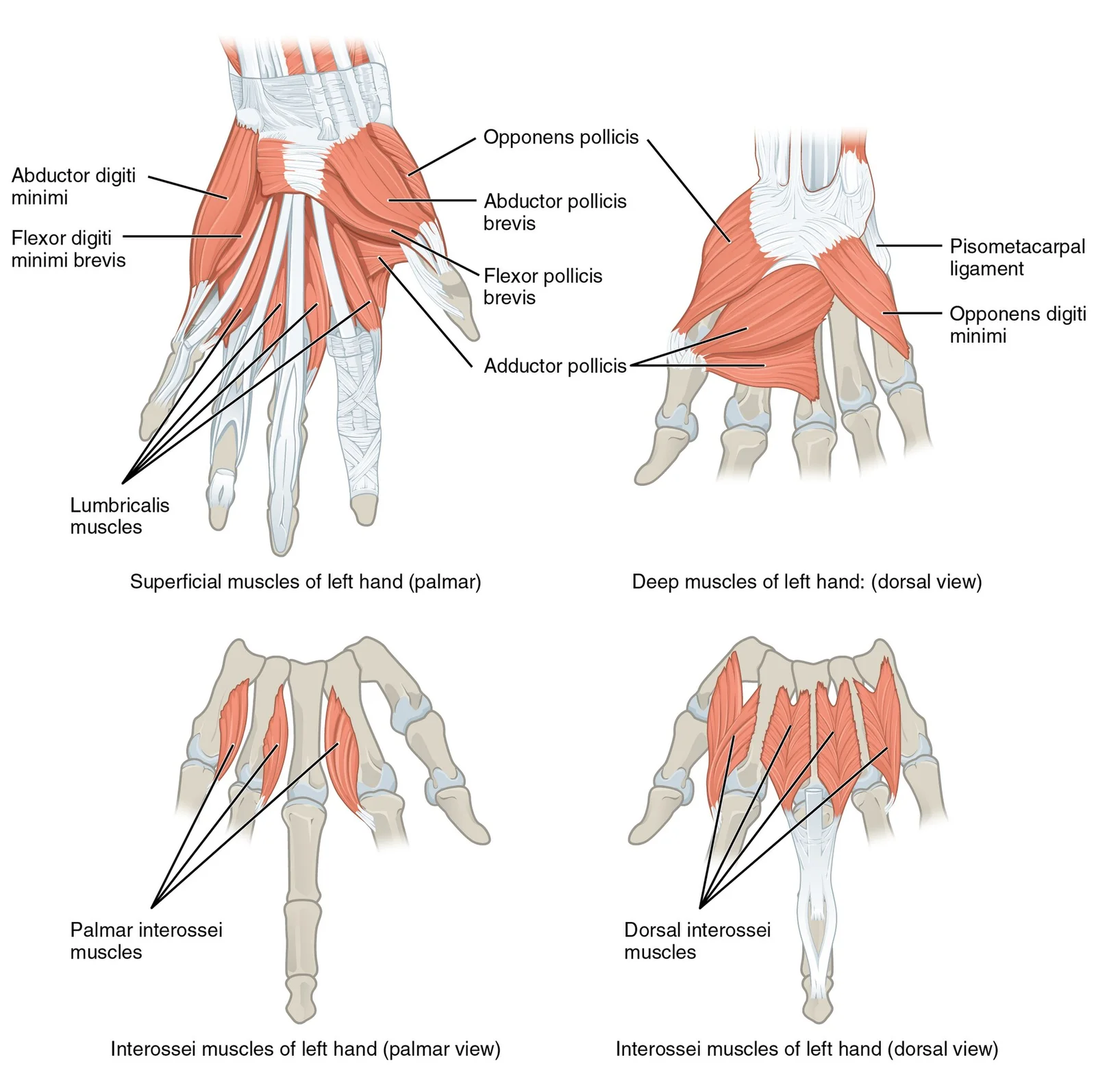
Figure 11121 Intrinsic Muscles of the Hand
Long description
Version 8.25 from the Textbook OpenStax Anatomy and Physiology Published May 18, 2016

Figure 1: Version 8.25 from the Textbook OpenStax Anatomy and Physiology Published May 18, 2016.
25 from the Textbook OpenStax Anatomy and Physiology Published May 18, 2016.
25 from the Textbook OpenStax Anatomy and Physiology Published May 18, 2016.
25 from the Textbook OpenStax Anatomy and Physiology Published May 18, 2016.
25 from the Textbook OpenStax Anatomy and Physiology Published May 18, 2016.
25 from the Textbook OpenStax Anatomy and Physiology Published May 18, 2016.
The hand is facilitated by two groups of muscles: extrinsic muscles originating in the forearm and intrinsic muscles originating in the palm. The tendons of these forearm muscles attach to the wrist and extend into the hand. At the wrist, fibrous bands called retinacula sheath these tendons. The flexor retinaculum extends over the palmar surface, while the extensor retinaculum extends over the dorsal surface. [1]
On the dorsum of the hand, the anatomy is organised into distinct layers. Kanavel described two main layers: a superficial fascia located over the extensor tendons and a deep fascia lining the interosseous muscles and metacarpals. The skin on the dorsal hand is thin and delicate, making it susceptible to injury. It contains numerous sensory receptors for touch, pressure, and temperature, which are vital for fine motor control. [2]
Further research by Anson et al. revealed that these fascial layers are divided into two layers each, separated by an areolar plane. This plane surrounds blood vessels and nerves. In the proximal two-thirds of the dorsum, anatomical layers are clearly defined. The superficial dorsal lamina lies less than 1 mm from the skin and is free of neurovascular structures. It is strongly compartmentalised by longitudinally oriented septa. In the distal third, these layers are less distinct. [2]
DefinitionDorsal Superficial Lamina
A space less than 1 mm from the skin, free of neurovascular structures, and compartmentalised by septa.
Palpable landmarks include the metacarpal bones and the extensor tendons. The thinness of the dorsal skin allows these structures to be easily visualised and palpated. Understanding this surface anatomy is crucial for practitioners performing procedures such as dermal filler injections, where accurate identification of layers prevents complications. The superficial lamina is the recommended site for volumizing materials. [2]
| Feature | Description |
|---|---|
| Skin | Thin, delicate, rich in sensory receptors |
| Superficial Fascia | Located over extensor tendons |
| Deep Fascia | Lines interosseous muscles and metacarpals |
| Areolar Plane | Separates fascial layers, contains vessels/nerves |
| Septa | Longitudinal, compartmentalises superficial lamina |
The arrangement of these structures supports the hand's dexterity. The extensor tendons glide smoothly over the dorsal surface, guided by the retinaculum. The underlying bones provide a rigid framework, while the superficial soft tissues allow for flexibility and sensation. This anatomical arrangement is consistent with textbook descriptions and remains accurate for clinical practice.
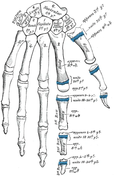
Figure 2Gray234
Long description
Plan of ossification of the hand. Labelled illustration from Wikimedia Commons, used under its open licence.

Figure 2: Plan of ossification of the hand.
The skeletal framework of the hand comprises twenty-seven bones divided into three distinct groups: the carpal bones of the wrist, the metacarpals of the palm, and the phalanges of the digits. Clinicians and anatomists examine the dorsum of the hand to assess volume loss, a frequent sign of aging. [2]
The eight carpal bones lie in two transverse rows proximally. The proximal row contains the scaphoid, lunate, triquetrum, and pisiform, while the distal row contains the trapezium, trapezoid, capitate, and hamate. Articulating distally with the carpal bones are the five metacarpal bones, numbered one through five starting from the radial thumb side to the ulnar little finger side. Each metacarpal consists of a proximal base, an intermediate shaft, and a distal head that forms the prominent knuckles. Extending from the metacarpal heads are the fourteen phalanges: two in the thumb and three in each of the remaining four fingers, designated as proximal, middle, and distal phalanges.
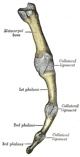
Figure 3Gray338
Long description
Metacarpophalangeal articulation and articulations of digit. Ulnar aspect.

Figure 3: Metacarpophalangeal articulation and articulations of digit (ulnar aspect).
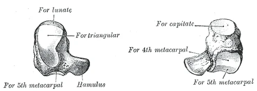
Figure 4Gray228
Long description
The left hamate bone. Labelled illustration from Wikimedia Commons, used under its open licence.

Figure 4: The left hamate bone.
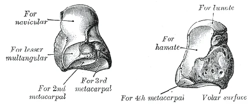
Figure 5Gray227
Long description
The left capitate bone. Labelled illustration from Wikimedia Commons, used under its open licence.

Figure 5: The left capitate bone.
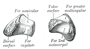
Figure 6Gray226
Long description
The left lesser multangular bone. Labelled illustration from Wikimedia Commons, used under its open licence.

Figure 6: The left lesser multangular bone.
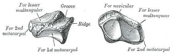
Figure 7Gray225
Long description
The left greater multangular bone. Labelled illustration from Wikimedia Commons, used under its open licence.

Figure 7: The left greater multangular bone.
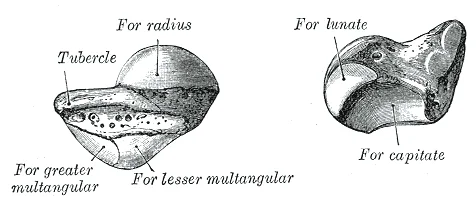
Figure 8Gray221
Long description
The left navicular bone. Labelled illustration from Wikimedia Commons, used under its open licence.

Figure 8: The left navicular bone.
Figure 9Gray233
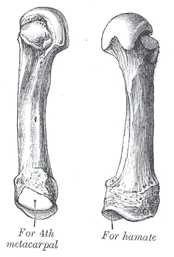
Long description
The fifth metacarpal. (Left.). Labelled illustration from Wikimedia Commons, used under its open licence.

Figure 9: The fifth metacarpal (Left.).
Figure 10Gray230
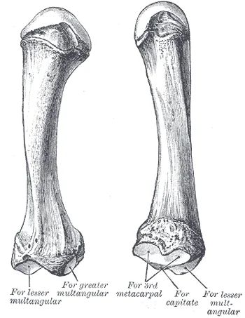
Long description
The second metacarpal. (Left.). Labelled illustration from Wikimedia Commons, used under its open licence.

Figure 10: The second metacarpal (Left.).
Figure 11Gray229
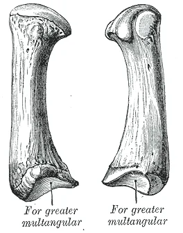
Long description
The first metacarpal. (Left.). Labelled illustration from Wikimedia Commons, used under its open licence.

Figure 11: The first metacarpal (Left.).
Figure 12Gray421
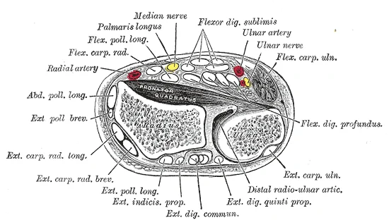
Long description
Transverse section across distal ends of radius and ulna.

Figure 12: Transverse section across distal ends of radius and ulna.
DefinitionDorsal Laminae
The dorsum of the hand consists of three fatty laminae, classified as superficial, intermediate, and deep, which are separated by three distinct fascial layers. [2]
Superficial to these skeletal structures lie specialized soft tissue layers. Histological and ultrasound evaluations reveal that the intermediate dorsal lamina is traversed by dorsal veins and sensory nerves, whereas the deep lamina is traversed by the extensor tendons. Furthermore, a fourth fascia has been identified in the dorsal intermediate lamina, located within the deep compartment that also houses the dorsal venous plexus and dorsal cutaneous nerves. Septal adhesions containing perforating vessels extend upward from the deep fascial arch toward the dermis. [2]
Figure 13Gray232
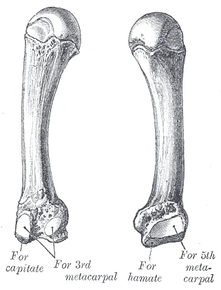
Long description
The fourth metacarpal. (Left.). Labelled illustration from Wikimedia Commons, used under its open licence.

Figure 13: The fourth metacarpal (Left.).
| Layer / Lamina | Structures Traversed or Contained | Associated Fascial Layers |
|---|---|---|
| Superficial lamina | Radial, middle, and ulnar fat compartments | Superficial fascia |
| Intermediate lamina | Dorsal veins and dorsal cutaneous nerves | Fourth fascia / intermediate fascia |
| Deep lamina | Extensor tendons and perforating vessels | Deep fascial arch |
Figure 14Gray219
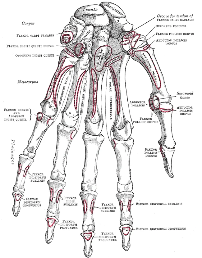
Long description
Bones of the left hand. Volar surface. Labelled illustration from Wikimedia Commons, used under its open licence.

Figure 14: Bones of the left hand (volar surface).
The joints of the hand comprise the radiocarpal, intercarpal, carpometacarpal, metacarpophalangeal, and interphalangeal joints. At the base, the radiocarpal joint lies between the distal radius and the proximal row of carpal bones, secured by strong collateral ligaments. Moving distally, the intercarpal joints connect the individual carpal bones, supported by palmar, dorsal, and interosseous intercarpal ligaments.
The carpometacarpal joints link the distal carpal row to the metacarpal bases. Notably, the carpometacarpal joint of the thumb is a saddle joint that allows for flexion, extension, abduction, adduction, circumduction, and opposition. The remaining carpometacarpal joints are plane synovial joints with limited gliding movements, reinforced by dorsal and palmar carpometacarpal ligaments.
The metacarpophalangeal joints are condyloid articulations situated between the rounded metacarpal heads and the concave bases of the proximal phalanges. Each metacarpophalangeal joint is enclosed by a joint capsule and reinforced by medial and lateral collateral ligaments, as well as the deep transverse metacarpal ligament, which unites the heads of the second through fifth metacarpals. These joints permit flexion, extension, abduction, adduction, and circumduction.
Finally, the interphalangeal joints, comprising the proximal and distal interphalangeal joints in the fingers and a single interphalangeal joint in the thumb, are hinge joints. They lie between the phalangeal heads and bases, allowing strictly uniaxial flexion and movement. Each interphalangeal joint features a fibrous joint capsule supported by robust collateral ligaments laterally and a palmar ligament anteriorly. Throughout the hand, these articular structures work alongside the intrinsic muscles, which both originate and insert within it. These intrinsic muscles allow your fingers to make precise movements for actions, such as typing or writing. The intrinsic muscles are divided into three groups: the thenar muscles on the radial aspect of the palm, the hypothenar muscles on the medial aspect of the palm, and the intermediate muscles in the midpalmar space. [1]
Figure 15Gray220
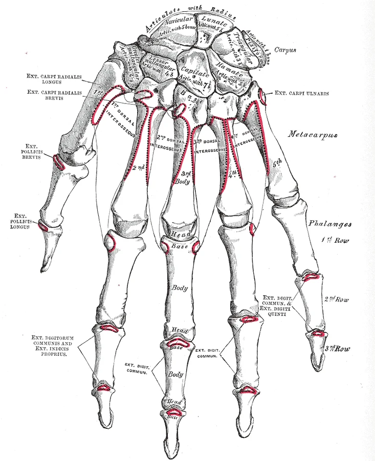
Long description
Bones of the right hand. Volar surface. Labelled illustration from Wikimedia Commons, used under its open licence.

Figure 15: Bones of the right hand (volar surface).
The extrinsic muscles of the hands originate along the forearm and insert into the hand in order to facilitate crude movements of the wrists, hands, and fingers. The superficial anterior compartment of the forearm produces flexion. These muscles are the flexor carpi radialis, palmaris longus, flexor carpi ulnaris, and the flexor digitorum superficialis. The deep anterior compartment produces flexion as well. These are the flexor pollicis longus and the flexor digitorum profundus. The rest of the compartments produce extension. The extensor carpi radialis longus, extensor carpi radialis brevis, extensor digitorum, extensor digiti minimi, and extensor carpi ulnaris are the muscles found in the superficial posterior compartment. The deep posterior compartment includes the abductor longus, extensor pollicis brevis, extensor pollicis longus, and the extensor indicis. [1]
Finally, the intrinsic muscles of the hands allow our fingers to make precise movements, such as typing and writing. They both originate and insert within the hand. The thenar muscles, which are located on the lateral part of the palm, are the abductor pollicis brevis, opponens pollicis, flexor pollicis brevis, and adductor pollicis. The hypothenar muscles, which are located on the medial part of the palm, are the abductor digiti minimi, flexor digiti minimi brevis, and opponens digiti minimi. The intermediate muscles, located in the middle of the palm, are the lumbricals, palmar interossei, and dorsal interossei. [1]
Figure 16Gray428
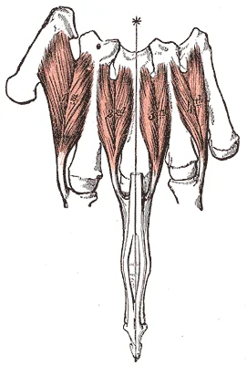
Long description
The Interossei dorsales of left hand. Labelled illustration from Wikimedia Commons, used under its open licence.

Figure 16: The Interossei dorsales of left hand.
The superficial lamina's distal portion is divided into four compartments situated in the space between the metacarpophalangeal joints. These compartments are structurally diverse from one another and possess distinctive features. [2]
DefinitionIntrinsic vs Extrinsic
Extrinsic muscles originate in the forearm to move the wrist and digits, whereas intrinsic muscles both originate and insert entirely within the hand for fine motor control.
Figure 17Gray425
Long description
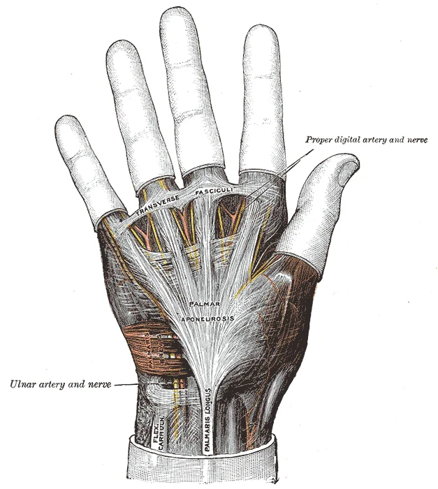
The palmar aponeurosis. Labelled illustration from Wikimedia Commons, used under its open licence.

Figure 17: The palmar aponeurosis.
Figure 18Gray424
Long description
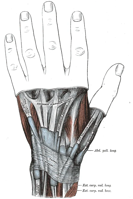
It enables the action of extensor muscles of forearm and hand by holding the tendons and allowing them to move freely.

Figure 18: It enables the action of extensor muscles of forearm and hand by holding the tendons and allowing them to move freely.
Figure 19Gray573
Long description
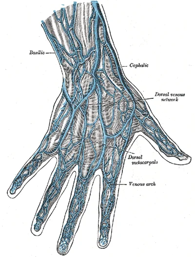
Diagram of the anatomy of the human hand

Figure 19: Diagram of the anatomy of the human hand.
The hand contains numerous intrinsic muscles that regulate precise finger movements. These are divided into compartments, including the thenar group, the hypothenar group, the lumbricals, and the interossei[1]. The hypothenar muscles include the abductor digiti minimi, flexor digiti minimi brevis, and the opponens digiti minimi, which form the rounded contour of the little finger and act on it[1]. The intermediate muscles act on all the fingers and include the lumbricals, the palmar interossei, and the dorsal interossei[1].
Figure 20Gray427
Long description
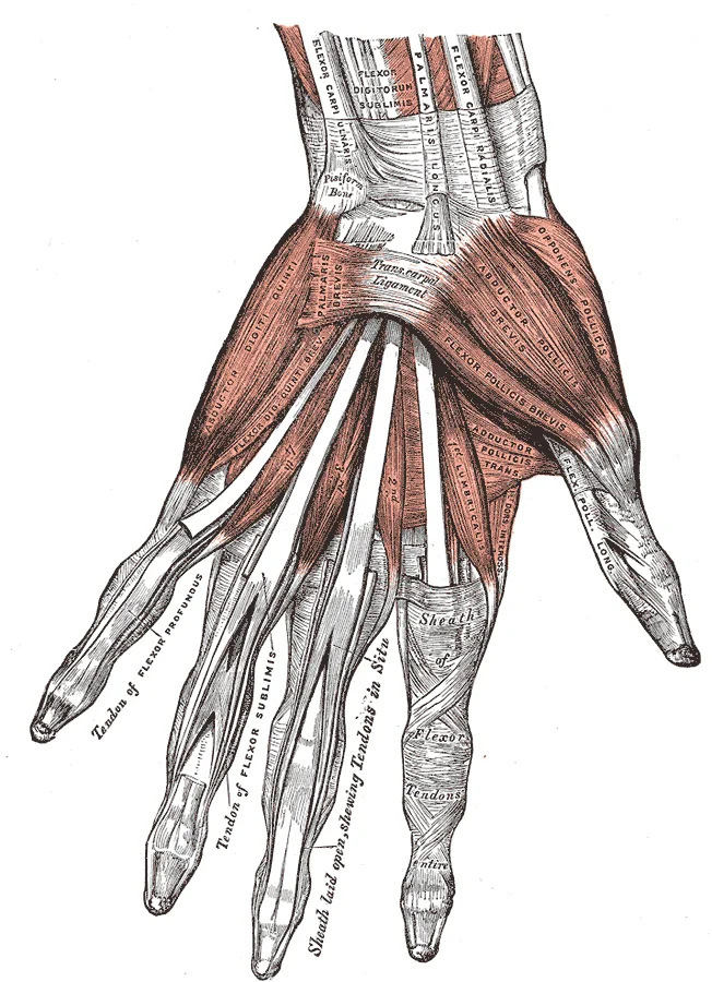
The muscles of the left hand. Palmar surface.

Figure 20: The muscles of the left hand (palmar surface).
| Muscle | Origin | Insertion | Action | Nerve Supply |
|---|---|---|---|---|
| Abductor digiti minimi | Pisiform | Base of proximal phalanx of little finger | Abducts little finger | Ulnar nerve[1] |
| Flexor digiti minimi brevis | Hook of hamate | Base of proximal phalanx of little finger | Flexes little finger | Ulnar nerve[1] |
| Opponens digiti minimi | Hook of hamate | 5th metacarpal shaft | Opposes little finger | Ulnar nerve[1] |
DefinitionIntrinsic muscles
Muscles originating and inserting entirely within the hand that control fine motor movements of the digits.
Ischaemic injury to the hand can occur due to compression or vascular compromise. Demyelination of the nerve develops in the location of compression and spreads to the intermodal segment where the axons are left intact. With continuous compression, blood flow to the endoneurial capillary system is interrupted, causing alterations in the blood-nerve barrier and the development of endoneurial edema. As a result, a vigorous cycle begins, which consists of venous congestion, ischemia, and local metabolic alterations. Ischemic injury is also noted as a significant element in CTS because of the assessment that symptoms rapidly resolve after carpal tunnel release surgery. Limb ischemia increases paraesthesias in carpal tunnel patients. This occurs in three phases, including increased intrafunicular pressure, injury to the capillary with leakage and edema, and obstruction of arterial flow in the patients. [3]
Figure 21Gray1237
Long description
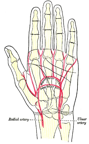
Palm of left hand, showing position of skin creases and bones, and surface markings for the volar arches.

Figure 21: Palm of left hand, showing position of skin creases and bones, and surface markings for the volar arches.
The arterial supply of the hand is derived primarily from the radial and ulnar arteries, which form superficial and deep palmar arches. These arches give rise to the common palmar digital arteries and proper palmar digital arteries, ensuring robust perfusion to the digits. Understanding this vascular network is critical for surgical planning and recognizing ischaemic complications.
The common palmar digital arteries arise from the superficial arch and run distally along the sides of the fingers.
The radial artery originates from the brachial artery and runs along the lateral aspect of the forearm. It passes deep to the tendons of the abductor pollicis longus and extensor pollicis brevis. In the hand, it contributes to the deep palmar arch, which lies on the metacarpal bones. The ulnar artery also originates from the brachial artery and travels along the medial aspect of the forearm. It enters the hand via Guyon’s canal, lying superficial to the flexor retinaculum. The ulnar artery forms the superficial palmar arch, which is located superficial to the flexor tendons.
Branches of these arteries include the princeps pollicis artery, which supplies the thumb, and the radialis indicis artery, which supplies the index finger. The common palmar digital arteries supply the second, third, and fourth digits. Each proper palmar digital artery supplies the adjacent sides of the fingers. The deep palmar arch gives off the palmar metacarpal arteries, which join the common palmar digital arteries.
Ischaemic injury to the hand can occur due to compression or vascular compromise. Demyelination of the nerve develops in the location of compression and spreads to the intermodal segment where the axons are left intact. With continuous compression, blood flow to the endoneurial capillary system is interrupted, causing alterations in the blood-nerve barrier and the development of endoneurial edema. As a result, a vigorous cycle begins, which consists of venous congestion, ischemia, and local metabolic alterations.
Clinical pearlVascular Anatomy
The superficial palmar arch is formed mainly by the ulnar artery, while the deep palmar arch is formed mainly by the radial artery.
| Structure | Origin | Course | Branches |
|---|---|---|---|
| Superficial Palmar Arch | Ulnar Artery | Superficial to flexor tendons | Common palmar digital arteries |
| Deep Palmar Arch | Radial Artery | On metacarpal bones | Palmar metacarpal arteries |
The venous system of the hand is organised into two distinct networks: the superficial dorsal venous network and the deep palmar venous plexus. These systems communicate extensively and ultimately drain into the major veins of the forearm. Understanding this arrangement is critical for venepuncture and understanding the spread of infection.
The network is formed by the union of the dorsal digital veins. These digital veins arise from the dorsal aspects of the fingers. They converge to form the dorsal metacarpal veins. The dorsal metacarpal veins run along the interosseous spaces. They anastomose with the palmar venous plexus. The cephalic vein arises from the radial side of this network. It runs along the lateral border of the forearm. The basilic vein arises from the ulnar side of the network. It runs along the medial border of the forearm. These two veins are the primary superficial collectors. They drain into the deep system via perforating veins.
The superficial dorsal venous network is located on the dorsum of the hand. It is situated just deep to the skin and superficial fascia. This network receives blood from the dorsal digital veins. The dorsal digital veins drain the dorsal aspects of the fingers. They also receive tributaries from the nail beds. The network is most prominent over the metacarpal region. It is often used for venepuncture due to its accessibility. The cephalic vein originates from the radial extremity of this network. The basilic vein originates from the ulnar extremity. These veins ascend the forearm to join the deep venous system.
The deep palmar venous plexus lies deep to the flexor tendons and the palmar aponeurosis. It is formed by the union of the digital veins from the palmar aspects of the fingers. These veins accompany the digital arteries. The plexus drains into the deep palmar veins. These veins accompany the radial and ulnar arteries. They ultimately drain into the brachial veins. The deep system is less accessible for clinical procedures. However, it is crucial for understanding the spread of deep space infections. The communication between the superficial and deep systems allows for collateral drainage. This redundancy protects against venous obstruction.
DefinitionSuperficial vs Deep
The superficial network is subcutaneous and visible; the deep plexus lies beneath the flexor tendons and is not visible.
| Structure | Location | Drainage Destination |
|---|---|---|
| Superficial Dorsal Network | Dorsum of hand, subcutaneous | Cephalic and Basilic veins |
| Deep Palmar Plexus | Palmar aspect, deep to tendons | Radial and Ulnar veins |
| Dorsal Digital Veins | Dorsal fingers | Superficial dorsal network |
| Palmar Digital Veins | Palmar fingers | Deep palmar plexus |
Understanding this spatial relationship is critical for localising sensory deficits.
The median nerve provides motor supply to the thenar muscles and the first two lumbricals. It supplies cutaneous sensation to the lateral three and a half digits, including the nail beds.
| Nerve | Motor Supply | Cutaneous Supply |
|---|---|---|
| Median | Thenar muscles, 1st and 2nd lumbricals | Lateral 3.5 digits, palmar aspect |
| Ulnar | Hypothenar, interossei, 3rd/4th lumbricals, adductor pollicis | Medial 1.5 digits, palmar and dorsal aspects |
On average, at least 3.8% of people who complain of aching, unresponsiveness, and an itchy feeling in their hands have CTS. Diagnosis for CTS is conducted through medical assessments and electrophysiological testing, although idiopathic CTS is the most typical method of diagnosis for patients suffering from these symptoms. In add [3]
Most western nations indicate a rise in the number of work-related musculoskeletal disorders (WMSDs). This is associated with increased strain and repetitive movements by individuals. Europe, in 1998, for instance, reported more than 60% of upper limb musculoskeletal disorders recognized as work-related being CTS incidences. The prevalence levels may also vary across the different occupations and industries, with industries, such as the fish processing industries reporting the occurrence of CTS in their workers estimated at 73%. [3]
Clinical pearlClinical Pearl
Remember that CTS symptoms often worsen at night or with wrist flexion, as this increases pressure within the carpal tunnel.
The lymphatic drainage of the hand is a critical pathway for immune surveillance and fluid balance, following a predictable pattern from the superficial tissues to the deep nodes. Understanding this route is essential for interpreting signs of infection or malignancy in the upper limb.
Specifically, vessels from the thumb, index, and middle fingers typically drain into the lateral group of axillary nodes. The vessels from the little finger and the medial aspect of the ring finger drain towards the epitrochlear nodes located above the medial epicondyle of the humerus. From the epitrochlear nodes, lymph flows into the axillary nodes. The axillary nodes are arranged in several groups, including the pectoral, subscapular, and humeral groups. The humeral group receives drainage from the upper limb. The final common pathway involves the subclavian trunk, which empties into the venous angle formed by the junction of the internal jugular and subclavian veins.
DefinitionEpitrochlear Node
A lymph node located above the medial epicondyle of the humerus, receiving drainage from the medial aspect of the forearm and hand.
The clinical significance of this drainage pattern cannot be overstated. Enlargement of the epitrochlear nodes is often a sign of infection in the hand or forearm, particularly involving the little finger. This is distinct from axillary lymphadenopathy, which may indicate pathology in the breast or lateral chest wall. When examining a patient with hand infections, always palpate the epitrochlear region. A tender, enlarged epitrochlear node suggests a local inflammatory process rather than a systemic condition.
| Region | Primary Drainage | Secondary Nodes | Clinical Significance |
|---|---|---|---|
| Lateral Hand (Thumb/Index) | Axillary Nodes | Subclavian Trunk | Breast pathology, arm infections |
| Medial Hand (Little Finger) | Epitrochlear Nodes | Axillary Nodes | Local hand/forearm infection |
| Palmar Surface | Axillary Nodes | Subclavian Trunk | General upper limb drainage |
These vessels drain into the deep axillary nodes. This deep pathway is less commonly involved in superficial infections but is crucial for understanding the spread of deep space infections in the hand. The convergence of superficial and deep drainage ensures efficient removal of interstitial fluid and pathogens.
Exam trapCommon Misconception
Do not assume all hand drainage goes directly to axillary nodes; the little finger drains to epitrochlear nodes first.
In summary, the lymphatic drainage of the hand is organized to protect the body from pathogens entering through the skin. The distinction between lateral and medial drainage pathways is key to localizing the source of infection. Always correlate physical findings with this anatomical map.
Figure 22Gray219 - Trapezium bone
Long description
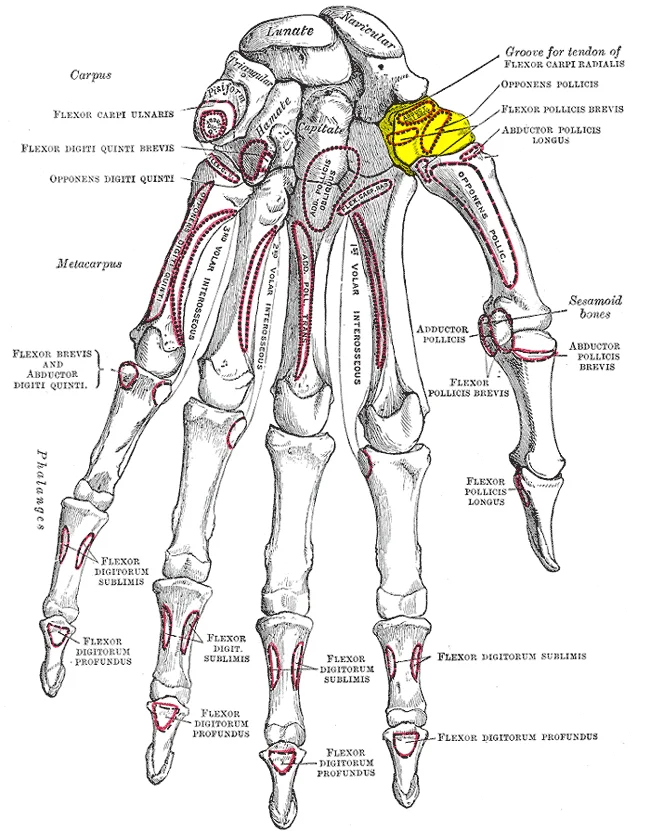
Bones of the left hand. Palmar surface. Trapezium is shown in yellow.

Figure 22: Bones of the left hand (palmar surface).
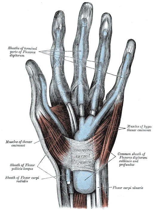
The hand contains distinct fascial spaces and synovial tendon sheaths that channel infection and structure movement. The midpalmar space lies deeply in the central palm, bounded by the third metacarpal bone anteriorly and the interosseous muscles posteriorly, while the thenar space sits lateral to it separated by the intermediate septum. Tendon sheaths enclose the long flexor tendons as they pass from the forearm into the digits, with the ulnar bursa wrapping the flexor digitorum superficialis and profundus tendons and often continuing into the little finger, and the radial bursa enveloping the flexor pollicis longus tendon into the thumb.
Figure 23Gray231
Long description
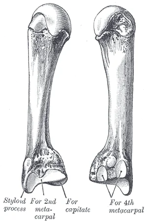
The third metacarpal. (Left.). Labelled illustration from Wikimedia Commons, used under its open licence.

Figure 23: The third metacarpal (Left.).
Figure 24Gray426
Long description
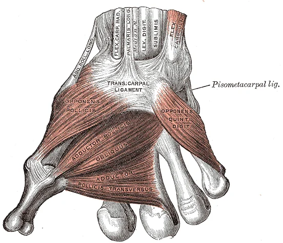
The muscles of the thumb. Labelled illustration from Wikimedia Commons, used under its open licence.

Figure 24: The muscles of the thumb.
Figure 25Gray423
Long description
The mucous sheaths of the tendons on the front of the wrist and digits.

Figure 25: The mucous sheaths of the tendons on the front of the wrist and digits.
Figure 26Gray422
Long description
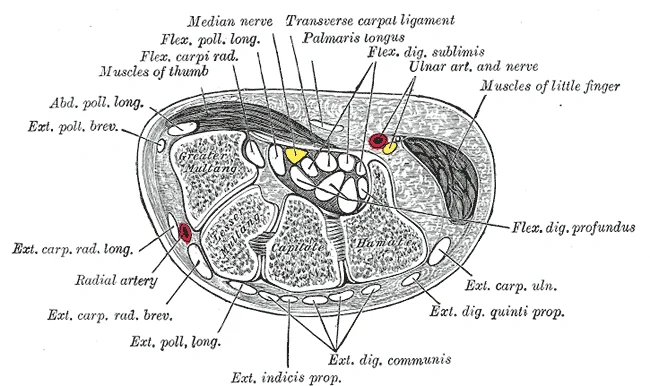
Transverse section across the wrist and digits.

Figure 26: Transverse section across the wrist and digits.
DefinitionFascial compartments
Closed anatomical spaces that restrict the spread of purulent exudate during deep hand infections.
Superficial to these spaces, the wrist transmits major neurovascular bundles.
Passing through this canal from proximal to distal are the ulnar nerve and the ulnar artery. The ulnar nerve enters the Guyon canal lateral to the pisiform bone and immediately divides into its superficial sensory branch and its deep motor branch.
| Component | Anatomical Structure |
|---|---|
| Medial boundary | Pisiform bone |
| Lateral boundary | Hook of the hamate |
| Floor | Flexor retinaculum and pisohamate ligament |
| Contents | Ulnar nerve and ulnar artery |
Radiological assessment of the hand relies on multiple imaging modalities, each offering distinct advantages for visualising osseous and soft-tissue structures from first principles.
X-rays provide the primary modality for evaluating cortical bone integrity, joint spaces, and radio-opaque foreign bodies.
Computed tomography delivers superior multiplanar bone definition, clearly delineating complex intra-articular fractures of the carpus and phalanges.
Magnetic resonance imaging provides unmatched soft-tissue contrast, allowing direct visualisation of the flexor and extensor tendons, the annular and cruciate pulley systems, the triangular fibrocartilage complex, and intrinsic muscle bellies within the palm.
Ultrasound offers dynamic, real-time assessment of tendon excursion, joint effusions, and peripheral nerve compression syndromes. For instance, therapeutic ultrasound is utilised alongside splinting, corticosteroids, physical therapy, and yoga as conventional treatment to encourage improved symptoms within two to six weeks, with the maximum benefit felt at three months. The management of carpal tunnel syndrome incidences in patients depends on the severity of the disease, and in minor and modest circumstances, a trial of conventional treatment is encouraged on the patients. [3]
Clinical pearlModalities
Combine plain radiography for initial bone screening with ultrasound or magnetic resonance imaging for soft-tissue pathology.
| Modality | Primary Indication | Key Advantage |
|---|---|---|
| X-ray | Osseous structures | Wide availability and high bone resolution |
| CT scan | Complex fractures | Multiplanar osseous reconstruction |
| MRI | Soft tissues and ligaments | Exceptional contrast for tendons and nerves |
| Ultrasound | Dynamic tendon evaluation | Real-time motion and cost-effectiveness |
Surface landmarks in the hand guide clinical examination and surgical approaches. The transverse carpal crease marks the proximal border of the carpal tunnel, whilst the distal palmar crease corresponds roughly to the metacarpophalangeal joints. Deep lacerations or blunt trauma can lead to specific injury patterns affecting tendons, nerves, and vessels. Flexor tendons in zone two, often termed no mans land, present particular surgical challenges due to the tight confines of the fibro-osseous sheath.
Nerve lesions produce characteristic postural deformities. High median nerve injury leads to the hand of benediction when attempting to make a fist, whereas an ulnar nerve lesion causes a claw hand deformity affecting the fourth and fifth digits. Carpal tunnel release is a common surgical procedure involving division of the transverse carpal ligament to decompress the median nerve.
DefinitionZone Two
The critical region of the flexor tendon sheath extending from the distal palmar crease to the middle of the middle phalanx.
Deep to this ligament lies the median nerve, positioned directly between the flexor tendons of the superficial and deep compartments. Lateral to the median nerve, locate the tendon of flexor carpi radialis within its own osseofibrous tunnel. Tracing proximally, the radial artery lies lateral to the flexor carpi radialis tendon at the wrist crease, serving as the primary site for pulse palpation.
| Nerve | Site of Lesion | Clinical Feature |
|---|---|---|
| Median | Wrist or palm | Ape hand deformity and thenar wasting |
| Ulnar | Elbow or wrist | Claw hand deformity of digits four and five |
| Radial | Spiral groove or wrist | Drop hand with loss of finger extension |
Clinical pearlSupination and pronation
In clinical anatomy, recognising this directional shift is essential for testing peripheral nerve lesions and understanding muscular compartments of the upper limb. [1]
Clinical pearlForearm flexors
These muscles act across the elbow joint to generate powerful lifting and pulling movements, receiving innervation from the musculocutaneous and radial nerves respectively. [1]
Clinical pearlForearm extensors
They counteract the flexor group to straighten the elbow joint, which is a vital component of pushing and stabilization tasks during physical examinations. [1]
Clinical pearlForearm pronators
The pronators of the forearm are the pronator teres and the pronator quadratus. Together they rotate the radius across the ulna, shifting the palm from an upward or forward-facing position to a downward-facing orientation. [1]
Clinical pearlElbow actions
Medical students must master these fundamental mechanical directions before assessing complex hand and wrist pathologies.
Exam trapConfusion of pronation
Remember that when the forearm faces posteriorly, it is pronated, distinct from the anteriorly facing supinated position. A common trap in practical exams is mixing up palm orientation with shoulder rotation. [1]
DefinitionAnterior facing
When the forearm faces anteriorly, it is defined as being supinated. This anatomical position places the radius and ulna parallel to one another.
DefinitionPosterior facing
When the forearm faces posteriorly, it is defined as being pronated. In this position, the radius crosses diagonally over the ulna bone.
Clinical pearlRedundant confirmation
This classification remains consistent across comparative texts when evaluating anterior compartment architecture.
| Muscle Group | Specific Muscles | Action |
|---|---|---|
| Flexors | Biceps brachii, brachialis, brachioradialis | Flexion at the elbow joint |
| Extensors | Triceps brachii, anconeus | Extension at the elbow joint |
| Pronators | Pronator teres, pronator quadratus | Pronation of the forearm |
| Supinator | Supinator | Turns the forearm anteriorly |
Forearm Actions
- Elbow Joint HingeExamines the radius and ulna bones.
- Question: Direction of forearm?
Anteriorly
- SupinationThe supinator turns the forearm anteriorly.
Posteriorly
- PronationThe forearm faces posteriorly.
Students often mistake the transverse carpal ligament for the entire flexor retinaculum, whereas it is only the dense, diminutive, and broad essential element of the flexor retinaculum. Another frequent error is confusing the thickness of this ligament; it ranges between 2 mm to 4 mm in thickness. [3]
Learners frequently miscalculate the spatial dimensions and landmarks of the carpal tunnel roof. The structure has an average width of 25 mm, and 31 mm in length. [3]
A common oversight in spatial orientation is misjudging the proximity of the transverse carpal ligament to the midcarpal joints. The average closeness to the central portion is 11 mm away from the capitate-lunate joint. [3]
Examiners frequently exploit confusion regarding the distal anatomical boundaries. The average distal limit of the distal portion is 10 mm distal to the carpometacarpal joint of the third metacarpal. [3]
Students often misattribute the epidemiological risk rates associated with median nerve compression at the wrist. In diabetic patients, the extent of incidence is 14% for patients without diabetes and 30% for patients with diabetic neuropathy, while the prevalence rate during pregnancy estimates at 2%. [3]
Learners occasionally fail to differentiate carpal tunnel syndrome from other upper limb neuropathies in clinical scenarios. Cervical radiculopathy presents with pain in the neck, numbness of the thumb and index finger, and positive results from the Spurling test, whereas de Quervain tendinopathy causes tenderness at the distal radial styloid.
Another diagnostic pitfall is confusing pronator syndrome with carpal tunnel entrapment. Pronator syndrome shows a history distinct from simple entrapment, featuring forearm pain, sensory loss over the thenar eminence, and weakness with thumb flexion, and wrist extension.
Finally, students mistake vasospastic disorders for nerve entrapments. Raynaud syndrome is characterized by patients showing symptoms associated with exposure to cold and typical change in color.
At the bedside, clinicians evaluate hand anatomy to differentiate nerve compressions from referred pain syndromes. Hand volume loss and aging changes are assessed using validated grading systems such as the five-category scale, which ranges from 0 to 4 based on the severity of volume loss. [2]
In the operating theatre, surgeons navigate the precise fascial borders and ligamentous attachments of the wrist to release compressed neural structures safely. Procedures for restoring volume loss in aging hands utilize autologous fat injection and injectable dermal fillers such as polymethylmethacrylate, calcium hydroxyapatite, hyaluronic acid, poly-L-lactic acid, polycaprolactone, collagen, and silicone. [2]
Hand Soft Tissue Management
- Assess volume lossScore on scale from 0 to 4
- Question: Intervention required?
Yes
- Choose autologous fat graft or dermal fillers
No
- Conservative monitoring
On imaging, clinicians examine the transverse carpal ligament, which spans from the distal part of the radius to the distal segment of the base of the third metacarpal. Radiological assessments must account for normal anatomical variations in ligament thickness and carpal architecture.
In examinations, students are tested on exact anatomical measurements, clinical thresholds, and differential diagnoses. Questions frequently target the specific differentiation between carpal tunnel syndrome, cervical radiculopathy, pronator syndrome, and de Quervain tendinopathy.
Clinical pearlDiagnostic accuracy
Always correlate sensory loss distribution with specific nerve entrapment zones rather than broad regional pain.
| Condition | Key Clinical Feature | Anatomical Landmark |
|---|---|---|
| Carpal tunnel syndrome | Paresthesia in median nerve distribution | Transverse carpal ligament |
| Cervical radiculopathy | Neck pain and positive Spurling test | Thumb and index finger |
| De Quervain tendinopathy | Tenderness at distal radial styloid | First dorsal compartment |
| Pronator syndrome | Forearm pain and thenar sensory loss | Proximal forearm |
- The transverse carpal ligament ranges between 2 mm to 4 mm in thickness.
- The average width of the transverse carpal ligament is 25 mm, and 31 mm in length.
- The average closeness to the central portion is 11 mm away from the capitate-lunate joint.
- The average distal limit of the distal portion is 10 mm distal to the carpometacarpal joint of the third metacarpal.
- The incidence of carpal tunnel syndrome is 14% for patients without diabetes and 30% for patients with diabetic neuropathy.
- The prevalence rate during pregnancy estimates at 2%.
- Aging hand volume loss is scored on a scale from 0 to 4.
- Options for restoring volume loss include autologous fat and various dermal fillers.
Answer each one in your head first, then open the answer.
What is the thickness range of the transverse carpal ligament?
Show answerHide answer
between 2 mm to 4 mmWhat is the average width of the transverse carpal ligament?
Show answerHide answer
25 mmWhat is the incidence of carpal tunnel syndrome in patients with diabetic neuropathy?
Show answerHide answer
30 percentWhat is the prevalence rate of carpal tunnel syndrome during pregnancy?
Show answerHide answer
2 percentWhat scale range is used for scoring hand volume loss?
Show answerHide answer
0 to 4
- Betts JG, Young KA, Wise JA, et al. Muscles of the Pectoral Girdle and Upper Limbs. OpenStax, Anatomy and Physiology 2e (CC BY-NC-SA 4.0). Accessed .
- Har-Shai L, Ofek SE, Lagziel T, et al. Revitalizing Hands: A Comprehensive Review of Anatomy and Treatment Options for Hand Rejuvenation. Cureus 2023;15(2):e35573. doi:10.7759/cureus.35573. Accessed .
- Genova A, Dix O, Saefan A, et al. Carpal Tunnel Syndrome: A Review of Literature. Cureus 2020;12(3):e7333. doi:10.7759/cureus.7333. Accessed .
Figure credits
- Figure 1: OpenStax, via Wikimedia Commons, CC BY 4.0, CC BY 4.0. resized.
- Figure 2: Henry Vandyke Carter, via Wikimedia Commons, Public domain, Public domain.
- Figure 3: Henry Vandyke Carter, via Wikimedia Commons, Public domain, Public domain.
- Figure 4: Henry Vandyke Carter, via Wikimedia Commons, Public domain, Public domain.
- Figure 5: Henry Vandyke Carter, via Wikimedia Commons, Public domain, Public domain.
- Figure 6: Henry Vandyke Carter, via Wikimedia Commons, Public domain, Public domain.
- Figure 7: Henry Vandyke Carter, via Wikimedia Commons, Public domain, Public domain.
- Figure 8: Henry Vandyke Carter, via Wikimedia Commons, Public domain, Public domain.
- Figure 9: Henry Vandyke Carter, via Wikimedia Commons, Public domain, Public domain.
- Figure 10: Henry Vandyke Carter, via Wikimedia Commons, Public domain, Public domain.
- Figure 11: Henry Vandyke Carter, via Wikimedia Commons, Public domain, Public domain.
- Figure 12: Henry Vandyke Carter, via Wikimedia Commons, Public domain, Public domain.
- Figure 13: Henry Vandyke Carter, via Wikimedia Commons, Public domain, Public domain.
- Figure 14: Henry Vandyke Carter, via Wikimedia Commons, Public domain, Public domain.
- Figure 15: Henry Vandyke Carter, via Wikimedia Commons, Public domain, Public domain.
- Figure 16: Henry Vandyke Carter, via Wikimedia Commons, Public domain, Public domain.
- Figure 17: Henry Vandyke Carter, via Wikimedia Commons, Public domain, Public domain.
- Figure 18: Henry Vandyke Carter, via Wikimedia Commons, Public domain, Public domain.
- Figure 19: Henry Vandyke Carter, via Wikimedia Commons, Public domain, Public domain.
- Figure 20: Henry Vandyke Carter, via Wikimedia Commons, Public domain, Public domain.
- Figure 21: Henry Vandyke Carter, via Wikimedia Commons, Public domain, Public domain.
- Figure 22: Henry Vandyke Carter, via Wikimedia Commons, Public domain, Public domain.
- Figure 23: Henry Vandyke Carter, via Wikimedia Commons, Public domain, Public domain.
- Figure 24: Henry Vandyke Carter, via Wikimedia Commons, Public domain, Public domain.
- Figure 25: Henry Vandyke Carter, via Wikimedia Commons, Public domain, Public domain.
- Figure 26: Henry Vandyke Carter, via Wikimedia Commons, Public domain, Public domain.
Educational content for medical students and doctors. It is not a substitute for clinical judgement or local guidelines.