Wrist joint
Anatomy of the wrist joint, carpal bones, and radioulnar mechanics.
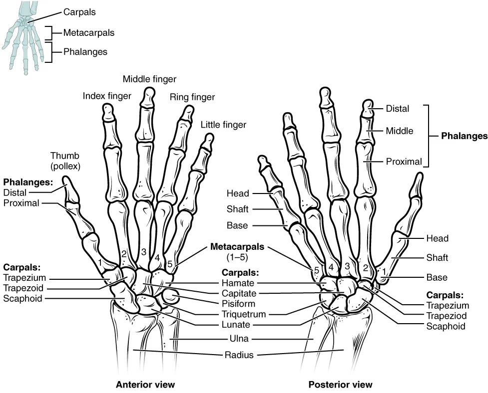
Figure 1806 Hand and Wrist
Long description
The eight carpal bones form the base of the hand. These are arranged into proximal and distal rows of four bones each. The metacarpal bones form the palm of the hand. The thumb and fingers consist of the phalanx bones.

Figure 1: The eight carpal bones form the base of the hand.
The wrist and base of the hand are formed by a series of eight small carpal bones. These bones are arranged in two rows, forming a proximal row of four carpal bones and a distal row of four carpal bones. The bones in the proximal row, running from the lateral (thumb) side to the medial side, are the scaphoid (“boat-shaped”), lunate (“moon-shaped”), triquetrum (“three-cornered”), and pisiform (“pea-shaped”) bones. The small, rounded pisiform bone articulates with the anterior surface of the triquetrum bone. The pisiform thus projects anteriorly, where it forms the bony bump that can be felt at the medial base of your hand. [1]
The carpal bones form the base of the hand. Within the carpal bones, the four proximal bones are united to each other by ligaments to form a unit. Only three of these bones, the scaphoid, lunate, and triquetrum, contribute to the radiocarpal joint. The scaphoid and lunate bones articulate directly with the distal end of the radius, whereas the triquetrum bone articulates with a fibrocartilaginous pad that spans the radius and styloid process of the ulna. The distal end of the ulna thus does not directly articulate with any of the carpal bones. [1]
The four distal carpal bones are also held together as a group by ligaments. The proximal and distal rows of carpal bones articulate with each other to form the midcarpal joint. Together, the radiocarpal and midcarpal joints are responsible for all movements of the hand at the wrist. The distal carpal bones also articulate with the metacarpal bones of the hand. [1]
At a condyloid joint (ellipsoid joint), the shallow depression at the end of one bone articulates with a rounded structure from an adjacent bone or bones. Another example is the radiocarpal joint of the wrist, between the shallow depression at the distal end of the radius bone and the rounded scaphoid, lunate, and triquetrum carpal bones. In this case, the articulation area has a more oval (elliptical) shape. Functionally, condyloid joints are biaxial joints that allow for two planes of movement. One movement involves the bending and straightening of the fingers or the anterior-posterior movements of the hand. [4]
Condyloid joints are found at the wrist (radiocarpal joint). Both condyloid and saddle joints are functionally classified as biaxial joints. Plane joints are found between the carpal bones (intercarpal joints) of the wrist. Plane joints are formed between the small, flattened surfaces of adjacent bones. These joints allow the bones to slide or rotate against each other, but the range of motion is usually slight and tightly limited by ligaments or surrounding bones. This type of joint is found at [4]
DefinitionRadiocarpal Joint
The primary wrist joint formed by the distal radius and the scaphoid, lunate, and triquetrum bones.
DefinitionMidcarpal Joint
The articulation between the proximal and distal rows of carpal bones.
Exam trapUlnar Articulation
The distal end of the ulna does not directly articulate with any of the carpal bones.
| Row | Bones (Lateral to Medial) |
|---|---|
| Proximal | Scaphoid, Lunate, Triquetrum, Pisiform |
| Distal | Trapezium, Trapezoid, Capitate, Hamate |
Pronation is the motion that moves the forearm from the supinated (anatomical) position to the pronated (palm backward) position. This motion is produced by rotation of the radius at the proximal radioulnar joint, accompanied by movement of the radius at the distal radioulnar joint. The proximal radioulnar joint is a pivot joint that allows for rotation of the head of the radius. Because of the slight curvature of the shaft of the radius, this rotation causes the distal end of the radius to cross over the distal ulna at the distal radioulnar joint. This crossing over brings the radius and ulna into an X-shape position. Supination is the opposite motion, in which rotation of the radius returns the bones to their parallel positions and moves the palm to the anterior facing (supinated) position. [3]
The palm of the hand contains five elongated metacarpal bones. These bones lie between the carpal bones of the wrist and the bones of the fingers and thumb. The proximal end of each metacarpal bone articulates with one of the distal carpal bones. Each of these articulations is a carpometacarpal joint. The expanded distal end of each metacarpal bone articulates at the metacarpophalangeal joint with the proximal phalanx bone of the thumb or one of the fingers. The distal end also forms the knuckles of the hand, at the base of the fingers. The metacarpal bones are numbered 1–5, beginning at the thumb. [1]
NoteClinical Note
Scaphoid fractures can lead to avascular necrosis due to its retrograde blood supply.
NoteClinical Note
Colles fracture involves a distal radius fracture with dorsal displacement.
The wrist joint is a synovial plane joint, meaning the articulating surfaces are flat or slightly curved and of approximately the same size, allowing the bones to slide against each other. This configuration makes the wrist functionally a multiaxial joint, capable of flexion, extension, abduction, and adduction, though the specific range is constrained by surrounding ligaments. The joint cavity is enclosed by an articular capsule, and the articulating bone surfaces are not directly connected by fibrous tissue or cartilage, which permits smooth movement. Friction is minimized by a thin layer of articular cartilage covering the bony surfaces and by synovial fluid secreted by the synovial membrane. [2] [4]
The primary weight-bearing component of the wrist is the distal end of the radius. The styloid process of the radius projects more distally than the styloid process of the ulna. This anatomical asymmetry is critical because it limits the range of movement for lateral deviations of the hand at the wrist joint. The ulna does not articulate directly with the carpal bones; instead, it is separated from the distal radius by the distal radioulnar joint. Between the distal ends of the radius and ulna, an articular disc is present. This disc serves to strongly unite the bones of the joint to each other, providing stability to the distal radioulnar articulation. [1] [4]
Ligaments are strong bands of fibrous connective tissue that connect the bones outside their articulating surfaces. They strengthen and support the joint by anchoring the bones together and preventing their separation. While they allow for normal movements, they limit the range of motion to prevent excessive or abnormal joint movements. Ligaments are classified based on their relationship to the fibrous articular capsule. An extrinsic ligament is located outside of the articular capsule, an intrinsic ligament is fused to or incorporated into the wall of the articular capsule, and an intracapsular ligament is located inside of the articular capsule. These structures provide the necessary stability for the multiaxial movements of the wrist. [4]
The smooth movement of the wrist relies on the integrity of the articular cartilage and the synovial membrane. Damage to the articular cartilage covering the bony surfaces can lead to painful conditions, such as arthritis. While this description often applies to the temporomandibular joint, the principle of cartilage wear affecting joint function is universal to synovial joints. The presence of a joint cavity is a key structural characteristic that distinguishes synovial joints from fibrous or c [4]
Similarly, it connects to the corresponding border of the ulna. This membrane unites the two forearm bones, providing stability and a surface for muscle attachment. Distally, the membrane extends towards the wrist joint, where it contributes to the structural integrity of the region. Medially, the ulnar notch of the radius is a shallow depression that articulates with the head of the ulna. This articulation forms the distal radioulnar joint, allowing rotation of the radius during pronation and supination. The smooth surface of the distal radius articulates with two carpal bones to form the radiocarpal joint, commonly known as the wrist joint. The synovial membrane lines the inner surface of the articular capsule, secreting synovial fluid to lubricate the joint. This fluid reduces friction between the bones, allowing smooth movement within the joint cavity. The articular cartilage covers the articulating surfaces of the bones, acting like a coating to prevent damage to the underlying bone tissue. The joint is functionally classified as a diarthrosis, permitting free movement. The capsule itself is a fibrous connective tissue structure attached to each bone just outside the area of the bone’s articulating surface. This arrangement ensures that the bones articulate within the joint cavity while maintaining structural support. This anatomical arrangement allows for both stability and mobility in the wrist and forearm. [1] [4]
Figure 2Gray336
Long description
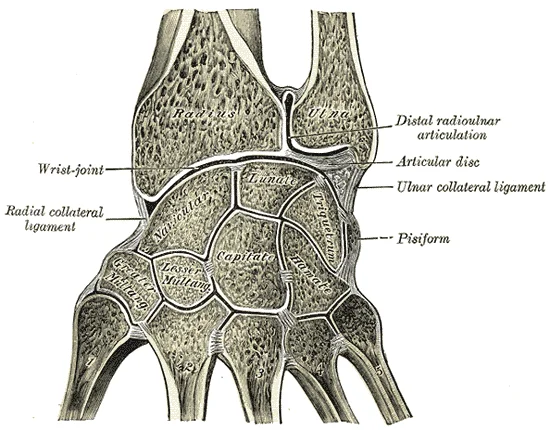
Vertical section through the articulations at the wrist, showing the synovial cavities.

Figure 2: Vertical section through the articulations at the wrist, showing the synovial cavities.
The wrist joint is reinforced by strong ligaments that help to strengthen the superior and anterior capsule walls. These intrinsic ligaments are crucial for maintaining the alignment of the carpal bones during the complex movements of the hand. The primary movements at the wrist involve flexion and extension, as well as radial and ulnar deviation. Flexion decreases the angle between the bones, bringing the palmar surface of the hand towards the anterior forearm. Extension increases this angle, straightening the joint and moving the hand posteriorly. Radial deviation involves moving the hand towards the thumb side, while ulnar deviation moves it towards the little finger side. The ligaments restrict these movements to prevent dislocation and ensure smooth articulation. [2]
Figure 3Gray335
Long description
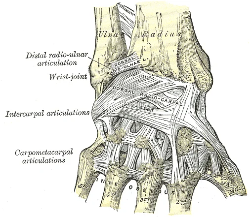
Ligaments of wrist. Posterior view. Labelled illustration from Wikimedia Commons, used under its open licence.

Figure 3: Ligaments of wrist (posterior view).
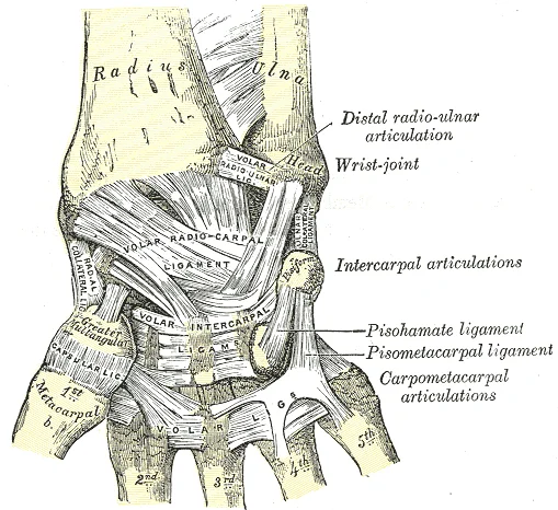
The stability of the wrist is derived from both bony congruence and ligamentous support. The distal radius articulates with the scaphoid and lunate, forming the radiocarpal joint. The ligaments surrounding this joint are thickened areas of the articular capsule. They arise from the margins of the radius and attach to the proximal row of carpal bones. This arrangement allows for a wide range of motion while maintaining joint integrity. The radioscaphocapitate ligament is particularly important in resisting excessive radial deviation and extension. The dorsal radiocarpal ligament prevents hyperextension, which could otherwise damage the anterior structures.
Figure 4Gray334
Long description
Ligaments of wrist. Anterior view. Labelled illustration from Wikimedia Commons, used under its open licence.

Figure 4: Ligaments of wrist (anterior view).
| Ligament | Attachments | Function |
|---|---|---|
| Radioscaphocapitate | Radial styloid to scaphoid and capitate | Stabilises proximal carpal row; resists radial deviation |
| Radiolunate | Radius to lunate | Central support for wrist joint |
| Dorsal Radiocarpal | Dorsal radius to scaphoid, lunate, triquetrum | Prevents hyperextension |
| Volar Radiocarpal | Volar radius to scaphoid, lunate, triquetrum | Prevents hyperflexion |
The ligaments are tightened by specific positions of the wrist. This mechanism ensures that the joint remains stable throughout its range of motion. The intrinsic ligaments provide passive stability, while the extrinsic muscles provide dynamic support. Together, they allow the wrist to perform fine motor tasks with precision and strength. The scaphoid and lunate are the key bones in this arrangement, and their alignment is maintained by the ligamentous network. Any disruption to this network can lead to instability and pain, highlighting the importance of these structures in wrist function.
The wrist joint is a complex synovial structure that facilitates a wide range of movements, including flexion, extension, abduction, and adduction. To understand its internal architecture, we must first consider the general principles of synovial joint protection. Synovial joints are lined with a capsule and contain lubricating fluid. However, additional structures are required to manage the high friction generated by tendons crossing the joint and skin moving over bony prominences. These structures are the bursae and tendon sheaths.
You can see these bursae situated between the skin and the distal ends of the radius and ulna.
Bursae are classified by their location relative to the surrounding tissues. A subcutaneous bursa is located between the skin and an underlying bone. It allows skin to move smoothly over the bone. Examples include the prepatellar bursa located over the kneecap and the olecranon bursa at the tip of the elbow. In the context of the wrist, subcutaneous bursae are found where the skin might rub against the bony prominences of the distal radius or ulna during movement. [4]
A submuscular bursa is found between a muscle and an underlying bone, or between adjacent muscles. These bursae protect muscles from rubbing against a bone or another muscle. While less commonly highlighted in basic wrist anatomy, they exist in regions where muscle bellies or tendons glide over bony surfaces. [4]
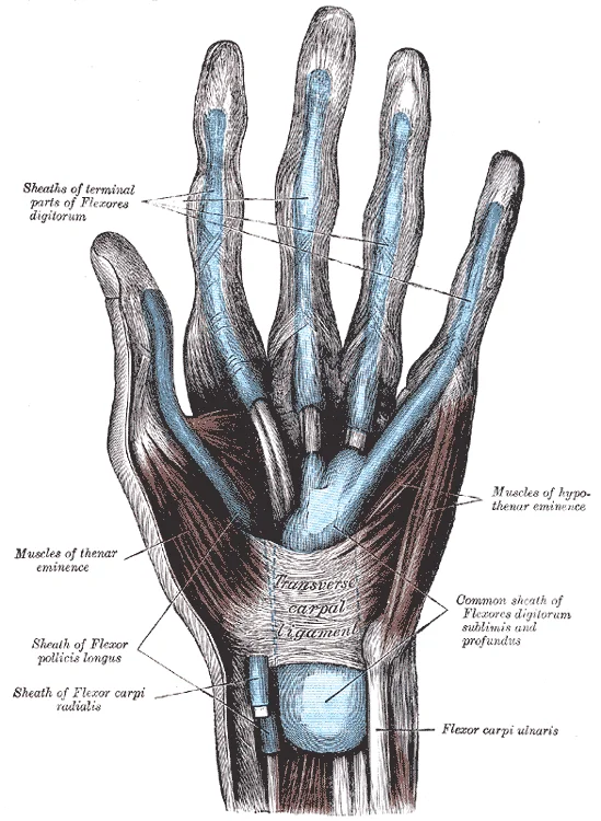
A subtendinous bursa prevents friction between bone and a muscle tendon. This is particularly relevant in the wrist, where numerous tendons cross the joint. The tendon sheath is similar in structure to a bursa, but smaller. It is a connective tissue sac that surrounds a muscle tendon at places where the tendon crosses a joint. It contains a lubricating fluid that allows for smooth motions of the tendon during muscle contraction and joint movements. This is crucial for the wrist, which requires precise and rapid movements of the fingers. [4]
The primary support for the wrist joint is provided by the ligaments and the arrangement of the carpal bones. However, the bursae and tendon sheaths are essential for reducing friction. Bursae contain a lubricating fluid that serves to reduce friction between structures. Subcutaneous bursae prevent friction between the skin and an underlying bone, submuscular bursae protect muscles from rubbing against a bone or another muscle, and a subtendinous bursa prevents friction between bone and a muscle tendon. Tendon sheaths contain a lubricating fluid and surround tendons to allow for smooth movement of the tendon as it crosses a joint. [4]
DefinitionBursa
A thin connective tissue sac filled with lubricating liquid, located in regions where skin, ligaments, muscles, or muscle tendons can rub against each other, usually near a body joint. [4]
DefinitionTendon Sheath
A connective tissue sac that surrounds a muscle tendon at places where the tendon crosses a joint, containing lubricating fluid for smooth motion.
| Type | Location | Function |
|---|---|---|
| Subcutaneous | Between skin and underlying bone | Allows skin to move smoothly over bone |
| Submuscular | Between muscle and bone, or between muscles | Protects muscles from rubbing against bone or other muscles |
| Subtendinous | Between bone and muscle tendon | Prevents friction between bone and tendon |
Figure 5Gray1233
Long description
The mucous sheaths of the tendons on the front of the wrist and digits.

Figure 5: The mucous sheaths of the tendons on the front of the wrist and digits.
The wrist joint, technically known as the radiocarpal joint, is a synovial condyloid joint that allows for a significant range of motion. It is formed by the articulation between the distal end of the radius and the articular disc, and the proximal row of carpal bones, specifically the scaphoid, lunate, and triquetrum. This structural arrangement permits movement in two primary planes: the sagittal plane and the coronal plane. [1]
The medial epicondyle is larger and more robust than the lateral epicondyle, reflecting the strength of the anterior forearm muscles compared to the weaker posterior forearm muscles.
The primary movements at the wrist are flexion, extension, abduction (radial deviation), and adduction (ulnar deviation). Flexion and extension are typically movements that take place within the sagittal plane and involve anterior or posterior movements of the limbs[1]. Flexion involves the anterior bending of the wrist, while extension involves a posterior-directed motion, such as straightening from a flexed position[1]. These movements are produced by the coordinated action of the flexor and extensor muscle groups. [3]
Abduction and adduction movements are seen at condyloid joints[1]. Abduction moves the hand laterally towards the thumb side, while adduction moves the hand medially towards the little finger side. These movements occur in the coronal plane. The joints of the upper limb provide for large ranges of motion, which give the upper limb great mobility, thus enabling actions such as the throwing of a ball or typing on a keyboard. [2][3]
| Muscle | Origin | Insertion | Action | Nerve Supply |
|---|---|---|---|---|
| Flexor Carpi Radialis | Medial epicondyle of humerus | Base of 2nd metacarpal | Flexion, Abduction | Median nerve |
| Flexor Carpi Ulnaris | Medial epicondyle of humerus | Pisiform, hook of hamate | Flexion, Adduction | Ulnar nerve |
| Extensor Carpi Radialis Longus | Lateral supracondylar ridge | Base of 2nd metacarpal | Extension, Abduction | Radial nerve |
| Extensor Carpi Ulnaris | Lateral epicondyle of humerus | Base of 5th metacarpal | Extension, Adduction | Radial nerve |
The muscles arise from the medial epicondyle, which is thus larger and more robust than the lateral epicondyle that gives rise to the weaker posterior forearm muscles. The roughened ridge of bone above the lateral epicondyle is the lateral supracondylar ridge, another attachment point for muscles that act on the forearm, wrist, and hand. The powerful grasping muscles of the anterior forearm arise from the medial epicondyle. [1]
DefinitionCondyloid Joint
A synovial joint where an oval condyle of one bone fits into an elliptical cavity of another, allowing movement in two planes.
The stability of the wrist is also supported by the articular disc, which separates the radiocarpal joint from the midcarpal joint. This disc allows for independent movement of the proximal carpal row while maintaining the integrity of the joint complex. While the shoulder is highly mobile, the wrist balances mobility with the stability needed for fine motor tasks. [2]
The wrist joint, or radiocarpal joint, is a synovial condyloid joint that permits movement in two planes. Its stability is not derived from a single structure but from the interplay of bony architecture, ligamentous constraints, and muscular tension. Understanding these factors requires examining the articulation between the distal radius and the proximal row of carpal bones.
The most prominent bony feature is the styloid process of the radius, which projects distally and laterally. It lies superior and lateral to the styloid process of the ulna. The ulnar styloid process is smaller and projects medially. Between these two processes lies the triangular fibrocartilage complex, which forms the articular disc. This disc separates the wrist joint from the distal radioulnar joint. The scaphoid and lunate bones articulate with the radius. The scaphoid is located laterally, while the lunate sits centrally. The stability of these bones depends on their tight fit within the concave surface of the radius and the supporting ligaments.
The primary bony constraint is the shape of the distal radius. This concavity cradles the convex proximal row of carpal bones. The radius is the lateral bone of the forearm and bears the majority of the load transmitted through the wrist. The ulna is the medial bone of the forearm and does not directly articulate with the carpal bones in the normal anatomical position. Instead, the triangular fibrocartilage complex acts as a cushion and stabilizer. The radial styloid process extends further distally than the ulnar styloid process. This difference in height prevents excessive radial deviation and contributes to the stability of the joint during grip activities. [1]
Ligamentous support is critical for maintaining alignment. The volar radiocarpal ligament is the strongest ligament of the wrist. It arises from the volar rim of the radius and inserts into the scaphoid, lunate, and triquetrum. This ligament prevents dorsal displacement of the carpal bones. The dorsal radiocarpal ligament is thinner and weaker. It arises from the dorsal rim of the radius and inserts into the proximal carpal row. It prevents excessive volar displacement. The ulnar collateral ligament arises from the ulnar styloid process and inserts into the triquetrum and pisiform. It stabilizes the medial aspect of the joint. The radial collateral ligament arises from the radial styloid process and inserts into the scaphoid. It stabilizes the lateral aspect of the joint.
Muscular tension also contributes to dynamic stability. The extrinsic muscles of the forearm cross the wrist joint. Their tendons pass through the carpal tunnel or over the dorsal aspect of the wrist. Contraction of these muscles compresses the carpal bones against the radius. This compression enhances joint stability during forceful activities. The intrinsic muscles of the hand provide fine control and additional support. They originate from the carpal bones and insert into the metacarpals and phalanges. Their action helps maintain the arches of the hand and wrist.
In summary, the stability of the wrist joint relies on the congruent fit of the radius and carpal bones, the strength of the volar radiocarpal ligament, and the dynamic support of the forearm muscles. Disruption of any of these factors can lead to instability and pain.
Hilton’s law states that the nerve supplying a joint also supplies the muscles moving it and the skin overlying it. This principle helps predict innervation patterns based on muscular and cutaneous distributions. The wrist joint receives its blood supply primarily from an anastomotic network surrounding the joint capsule. This network is formed by branches from the radial and ulnar arteries. These vessels wrap around the distal radius and ulna, sending articular branches into the synovial membrane. The rich vascular network ensures adequate perfusion for the cartilage and surrounding structures during movement.
The ulnar artery runs medially, contributing to the dorsal carpal arch. These arches connect to form a vascular ring around the carpal bones. Its blood supply enters primarily from the distal pole and the radial aspect. This retrograde flow pattern is critical, as the proximal pole receives blood last. Disruption of this supply can lead to avascular necrosis.
The carpal bones form a U-shaped grouping in the articulated hand. A strong ligament called the flexor retinaculum spans the top of this U-shaped area to maintain this grouping. The flexor retinaculum is attached laterally to the trapezium and scaphoid bones, and medially to the hamate and pisiform bones. Together, the carpal bones and the flexor retinaculum form a passageway called the carpal tunnel. The carpal bones form the walls and floor, while the flexor retinaculum forms the roof. The tendons of nine muscles of the anterior forearm and an important nerve pass through this narrow tunnel. Overuse of the muscle tendons or wrist injury can produce inflammation and swelling within this space. This produces compression of the nerve, resulting in carpal tunnel syndrome, which is characterized by pain or numbness, and muscle weakness in those areas of the hand supplied by this nerve.
The scaphoid bone is the largest of the proximal carpal bones. It lies between the radius and the distal row of carpals. Its blood supply is tenuous, entering from the distal two-thirds. The proximal one-third receives blood via retrograde flow from the distal segment. This anatomical arrangement makes the proximal pole vulnerable to ischemia if the distal supply is compromised. Fractures of the scaphoid waist are common. They may disrupt the vascular channel, leading to non-union or avascular necrosis of the proximal fragment. Understanding this vascular anatomy is essential for diagnosis and management. The ligaments stabilizing the scaphoid include the scapholunate and radioscaphoid ligaments. These ligaments maintain the alignment of the proximal carpal row during wrist movements.
Clinical pearlScaphoid Vascular Risk
The scaphoid has a retrograde blood supply; fractures through the waist risk avascular necrosis of the proximal pole.
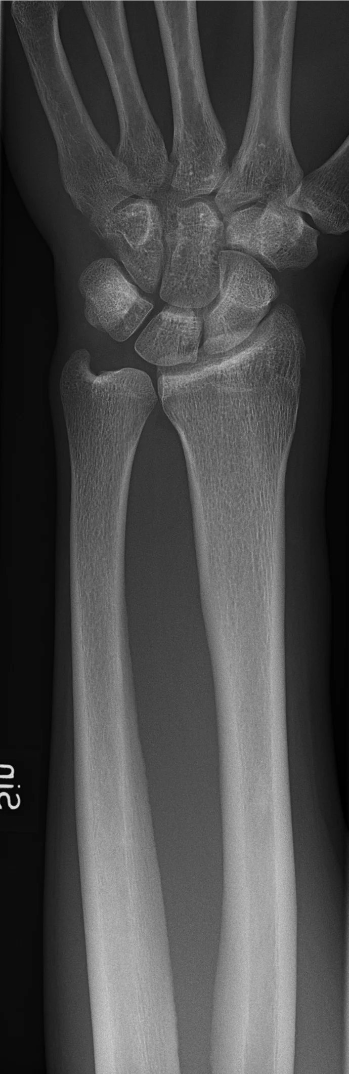
Figure 6X-ray of normal wrist by dorsoplantar projection
Long description
edit X-ray on the left side of a 31 year old male, showing normal anatomy and no injuries. Taken at Uddevalla Hospital.

Figure 6: Edit X-ray on the left side of a 31 year old male, showing normal anatomy and no injuries (taken at uddevalla hospital).
The carpal bones are arranged in two rows. The proximal row, from lateral to medial, consists of the scaphoid, lunate, triquetrum, and pisiform. The distal row comprises the trapezium, trapezoid, capitate, and hamate. The scaphoid is the largest bone in the proximal row and is often the first to fracture. The lunate articulates with the radius and is central to wrist stability. The capitate is the largest bone in the distal row and articulates with the lunate. The radiocarpal joint is formed by the distal radius and the articular disc of the distal radioulnar joint articulating with the proximal row of carpal bones. The intercarpal joints allow for complex movements including flexion, extension, abduction, and adduction. The scaphoid and lunate are particularly important in radiological assessment due to their susceptibility to injury and avascular necrosis. The pisiform is a sesamoid bone embedded in the flexor carpi ulnaris tendon. The hamate has a distinctive hook that serves as an attachment for ligaments. The trapezium articulates with the first metacarpal, allowing for the opposition of the thumb. The radiological anatomy provides critical information for diagnosing fractures, dislocations, and degenerative changes. The alignment of the carpal bones is essential for normal wrist function. Any disruption in the normal alignment, such as a scapholunate dissociation, can lead to chronic instability. The articular surfaces must be smooth to allow for pain-free movement. The radiocarpal joint is a condyloid joint, allowing movement in two planes. The intercarpal joints are plane joints, allowing gliding movements. The distal radioulnar joint allows for pronation and supination of the forearm. The ligaments surrounding the wrist provide stability while allowing for flexibility. The scapholunate ligament is the most important ligament in maintaining carpal stability. The radioscaphocapitate ligament also plays a key role in wrist stability. The ulnocarpal ligaments stabilize the ulnar side of the wrist. The volar and dorsal radiocarpal ligaments stabilize the joint in flexion and extension respectively. The radiological appearance of these structures is crucial for accurate diagnosis. The bone density and cortical integrity should be assessed. The joint spaces should be uniform and symmetric. Any narrowing of the joint space may indicate osteoarthritis. The presence of osteophytes is a sign of degenerative change. The alignment of the carpal bones should be normal. The scaphoid should not be rotated or displaced. The lunate should be aligned with the radius and capitate. Any abnormality in this alignment requires further investigation. The radiological anatomy of the wrist is complex but essential for understanding wrist pathology. [1]
DefinitionRadiocarpal Joint
The primary joint of the wrist, formed by the articulation between the distal radius and the proximal row of carpal bones, specifically the scaphoid and lunate.
The wrist joint is a synovial joint that allows for a wide range of motion. It is composed of the distal radius, the articular disc, and the proximal carpal bones. The joint capsule is thin and loose, allowing for flexibility. The ligaments provide stability and limit excessive movement. The blood supply to the wrist is rich, but some bones, such as the scaphoid, have a tenuous blood supply. This makes them prone to avascular necrosis if fractured. The nerves supplying the wrist are derived from the median, ulnar, and radial nerves. These nerves provide sensory and motor innervation to the wrist and hand. The radiological anatomy is essential for diagnosing fractures, dislocations, and other pathologies. The anteroposterior and lateral views are the standard projections. Additional views may be required for specific injuries. The interpretation of wrist radiographs requires a systematic approach. The bones, joints, and soft tissues should be assessed in order. Any abnormality should be noted and correlated with clinical findings. The radiological anatomy of the wrist is a fundamental topic for medical students. Understanding the normal anatomy is key to recognizing pathology. The wrist is a complex joint that is frequently injured. Radiological imaging is the primary tool for diagnosis. The accuracy of the diagnosis depends on the quality of the images and the expertise of the radiologist. The treatment of wrist injuries depends on the type and severity of the injury. Some injuries can be treated conservatively, while others require surgical intervention. The radiological anatomy plays a crucial role in determining the appropriate treatment.
| Bone | Row | Key Feature |
|---|---|---|
| Scaphoid | Proximal | [2] |
| Lunate | Proximal | Central to wrist stability |
| Triquetrum | Proximal | Articulates with pisiform |
| Pisiform | Proximal | Sesamoid bone in FCU tendon |
| Trapezium | Distal | Articulates with 1st metacarpal |
| Trapezoid | Distal | Smallest bone in distal row |
| Capitate | Distal | Largest carpal bone |
| Hamate | Distal | Has a hook of hamate |
The wrist joint, or radiocarpal joint, is a condyloid synovial joint formed by the distal end of the radius and the articular disc of the distal radioulnar joint articulating with the proximal row of carpal bones (scaphoid, lunate, and triquetrum). Understanding its anatomy is critical for managing common injuries, assessing function, and performing diagnostic procedures.
Wrist injuries frequently involve ligamentous disruption rather than frank dislocation of the carpal bones, although perilunate and lunate dislocations are severe traumatic events. The scaphoid bone is the most commonly fractured carpal bone, often resulting from a fall on an outstretched hand with the wrist extended. Due to its retrograde blood supply originating from the distal end, fractures of the scaphoid waist are prone to avascular necrosis and non-union. [2]
Ligamentous injuries typically affect the scapholunate interval. The scapholunate ligament is the primary stabilizer between the scaphoid and lunate. Disruption leads to scapholunate dissociation, where the scaphoid flexes and the lunate extends, creating a gap visible on radiographs known as the 'Terry Thomas sign' or 'David Letterman sign'. This instability can progress to carpal collapse patterns such as dorsal intercalated segment instability (DISI).
Clinical examination focuses on identifying pain, instability, and neurological compromise. Palpation of the anatomical snuffbox is essential; tenderness here strongly suggests a scaphoid fracture, even if initial radiographs are negative.
Specific dynamic tests assess ligamentous integrity. The scaphoid shift test (Watson’s test) evaluates scapholunate instability. The examiner stabilizes the distal radius while shifting the scaphoid dorsally and volarly; a painful clunk indicates ligamentous disruption. The lunate shift test assesses lunotriquetral instability. Neurovascular assessment is mandatory, particularly for the median nerve, as acute carpal tunnel syndrome can result from swelling or fracture displacement.
Joint aspiration is rarely performed directly into the radiocarpal joint due to its small volume and complex anatomy, but it may be indicated for septic arthritis or large effusions. The safest approach is dorsal. The patient’s wrist is flexed slightly to tense the dorsal capsule. The needle is inserted through the scapholunate interval or just radial to the extensor pollicis longus tendon, avoiding the radial artery which lies volar to the radius.
Ultrasound guidance is preferred to identify fluid collections and avoid neurovascular structures. If infection is suspected, synovial fluid should be sent for cell count, Gram stain, and culture. For diagnostic purposes, arthrography may be used to visualize ligamentous tears not seen on static imaging.
The wrist joint is a complex synovial articulation that facilitates a wide range of movements essential for hand function. Understanding its anatomy is critical for diagnosing injuries and planning surgical interventions.
The radiocarpal joint is formed by the distal end of the radius and the articular disc, which articulate with the proximal row of carpal bones. Specifically, the scaphoid, lunate, and triquetrum bones form the concave surface that receives the convex distal radius. The ulna does not directly articulate with the carpal bones; instead, it is separated by the triangular fibrocartilage complex (TFC). This arrangement allows the wrist to bear weight primarily through the radius. The midcarpal joint lies between the proximal and distal rows of carpal bones, contributing significantly to overall wrist mobility.
Stability of the wrist is maintained by strong ligamentous structures. The palmar radiocarpal ligament is the strongest ligament of the wrist, preventing hyperextension. The dorsal radiocarpal ligament also provides significant support. The ulnar collateral ligament and radial collateral ligament reinforce the sides of the joint. The TFC is crucial for stabilizing the distal radioulnar joint and absorbing shock across the wrist. Damage to these ligaments can lead to chronic instability and pain.
The median nerve passes through the carpal tunnel, alongside the flexor tendons. It is susceptible to compression in carpal tunnel syndrome. Both nerves provide sensory innervation to specific areas of the hand. The dorsal carpal branch of the radial artery supplies the dorsal aspect of the wrist.
The wrist joint allows for flexion, extension, abduction (radial deviation), and adduction (ulnar deviation). Flexion and extension occur primarily in the sagittal plane. Radial and ulnar deviation occur in the frontal plane. These movements are coordinated by muscles acting across the joint. The range of motion is limited by bony congruence and ligamentous tension.
Fractures of the scaphoid are common and can lead to avascular necrosis due to its retrograde blood supply. The scaphoid is the most frequently fractured carpal bone. Non-union of scaphoid fractures is a significant complication. Carpal tunnel syndrome is the most common entrapment neuropathy of the upper limb. It presents with numbness and tingling in the median nerve distribution.
Exam trapUlnar Articulation
The ulna does not articulate directly with the carpal bones; the articular disc intervenes.
Clinical pearlScaphoid Blood Supply
Fractures risk avascular necrosis because blood flows from distal to proximal.
DefinitionRadiocarpal Joint
The main wrist joint between the radius, articular disc, and proximal carpal row.
Students frequently confuse the carrying angle with the anatomical alignment of the upper limb. A common error is assuming the arm and forearm form a straight line in the anatomical position. In reality, the forearm deviates laterally by 5–15 degrees from the line of the arm. This deviation is called the carrying angle. It allows the forearm and hand to swing freely or to carry an object without hitting the hip. The carrying angle is larger in females to accommodate their wider pelvis. Failing to recognise this lateral deviation leads to incorrect descriptions of limb posture. [1]
Another frequent mistake involves the orientation of the radius and ulna during forearm rotation. Students often believe the bones remain parallel during pronation. In the anatomical position, the upper limb is held next to the body with the palm facing forward. This is the supinated position of the forearm. In this position, the radius and ulna are parallel to each other. When the palm of the hand faces backward, the forearm is in the pronated position. In pronation, the radius and ulna form an X-shape. Confusing these positions results in errors when describing movements such as scooping soup, which is supination. [3]
Misidentifying the stabilising ligaments of the elbow is also prevalent. The articular capsule of the elbow is thin on its anterior and posterior aspects. It is thickened along its outside margins by strong intrinsic ligaments. These ligaments prevent side-to-side movements and hyperextension. On the medial side is the triangular ulnar collateral ligament. This arises from the medial epicondyle of the humerus and attaches to the medial side of the proximal ulna. The strongest part of this ligament is the anterior portion, which resists hyperextension of the elbow. Injury to this ligament is associated with frequent, forceful extensions of the forearm, as is seen in baseball pitchers. [2]
Exam trapHamate Hook
The hook of the hamate is a prominent bony extension on the anterior side of the hamate bone. Do not confuse this with the styloid process of the ulna, which projects from the posterior side of the ulnar head. [1]
DefinitionCarrying Angle
The carrying angle is the lateral deviation of the forearm from the line of the arm, measuring 5–15 degrees.
This knowledge is applied at the bedside when assessing limb alignment and joint stability. The carrying angle is observed during physical examination to detect abnormalities in the elbow joint. Clinicians assess the ulnar collateral ligament in patients with medial elbow pain, particularly those with a history of throwing sports. The integrity of this ligament is crucial for preventing hyperextension and side-to-side instability.
In the operating theatre, understanding the attachment points of ligaments is vital for reconstruction. Reconstructive surgical repair of the ulnar collateral ligament is referred to as Tommy John surgery. This procedure is named for the former major league pitcher who was the first person to have this treatment. Surgeons must accurately identify the origin from the medial epicondyle of the humerus and the attachment to the medial side of the proximal ulna. [2]
On imaging, radiologists evaluate the relationship between the radius and ulna. The interosseous border of the ulna forms a ridge on the lateral side of the shaft. This is the line of attachment for the interosseous membrane of the forearm. This membrane is a sheet of dense connective tissue that unites the ulna and radius bones. The small, rounded area that forms the distal end is the head of the ulna. Projecting from the posterior side of the ulnar head is the styloid process of the ulna. This serves as an attachment point for a connective tissue structure that unites the distal ends of the ulna and radius. [1]
Forearm Rotation Analysis
- Observe Palm PositionIs the palm facing forward or backward?
- Question: Palm Forward?
Yes
- SupinationRadius and ulna are parallel
No
- PronationRadius and ulna form an X-shape
In examinations, students must distinguish between different types of limb movements. Rotation occurs at the ball-and-socket joints of the shoulder and hip. Here, the humerus and femur rotate around their long axis. This moves the anterior surface of the arm or thigh either toward or away from the midline of the body. Movement that brings the anterior surface of the limb toward the midline of the body is called medial (internal) rotation. Conversely, rotation of the limb so that the anterior surface moves away from the midline is lateral (external) rotation. Be sure to distinguish medial and lateral rotation, which can only occur at the multiaxial shoulder and hip joints, from circumduction, which can occur at either biaxial or multiaxial joints.
| Structure | Location/Attachment | Function |
|---|---|---|
| Ulnar Collateral Ligament | Medial epicondyle to proximal ulna | Resists hyperextension and side-to-side movement |
| Interosseous Membrane | Interosseous border of ulna to radius | Unites ulna and radius bones |
| Styloid Process of Ulna | Posterior side of ulnar head | Attachment for distal radioulnar ligament |
NoteSupination Mnemonic
Supination is the motion you use when scooping up soup with a spoon. [3]
The carrying angle represents a lateral deviation of 5–15 degrees. The ulnar collateral ligament arises from the medial epicondyle of the humerus. The anterior portion of the ulnar collateral ligament resists hyperextension. Supination places the radius and ulna in a parallel position. Pronation causes the radius and ulna to form an X-shape. The hook of the hamate is a bony extension on the anterior side. The styloid process of the ulna projects from the posterior side of the ulnar head. The interosseous membrane attaches to the interosseous border of the ulna.
Answer each one in your head first, then open the answer.
What is the typical range of the carrying angle?
Show answerHide answer
5–15 degreesWhich ligament resists hyperextension of the elbow?
Show answerHide answer
Ulnar collateral ligamentWhat shape do the radius and ulna form during pronation?
Show answerHide answer
X-shapeWhere is the hook of the hamate located?
Show answerHide answer
Anterior side of the hamate boneWhat is the attachment point for the interosseous membrane on the ulna?
Show answerHide answer
Interosseous border of the ulna
- Betts JG, Young KA, Wise JA, et al. Bones of the Upper Limb. OpenStax, Anatomy and Physiology 2e (CC BY-NC-SA 4.0). Accessed .
- Betts JG, Young KA, Wise JA, et al. Anatomy of Selected Synovial Joints. OpenStax, Anatomy and Physiology 2e (CC BY-NC-SA 4.0). Accessed .
- Betts JG, Young KA, Wise JA, et al. Types of Body Movements. OpenStax, Anatomy and Physiology 2e (CC BY-NC-SA 4.0). Accessed .
- Betts JG, Young KA, Wise JA, et al. Synovial Joints. OpenStax, Anatomy and Physiology 2e (CC BY-NC-SA 4.0). Accessed .
Figure credits
- Figure 1: OpenStax College, via Wikimedia Commons, CC BY 3.0, CC BY 3.0.
- Figure 2: Henry Vandyke Carter, via Wikimedia Commons, Public domain, Public domain.
- Figure 3: Henry Vandyke Carter, via Wikimedia Commons, Public domain, Public domain.
- Figure 4: Henry Vandyke Carter, via Wikimedia Commons, Public domain, Public domain.
- Figure 5: Henry Vandyke Carter, via Wikimedia Commons, Public domain, Public domain.
- Figure 6: Mikael Häggström, M.D., via Wikimedia Commons, CC0, CC0.
Educational content for medical students and doctors. It is not a substitute for clinical judgement or local guidelines.