Forearm compartments
Anatomical layout of the forearm’s four fascial compartments and their neurovascular contents.
Figure 1Gray333
Long description
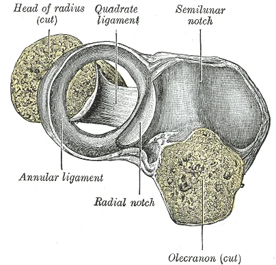
Annular ligament of radius, from above. The head of the radius has been sawn off and the bone dislodged from the ligament.

Figure 1: Annular ligament of radius, from above.
These bones lie between the carpal bones of the wrist and the bones of the fingers and thumb. [2]
To facilitate understanding of its anatomy, the forearm is usually divided into fascial compartments. The forearm is a region of high complexity, with 20 muscles and an extensive network of vessels and nerves. The forearm, made of the radius and ulna bones, has four main types of action at the hinge of the elbow joint: flexion, extension, pronation, and supination. The pronators are the pronator teres and the pronator quadratus. When the forearm faces anteriorly, it is supinated. When the forearm faces posteriorly, it is pronated. [3] [1]
Figure 2Gray417
Long description
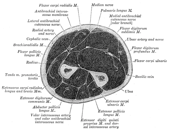
Cross-section through the middle of the forearm. (Eycleshymer and Schoemaker.)

Figure 2: Cross-section through the middle of the forearm (Eycleshymer and Schoemaker.).
DefinitionPronator Teres
A key muscle in the anterior compartment that gives rise to the pronator syndrome when it compresses adjacent structures.
Exam trapVolkmann Ischaemic Contracture
A permanent flexion contracture of the hand at the wrist, with the fingers flexed and the thumb adducted, resulting from untreated compartment syndrome.
As depicted in , the radial artery is usually smaller than the ulnar artery and is a more direct terminal branch of the brachial artery. The superficial branch of the radial nerve is located near the radial artery, their proximity being greatest in the middle third of the forearm. In the distal third of the forearm, there is a relative separation of the radial artery from the superficial branch of the radial nerve. The radial artery continues on an anterolateral course and the superficial branch of the radial nerve turns dorsally, piercing the fascia between the tendons of the brachioradialis and extensor carpi radialis longus muscles. [4]
| Structure | Relation |
|---|---|
| Ulnar Artery | Passes below the ulnar head of the pronator teres |
| Median Nerve | Passes above the ulnar head of the pronator teres [4] |
| Radial Artery | Crosses between brachioradialis (lateral) and pronator teres (medial) |
The forearm itself contains no true synovial joints; its two bones, the radius and ulna, are connected by the proximal and distal radioulnar joints, which are synovial pivot joints, and by the interosseous membrane, a fibrous syndesmosis. The proximal radioulnar joint lies within the elbow joint capsule, while the distal radioulnar joint is separate, near the wrist. Both allow rotation of the radius around the ulna, producing pronation and supination of the hand. The interosseous membrane, a strong sheet of fibrous tissue spanning the space between the radial and ulnar shafts, transmits forces from the radius to the ulna during weight-bearing and provides attachment for deep forearm muscleshebdomadal.
The functional movements of the forearm are therefore pronation and supination, which occur at these radioulnar joints. Pronation rotates the palm to face posteriorly (or inferiorly when the arm is at the side); supination returns it to face anteriorly (or superiorly). The range of motion is approximately 180 degrees in total, with about 85 degrees of pronation and 90 degrees of supination from the neutral position. These movements are produced by the muscles of the forearm compartments: the dorsal compartment muscles function in wrist extension, digit extension, and forearm supination.
The elbow joint itself, a hinge-type synovial joint, allows flexion and extension of the forearm on the arm. Its primary ligaments are the ulnar collateral ligament (medial) and the radial collateral ligament (lateral), which stabilise the joint during movement. The annular ligament of the radius encircles the radial head, holding it against the ulna and permitting rotation during pronation and supination. The wrist joint, a condyloid (ellipsoid) synovial joint, connects the distal radius and the articular disc of the distal radioulnar joint to the proximal carpal row, allowing flexion, extension, abduction and adduction of the hand.
The fascial compartments of the forearm are defined by the deep fascia and the interosseous membrane, not by synovial joints. The volar (flexor-pronator) compartment, dorsal (extensor-supinator) compartment and lateral (mobile wad of Henry) compartment are separated by fascial septa. The lateral compartment has a common fascial envelope that allows broader group sliding during pronation and supination, which is why its muscles can be retracted more easily during surgery. The interosseous me
The distal radioulnar joint is stabilised by the triangular fibrocartilage complex (TFCC), which includes the articular disc, dorsal and volar radioulnar ligaments, and the ulnar collateral ligament. This complex allows the radius to rotate around the fixed ulna during pronation and supination while maintaining joint stability. The proximal radioulnar joint is stabilised by the annular ligament and the quadrate ligament, which prevent excessive separation of the radial head from the ulna during rotation.
In clinical practice, the integrity of these joints is assessed by specific movements: resisted pronation and supination test the radioulnar joints and their associated muscles; flexion and extension test the elbow; and the TFCC is stressed by ulnar deviation and rotation of the wrist. The fascial compartments, however, are evaluated by palpation, measurement of intracompartmental pressure (normal resting pressure is under 10 mmHg; compartment syndrome is suspected above 30 mmHg), and imaging su
The anterior compartment, also called the volar or flexor-pronator compartment, lies on the palmar aspect. It contains the muscles responsible for wrist flexion, digit flexion, and forearm pronation. Superficially, the biceps brachii tendon is palpable in the antecubital fossa. The biceps brachii is a two-headed muscle that crosses both the shoulder and elbow joints to flex the forearm. It also participates in supinating the forearm at the radioulnar joints and flexing the arm at the shoulder jo [2][3]
The lateral compartment, or mobile wad of Henry, is situated on the radial side. The muscles here share a common fascial envelope that allows broader group sliding during pronation and supination. This feature allows the muscles to be retracted more easily during surgical procedures. The brachioradialis is a key component of this wad. [3]
The posterior compartment, or dorsal extensor-supinator compartment, lies on the dorsal surface. These include the abductor pollicis longus, extensor pollicis brevis, extensor pollicis longus, and extensor indicis.
At the wrist, the tendons of the forearm muscles attach and extend into the hand. Fibrous bands called retinacula sheath the tendons at the wrist. The flexor retinaculum extends over the palmar surface of the hand, while the extensor retinaculum extends over the dorsal surface. The cutaneous branches of the median and ulnar nerves, which originate in the forearm, are usually relatively easy to identify. These cutaneous nerves are better visualized in the proximal portion of the forearm, where they a [1][3]
DefinitionCarrying Angle
The lateral deviation of the forearm from the arm in the anatomical position, measuring 5–15 degrees, which prevents the forearm from striking the hip during movement.
| Muscle | Action | Nerve Supply |
|---|---|---|
| Biceps brachii | Flexion, supination | Musculocutaneous |
| Brachialis | Flexion | Musculocutaneous |
| Brachioradialis | Flexion | Radial |
Figure 3Gray213
Long description
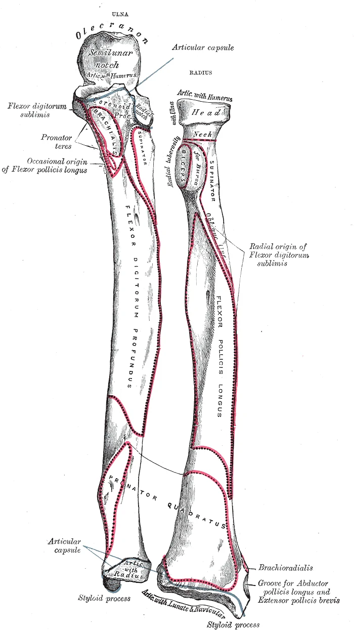
Bones of left forearm. Anterior aspect. Labelled illustration from Wikimedia Commons, used under its open licence.

Figure 3: Bones of left forearm (anterior aspect).
The medial bone is the ulna, and the lateral bone is the radius. The ulna is the medial bone of the forearm. It runs parallel to the radius, which is the lateral bone of the forearm. [2]
More distal is the shaft of the ulna. The lateral side of the shaft forms a ridge called the interosseous border of the ulna. This is the line of attachment for the interosseous membrane of the forearm, a sheet of dense connective tissue that unites the ulna and radius bones. The small, rounded area that forms the distal end is the head of the ulna. Projecting from the posterior side of the ulnar head is the styloid process of the ulna, a short bony projection. This serves as an attachment point for a connective tissue structure that unites the distal ends of the ulna and radius. [2]
The radius runs parallel to the ulna, on the lateral (thumb) side of the forearm. The head of the radius is a disc-shaped structure that forms the proximal end. The small depression on the surface of the head articulates with the capitulum of the humerus as part of the elbow joint, whereas the smooth, outer margin of the head articulates with the radial notch of the ulna at the proximal radioulnar joint. The neck of the radius is the narrowed region immediately below the expanded head. Inferior [2]
This ridge forms the interosseous border of the radius, which, like the similar border of the ulna, is the line of attachment for the interosseous membrane that unites the two forearm bones. The distal end of the radius has a smooth surface for articulation with two carpal bones to form the radiocarpal joint or wrist joint. On the medial side of the distal radius is the ulnar notch of the radius. This shallow depression articulates with the head of the ulna, which together form the distal radioulnar joint. The lateral end of the radius has a pointed projection called the styloid process of the radius. This provides attachment for ligaments that support the lateral side of the wrist joint. [2]
The central region is dominated by the flexor digitorum superficialis. The deep structures lie beneath these superficial layers, separated by intermuscular septa. The fascia encloses the muscles, creating distinct compartments that house specific groups of muscles and nerves.
The forearm contains two main groups of muscles: extrinsic and intrinsic. The extrinsic muscles originate in the forearm and insert into the hand, facilitating crude movements of the wrists, hands, and fingers. The intrinsic muscles originate in the palm and act on the hand itself. The extrinsic muscles are divided into anterior (flexor) and posterior (extensor) compartments. The anterior compartment produces flexion. It includes superficial muscles such as the flexor carpi radialis, palmaris longus, flexor carpi ulnaris, and flexor digitorum superficialis. The deep anterior compartment includes the flexor pollicis longus and flexor digitorum profundus. The posterior compartments produce extension. The superficial posterior compartment contains the extensor carpi radialis longus, extensor carpi radialis brevis, extensor digitorum, extensor digiti minimi, and extensor carpi ulnaris. [1]
The volar and dorsal compartments can be subdivided into superficial and deep layers. Some authors subdivide the volar compartment into three layers, with the flexor digitorum superficialis as the intermediate layer. The muscles in the anterior compartment originate on the humerus and insert onto different parts of the hand. They make up the bulk of the forearm. The powerful grasping muscles of the anterior forearm arise from the medial epicondyle of the humerus. This epicondyle is larger and more robust than the lateral epicondyle, which gives rise to the weaker posterior forearm muscles. The lateral epicondyle is smaller and located on the lateral side of the distal humerus. The roughened ridge above it is the lateral supracondylar ridge. [3]
DefinitionCarpal Tunnel Syndrome
Irritation of tendons in the carpal tunnel can pinch the median nerve, causing Carpal Tunnel Syndrome.
| Muscle | Innervation |
|---|---|
| Flexor carpi ulnaris | Ulnar nerve |
| Flexor digitorum profundus (medial part) | Ulnar nerve |
| Flexor digitorum profundus (lateral part) | Anterior interosseous nerve |
| Superficial layer muscles | Median nerve |
| Deep layer muscles | Anterior interosseous nerve |
The anterior interosseous nerve accompanies the anterior interosseous artery near the interosseous membrane. These structures cross the forearm between the edges of the flexor pollicis longus and flexor digitorum profundus muscles. The median artery is a fetal artery that normally regresses as the radial and ulnar arteries develop. In approximately 30% of individuals, it persists into adulthood, following the course of the median nerve. [4]
NotePersistent Median Artery
In 30% of people, the median artery persists into adulthood.
Cutaneous sensory innervation of the forearm is mainly supplied by three nerves. These are the lateral antebrachial cutaneous nerve, a terminal branch of the musculocutaneous nerve; the posterior antebrachial cutaneous nerve, a branch of the radial nerve; and the medial antebrachial cutaneous nerve, a direct branch of the brachial plexus. [4]
Figure 4815 The Carpal Tunnel
Long description
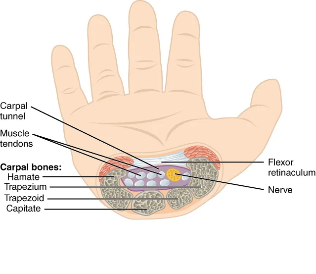
The carpal tunnel is the passageway by which nine muscle tendons and a major nerve enter the hand from the anterior forearm. The walls and floor of the carpal tunnel are formed by the U-shaped grouping of the carpal bones, and the roof is formed by the flexor retinaculum, a strong ligament that ante

Figure 4: The carpal tunnel is the passageway by which nine muscle tendons and a major nerve enter the hand from the anterior forearm.
The forearm is divided into anterior and posterior compartments, separated by the interosseous membrane and the radius and ulna. This illustration highlights the superficial, intermediate, and deep flexor muscles, as well as the extensor group, and indicates the regions susceptible to compartment syndrome.
Figure 5Gray419
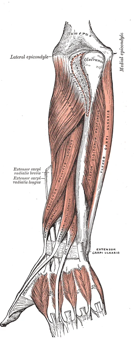
Long description
Posterior surface of the forearm. Deep muscles.

Figure 5: Posterior surface of the forearm (deep muscles).
Figure 6Gray418
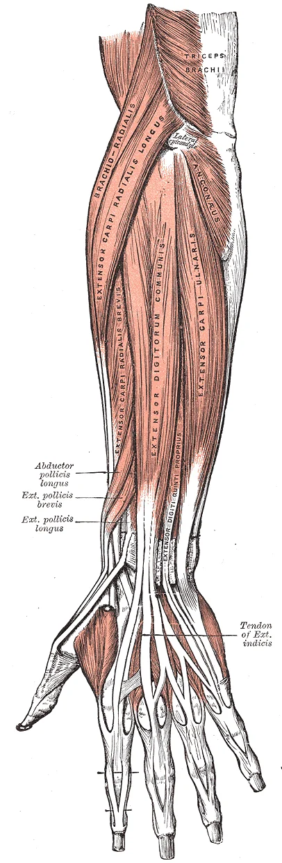
Long description
Posterior surface of the forearm. Superficial muscles.

Figure 6: Posterior surface of the forearm (superficial muscles).
Figure 7Gray416
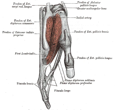
Long description
Tendons of forefinger and vincula tendina.

Figure 7: Tendons of forefinger and vincula tendina.
Figure 8Gray415
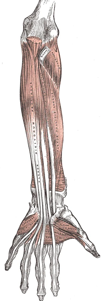
Long description
Front of the left forearm. Deep muscles.

Figure 8: Front of the left forearm (deep muscles).
The anterior compartment contains the flexor muscles, which are innervated by the median, ulnar, and anterior interosseous nerves. These muscles produce flexion and bend fingers to make a fist[1]. They are arranged in three layers: superficial, intermediate, and deep.
| Muscle | Origin | Insertion | Action | Nerve Supply |
|---|---|---|---|---|
| Pronator teres | Humerus, Ulna | Radius | Pronation | Median |
| Flexor carpi radialis | Humerus | Metacarpals II, III | Wrist flexion, abduction | Median |
| Palmaris longus | Humerus | Palmar aponeurosis | Wrist flexion | Median |
| Flexor carpi ulnaris | Humerus, Ulna | Pisiform, Metacarpal V | Wrist flexion, adduction | Ulnar |
| Flexor digitorum superficialis | Humerus, Ulna, Radius | Middle phalanges II-V | Finger flexion | Median |
| Flexor pollicis longus | Radius | Distal phalanx thumb | Thumb flexion | Anterior interosseous |
| Flexor digitorum profundus | Ulna, Interosseous membrane | Distal phalanges II-V | Finger flexion | Ulnar, Anterior interosseous |
| Pronator quadratus | Ulna | Radius | Pronation | Anterior interosseous |
The superficial layer includes the pronator teres, flexor carpi radialis, palmaris longus, and flexor carpi ulnaris. The intermediate layer consists of the flexor digitorum superficialis. The deep layer contains the flexor pollicis longus, flexor digitorum profundus, and pronator quadratus. The median nerve supplies most of these muscles, while the ulnar nerve supplies the flexor carpi ulnaris and the medial half of the flexor digitorum profundus. The anterior interosseous nerve, a branch of the median nerve, supplies the flexor pollicis longus, the lateral half of the flexor digitorum profundus, and the pronator quadratus. [3]
The posterior compartment contains the extensor muscles, which are innervated by the deep branch of the radial nerve and its continuation, the posterior interosseous nerve. The exception is the anconeus muscle, which is innervated directly by the radial nerve. In the lateral compartment, the brachioradialis and extensor carpi radialis longus are directly innervated by the radial nerve. [3]
| Muscle | Origin | Insertion | Action | Nerve Supply |
|---|---|---|---|---|
| Brachioradialis | Humerus | Radius | Elbow flexion | Radial |
| Extensor carpi radialis longus | Humerus | Metacarpal II | Wrist extension, abduction | Radial |
| Extensor carpi radialis brevis | Humerus | Metacarpal III | Wrist extension, abduction | Posterior interosseous |
| Extensor digitorum | Humerus | Digits II-V | Finger extension | Posterior interosseous |
| Extensor digiti minimi | Humerus | Digit V | Little finger extension | Posterior interosseous |
| Extensor carpi ulnaris | Humerus, Ulna | Metacarpal V | Wrist extension, adduction | Posterior interosseous |
| Supinator | Humerus, Ulna | Radius | Forearm supination | Posterior interosseous |
| Abductor pollicis longus | Ulna, Radius, Interosseous membrane | Metacarpal I | Thumb abduction | Posterior interosseous |
| Extensor pollicis brevis | Radius | Proximal phalanx thumb | Thumb extension | Posterior interosseous |
| Extensor pollicis longus | Ulna, Interosseous membrane | Distal phalanx thumb | Thumb extension | Posterior interosseous |
| Extensor indicis | Ulna, Interosseous membrane | Index finger | Index finger extension | Posterior interosseous |
| Anconeus | Humerus | Ulna | Elbow extension | Radial |
Compartment syndrome can occur in these tight fascial compartments, leading to increased pressure and compromised blood flow. The deep anterior compartment produces flexion and bends fingers to make a fist[1]. The intrinsic muscles of the hand both originate and insert within it[1]. These muscles allow your fingers to also make precise movements for actions, such as typing or writing[1]. The thenar muscles are on the radial aspect of the palm. The hypothenar muscles are on the medial aspect of the palm, and the intermediate muscles are midpalmar[1]. The hypothenar muscles include the abductor digiti minimi, flexor digiti minimi brevis, and the opponens digiti minimi[1]. These muscles form the hypothenar eminence, the rounded contour of the little finger, and as such, they all act on the little finger[1]. Finally, the intermediate muscles act on all the fingers and include the lumbrical, the palmar interossei, and the dorsal interossei[1].
The arterial supply to the forearm is derived from the brachial artery, which bifurcates in the cubital fossa to form the two principal vessels: the radial and ulnar arteries. [1] [4]
The radial artery arises from the brachial artery in the cubital fossa. It courses laterally, initially lying deep to the brachioradialis muscle. As it descends, it passes anterior to the supinator muscle and then runs along the lateral aspect of the radius.
It then runs along the medial aspect of the forearm, deep to the flexor digitorum superficialis. Its branches include the anterior and posterior interosseous arteries, which supply the deep anterior and posterior compartments respectively, and the common interosseous artery. The ulnar artery terminates in the hand by forming the superficial palmar arch.
The common interosseous artery arises from the ulnar artery shortly after its origin. It divides into the anterior and posterior interosseous arteries. The anterior interosseous artery runs on the interosseous membrane, supplying the deep muscles of the anterior compartment. The posterior interosseous artery runs on the interosseous membrane, supplying the deep muscles of the posterior compartment. These arteries anastomose with each other and with branches of the radial and ulnar arteries, ensuring a rich collateral circulation. [4]
DefinitionForearm Compartments
The forearm is divided into anterior (flexor) and posterior (extensor) compartments, separated by the interosseous membrane. Each compartment contains specific muscles, nerves, and vessels.
| Vessel | Origin | Course | Branches | Termination |
|---|---|---|---|---|
| Radial Artery | Brachial Artery | Lateral forearm, deep to brachioradialis | Radial recurrent, anterior/posterior interosseous | Deep palmar arch |
| Ulnar Artery | Brachial Artery | Medial forearm, deep to flexor carpi radialis | Common interosseous, anterior/posterior interosseous | Superficial palmar arch |
| Common Interosseous | Ulnar Artery | Proximal forearm, divides into anterior/posterior | Anterior and posterior interosseous arteries | Interosseous membrane |
The venous system of the forearm mirrors the arterial supply, forming a dual network of deep and superficial channels that facilitate efficient blood return to the heart. Understanding this drainage is critical for interpreting clinical signs of vascular compromise or trauma.
The basilic vein lies medially, piercing the deep fascia in the mid-arm region. Deeply, the radial and ulnar veins accompany their respective arteries. The interosseous veins are situated between the radius and ulna, draining the deep posterior compartment.
The superficial veins lie within the subcutaneous tissue and are not accompanied by arteries. They are clinically significant for venipuncture and assessing volume status.
The cephalic vein originates from the lateral side of the dorsal venous network of the hand. It ascends along the lateral border of the forearm, superficial to the brachioradialis. It pierces the deltopectoral fascia to join the axillary vein.
In the mid-arm, it pierces the deep fascia to join the brachial veins, forming the axillary vein.
The deep veins accompany the major arteries of the forearm and are often paired as venae comitantes. They carry the majority of the venous return from the forearm muscles.
The radial veins are paired vessels that accompany the radial artery. They originate from the deep palmar venous arch. They ascend along the lateral aspect of the forearm, deep to the brachioradialis and supinator muscles. They unite with the ulnar veins to form the brachial veins.
The ulnar veins are paired vessels accompanying the ulnar artery. They begin in the deep palmar venous arch. They ascend along the medial aspect of the forearm, deep to the flexor muscles. They are larger than the radial veins and contribute more significantly to the formation of the brachial veins.
The interosseous veins drain the deep posterior compartment. They accompany the anterior and posterior interosseous arteries. They pass through the interosseous membrane to join the ulnar veins.
DefinitionVenous Drainage
The process by which deoxygenated blood is returned from the forearm tissues to the central circulation via superficial and deep venous networks.
| Structure | Origin | Course | Termination |
|---|---|---|---|
| Cephalic Vein | Dorsal venous network | Lateral forearm, superficial | Axillary vein |
| Basilic Vein | Dorsal venous network | Medial forearm, superficial | Brachial veins |
| Radial Veins | Deep palmar arch | Lateral forearm, deep | Brachial veins |
| Ulnar Veins | Deep palmar arch | Medial forearm, deep | Brachial veins |
Clinical pearlClinical Pearl
Superficial veins are preferred for venipuncture due to their accessibility, while deep veins are less visible but carry the bulk of venous return.
The forearm is supplied by three major peripheral nerves: the median, ulnar, and radial nerves. These nerves originate from the brachial plexus and descend through the arm before entering the forearm. They provide both motor innervation to the muscles of the anterior and posterior compartments and cutaneous sensation to the skin of the forearm and hand. Understanding their course and distribution is essential for localising lesions.
The median nerve lies centrally, deep to the flexor digitorum superficialis. The ulnar nerve is positioned medially, running between the flexor carpi ulnaris and the flexor digitorum profundus. The radial nerve, specifically its deep branch (posterior interosseous nerve), is located laterally and posteriorly, emerging from the supinator muscle.
DefinitionAnterior Interosseous Nerve
A pure motor branch of the median nerve that supplies the deep muscles of the anterior forearm.
The median nerve provides motor supply to most of the flexor muscles in the anterior compartment. It innervates the pronator teres, flexor carpi radialis, palmaris longus, flexor digitorum superficialis, and the lateral half of the flexor digitorum profundus. It also supplies the flexor pollicis longus and pronator quadratus via its anterior interosseous branch. Cutaneously, the median nerve supplies the palmar aspect of the lateral three and a half digits.
The ulnar nerve supplies the medial half of the flexor digitorum profundus and the flexor carpi ulnaris. It also innervates the intrinsic muscles of the hand, including the hypothenar muscles, the interossei, and the medial two lumbricals. Cutaneously, it supplies the palmar and dorsal aspects of the medial one and a half digits.
The radial nerve divides into superficial and deep branches. The superficial branch is primarily sensory, supplying the dorsal aspect of the lateral three and a half digits.
| Nerve | Motor Supply | Cutaneous Supply |
|---|---|---|
| Median | Pronator teres, FCR, PL, FDS, lateral FDP, FPL, PQ | Lateral 3.5 digits (palmar) |
| Ulnar | FCU, medial FDP, intrinsic hand muscles | Medial 1.5 digits (palmar/dorsal) |
| Radial | Brachioradialis, ECRB, supinator, extensors | Lateral 3.5 digits (dorsal) |
The lymphatic drainage of the forearm is a critical pathway for immune surveillance and fluid homeostasis, mirroring the vascular supply but with distinct nodal targets. Understanding this system requires visualising the deep and superficial networks that converge towards the axilla. This diagram presents an anterior view of the right forearm, with the medial side on the left and the lateral side on the right. The image highlights the superficial lymphatic vessels running along the cephalic and basilic veins, as well as the deep vessels accompanying the radial, ulnar, and interosseous arteries.
DefinitionSuperficial Lymphatics
Vessels that accompany the superficial veins and drain the skin and subcutaneous tissues of the forearm.
Each of these articulations is a carpometacarpal joint [2]
Exam trapCubital Nodes
These nodes are often overlooked but are the first filter for medial forearm infections. Swelling here indicates pathology in the medial forearm or hand.
The deep lymphatic network accompanies the major arteries. Lymphatics from the radial artery run along its course. They drain the lateral compartment muscles. These vessels converge towards the lateral axillary nodes. Lymphatics from the ulnar artery follow a similar path. They drain the medial compartment muscles. These vessels also contribute to the cubital nodes before proceeding to the axilla. The interosseous arteries have their own lymphatic companions. These vessels drain the deep posterior compartment. They drain towards the central and subscapular axillary nodes.
| Structure | Accompanying Vessel | Primary Nodal Target |
|---|---|---|
| Lateral Skin | Cephalic Vein | Lateral Axillary Nodes |
| Medial Skin | Basilic Vein | Cubital Nodes |
| Lateral Muscles | Radial Artery | Lateral Axillary Nodes |
| Medial Muscles | Ulnar Artery | Cubital / Central Axillary Nodes |
| Deep Posterior | Interosseous Arteries | Central / Subscapular Nodes |
Clinical relevance is paramount. Infections of the hand or forearm can spread rapidly via these channels. A cut on the lateral thumb may lead to axillary lymphadenopathy. A wound on the medial forearm may cause tender swelling above the elbow. Recognising the anatomical routes helps in diagnosing the source of infection. The lymphatic system acts as a filter, trapping pathogens and initiating an immune response. This protective mechanism is essential for maintaining health in the upper limb.
The forearm is divided into anterior and posterior compartments by the interosseous membrane and the radius and ulna. The image displays the transverse plane, with the anterior aspect at the top and posterior at the bottom. The radius lies laterally and the ulna medially. The interosseous membrane spans between the two bones, creating the central space that separates the compartments.
DefinitionInterosseous Membrane
A strong fibrous sheet connecting the shafts of the radius and ulna, providing attachment for muscles and stabilising the interosseous space. [1]
These are the flexor pollicis longus and the flexor digitorum profundus. [1]
| Muscle | Origin | Insertion | Action | Nerve Supply |
|---|---|---|---|---|
| Pronator Teres | Medial epicondyle of humerus; coronoid process of ulna | Mid-shaft of radius | Pronation of forearm | Median nerve |
| Flexor Carpi Radialis | Medial epicondyle of humerus | Base of 2nd and 3rd metacarpals | Wrist flexion and abduction | Median nerve |
| Palmaris Longus | Medial epicondyle of humerus | Palmar aponeurosis | Wrist flexion | Median nerve |
| Flexor Carpi Ulnaris | Medial epicondyle of humerus; Olecranon and posterior ulna | Pisiform, hook of hamate, 5th metacarpal | Wrist flexion and adduction | Ulnar nerve |
| Flexor Digitorum Superficialis | Medial epicondyle of humerus; Radius; Ulna | Middle phalanges of digits 2-5 | Flexion of proximal interphalangeal joints | Median nerve |
| Flexor Digitorum Profundus | Proximal ulna and interosseous membrane | Distal phalanges of digits 2-5 | Flexion of distal interphalangeal joints | Ulnar and Median nerves |
| Flexor Pollicis Longus | Anterior radius and interosseous membrane | Distal phalanx of thumb | Flexion of interphalangeal joint of thumb | Anterior interosseous nerve |
The posterior compartment contains the extensor muscles. The superficial layer includes the brachioradialis, extensor carpi radialis longus, extensor carpi radialis brevis, extensor digitorum, extensor digiti minimi, and extensor carpi ulnaris. The deep layer comprises the supinator, abductor pollicis longus, extensor pollicis brevis, extensor pollicis longus, and extensor indicis. The radial nerve divides into the superficial and deep branches. The deep branch pierces the supinator muscle to become the posterior interosseous nerve. This nerve supplies all muscles in the posterior compartment except the brachioradialis and extensor carpi radialis longus. The posterior interosseous artery accompanies this nerve.
Figure 9Gray420
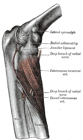
Long description
The Supinator. Labelled illustration from Wikimedia Commons, used under its open licence.

Figure 9: The Supinator.
Exam trapNerve Supply
The radial nerve supplies the brachioradialis and extensor carpi radialis longus before dividing. The posterior interosseous nerve does not supply these muscles.
The neurovascular bundles are critical landmarks. The median nerve is central in the anterior compartment. The ulnar nerve passes behind the medial epicondyle and enters the forearm between the two heads of the flexor carpi ulnaris. The radial artery lies lateral to the flexor carpi radialis tendon. The ulnar artery lies medial to the flexor carpi ulnaris tendon. These vessels anastomose around the elbow and wrist.
Understanding the forearm requires correlating surface landmarks with deep compartmental structures. The forearm is divided into anterior (flexor) and posterior (extensor) compartments by the interosseous membrane and the radius and ulna. Radiological imaging allows us to visualise these compartments, their contents, and the boundaries that separate them.
The radius is located laterally (on the left in standard anatomical position views) and the ulna medially. Between them lies the interosseous membrane, which appears as a thin, dense line. The anterior compartment contains the flexor muscles, including the flexor carpi radialis and palmaris longus superficially, and the flexor digitorum superficialis and profundus deeper. The posterior compartment contains the extensor muscles, such as the extensor carpi radialis longus and brevis, and the extensor digitorum. The neurovascular bundles, including the radial and ulnar arteries and the median and ulnar nerves, are situated within these compartments, often adjacent to the bones.
X-ray imaging is the first-line modality for assessing bony anatomy. It clearly delineates the radius and ulna, the interosseous space, and any fractures or dislocations. While X-rays do not show soft tissue compartments directly, the alignment of the bones provides indirect evidence of compartmental integrity.
CT scans offer superior detail of the bony architecture and can reveal subtle fractures. In soft tissue windows, CT can distinguish between muscle groups and identify haematomas or fluid collections within the compartments. It is particularly useful in trauma settings where complex fractures are suspected.
MRI is the gold standard for evaluating soft tissue structures. It provides excellent contrast resolution, allowing clear differentiation between muscle, tendon, nerve, and fat. MRI can visualise the interosseous membrane, ligaments, and neurovascular bundles in high detail. It is essential for diagnosing compartment syndrome, tendon tears, and nerve entrapments. The high spatial resolution of modern MRI sequences allows for precise localisation of pathology within specific compartments.
Ultrasound is a dynamic, real-time imaging modality. Transducers that operate in the frequency range between 1 and 70 MHz are available in clinical practice, and new transducers have been introduced to the market more quickly than in previous decades(,). The new high-frequency ultrasound (HFUS) and ultra-high-frequency ultrasound (UHFUS) transducers have provided significant improvements in image spatial resolution. However, their widespread use is limited by their low penetration capability(,). Ultrasound is particularly useful for assessing tendons and nerves, allowing for dynamic evaluation during movement. It can also guide interventions, such as injections or biopsies, within the forearm compartments. [3]
DefinitionForearm Compartments
The forearm is divided into anterior and posterior compartments by the interosseous membrane and the radius and ulna, containing distinct muscle groups and neurovascular structures.
| Compartment | Primary Muscles | Key Neurovascular Structures |
|---|---|---|
| Anterior | Flexor carpi radialis, Palmaris longus, Flexor digitorum superficialis, Flexor digitorum profundus | Median nerve, Ulnar artery |
| Posterior | Extensor carpi radialis longus, Extensor carpi radialis brevis, Extensor digitorum | Radial nerve, Posterior interosseous nerve |
The forearm is divided into anterior and posterior compartments by the interosseous membrane and the antebrachial fascia. These compartments contain muscles, nerves, and vessels that are susceptible to compression and injury. Understanding the spatial relationships is critical for diagnosing nerve palsies and managing compartment syndrome.
In the anterior compartment, the flexor digitorum superficialis lies superficially, covering the deeper flexor digitorum profundus. The median nerve runs between these two muscle layers, making it vulnerable to compression within this space. The ulnar nerve is situated more medially, adjacent to the flexor carpi ulnaris. In the posterior compartment, the extensor digitorum is the most prominent muscle, with the radial nerve and posterior interosseous nerve coursing through the supinator muscle. Recognising these relationships allows clinicians to predict which nerve is affected by swelling in a specific compartment. [4]
The anterior compartment contains muscles responsible for wrist and finger flexion. These muscles originate from the medial epicondyle of the humerus and the adjacent parts of the radius and ulna. They insert onto the metacarpals and phalanges. The primary nerve supply is the median nerve, with the ulnar nerve supplying the medial half of the flexor digitorum profundus and the flexor carpi ulnaris.
| Muscle | Origin | Insertion | Action | Nerve Supply |
|---|---|---|---|---|
| Flexor carpi radialis | Medial epicondyle | Base of 2nd metacarpal | Wrist flexion and abduction | Median nerve |
| Palmaris longus | Medial epicondyle | Palmar aponeurosis | Wrist flexion | Median nerve |
| Flexor carpi ulnaris | Medial epicondyle, olecranon | Pisiform, hook of hamate | Wrist flexion and adduction | Ulnar nerve |
| Flexor digitorum superficialis | Medial epicondyle, radius | Middle phalanges | Finger flexion | Median nerve |
| Flexor digitorum profundus | Ulna, interosseous membrane | Distal phalanges | Distal finger flexion | Ulnar and Median nerves |
The posterior compartment contains extensor muscles. These originate from the lateral epicondyle and the posterior surfaces of the radius and ulna. They insert onto the dorsal aspects of the hand and fingers. The radial nerve, specifically its deep branch becoming the posterior interosseous nerve, supplies these muscles.
| Muscle | Origin | Insertion | Action | Nerve Supply |
|---|---|---|---|---|
| Extensor carpi radialis longus | Lateral supracondylar ridge | Base of 2nd metacarpal | Wrist extension and abduction | Radial nerve |
| Extensor carpi radialis brevis | Lateral epicondyle | Base of 3rd metacarpal | Wrist extension and abduction | Posterior interosseous nerve |
| Extensor digitorum | Lateral epicondyle | Dorsal digital expansions | Finger extension | Posterior interosseous nerve |
| Extensor digiti minimi | Lateral epicondyle | Dorsal expansion of 5th finger | Little finger extension | Posterior interosseous nerve |
| Extensor carpi ulnaris | Lateral epicondyle, ulna | Base of 5th metacarpal | Wrist extension and adduction | Posterior interosseous nerve |
Compartment syndrome arises when pressure within a closed fascial space exceeds perfusion pressure, leading to ischaemia. The anterior compartment is the most common site due to its larger muscle mass. Clinical signs include pain out of proportion to injury, pain on passive stretch, and paresthesia. Early recognition prevents permanent nerve and muscle damage. Surface landmarks such as the medial and lateral epicondyles help locate the compartment boundaries during examination.
The forearm is divided into two distinct compartments, each containing specific muscles, nerves, and vessels that are critical for upper limb function. Understanding the anatomical boundaries and contents is essential for diagnosing compartment syndrome and nerve injuries.
The anterior compartment lies deep to the fascia and is bounded by the interosseous membrane laterally. It primarily contains flexor muscles of the wrist and fingers.
| Muscle | Origin | Insertion | Action | Nerve Supply |
|---|---|---|---|---|
| Flexor carpi radialis | Medial epicondyle | Base of 2nd metacarpal | Wrist flexion and abduction | Median nerve |
| Flexor carpi ulnaris | Medial epicondyle and olecranon | Pisiform and 5th metacarpal | Wrist flexion and adduction | Ulnar nerve |
| Palmaris longus | Medial epicondyle | Palmar aponeurosis | Wrist flexion | Median nerve |
A common trap is confusing the nerve supply. The median nerve supplies most anterior muscles, but the ulnar nerve supplies the flexor carpi ulnaris and the medial half of the flexor digitorum profundus. The surgical neck of the humerus is a common site for fractures, which can injure the axillary nerve, but this is proximal to the forearm compartments. [2]
The posterior compartment contains extensor muscles. These muscles are generally innervated by the radial nerve.
| Muscle | Origin | Insertion | Action | Nerve Supply |
|---|---|---|---|---|
| Extensor carpi radialis longus | Lateral supracondylar ridge | Base of 2nd metacarpal | Wrist extension and abduction | Radial nerve |
| Extensor carpi ulnaris | Lateral epicondyle | Base of 5th metacarpal | Wrist extension and adduction | Radial nerve |
| Brachioradialis | Lateral supracondylar ridge | Styloid process of radius | Elbow flexion | Radial nerve |
This landmark is proximal to the elbow and does not directly relate to forearm compartment contents, but it is a key surface anatomy feature for identifying the humerus. [2]
Compartment syndrome is a surgical emergency caused by increased pressure within a closed fascial space. The five Ps are pain, pallor, paresthesia, paralysis, and pulselessness. Pain out of proportion to injury is the earliest sign.
The metacarpal bones are five elongated bones in the palm, numbered 1 to 5 from the thumb. Each metacarpal articulates proximally with a carpal bone at the carpometacarpal joint and distally with a phalanx at the metacarpophalangeal joint. Fractures here are common but do not typically cause compartment syndrome due to the lack of tight fascial compartments in the palm compared to the forearm. [2]
Students frequently confuse the anatomical landmarks of the wrist, leading to errors in clinical examination and imaging interpretation. A primary error involves the relative positioning of the styloid processes. The styloid process of the radius projects more distally than the styloid process of the ulna. This anatomical fact limits the range of movement for lateral deviations of the hand at the wrist joint. When students assume the ulnar styloid is more prominent distally, they misinterpret the mechanics of wrist deviation. [2]
Another frequent mistake concerns the carpal bones. Learners often struggle with the arrangement of these eight small bones. A helpful mnemonic for remembering the arrangement of the carpal bones is “So Long To Pinky, Here Comes The Thumb”. This mnemonic starts on the lateral side and names the proximal bones from lateral to medial (scaphoid, lunate, triquetrum, pisiform), then makes a U-turn to name the distal bones from medial to lateral (hamate, capitate, trapezoid, trapezium). Thus, it starts and finishes on the lateral side. Without this structured approach, students may misidentify the hook of the hamate or the scaphoid, leading to incorrect diagnosis of fractures. [2]
Students also often overlook the specific mobility characteristics of the metacarpals. The first metacarpal bone, at the base of the thumb, is separated from the other metacarpal bones. This allows it a freedom of motion that is independent of the other metacarpal bones, which is very important for thumb mobility. In contrast, the second and third metacarpal bones are firmly anchored in place and are immobile. The fourth and fifth metacarpal bones have limited anterior-posterior mobility, a motion that is greater for the fifth bone. This mobility is important during power gripping with the hand. The anterior movement of these bones, particularly the fifth metacarpal bone, increases the strength of contact for the medial hand during gripping actions. Confusing the mobility of the first metacarpal with the stability of the second and third is a common conceptual error. [2]
Finally, students may misinterpret ultrasound findings in tendon pathology. Tendinosis, which usually appears as tendon thickening and fibrillar pattern loss, is typically accompanied by hypoechoic changes. Doppler may help in the detection of associated hyperemia. Partial and complete tears are characterized by areas of discontinuity of fibers, usually filled with fluid. Dynamic evaluation can facilitate the differential diagnosis between tendinosis and a torn tendon. Assuming that hypoechoic changes alone indicate a tear, without considering the fibrillar pattern or dynamic assessment, leads to over-diagnosis of structural failure. [3]
Exam trapStyloid Position
The radial styloid is more distal than the ulnar styloid, limiting lateral wrist deviation.
DefinitionCarpal Mnemonic
“So Long To Pinky, Here Comes The Thumb” describes the proximal then distal carpal arrangement.
This knowledge is used at the bedside to assess wrist stability and grip strength. Clinicians evaluate the distal radius and ulna to determine the extent of lateral deviation possible at the wrist joint. The fact that the styloid process of the radius projects more distally than the styloid process of the ulna is critical when assessing ligamentous integrity. If the ulnar styloid is fractured, the relative position remains unchanged, but the stability of the distal radioulnar joint may be compromised. Understanding that the styloid process of the radius projects more distally, thereby limiting the range of movement for lateral deviations of the hand at the wrist joint helps in predicting the direction of instability.
In the operating theatre, precise identification of carpal bones is essential. Surgeons must locate the hook of the hamate bone, which is characterized by a prominent bony extension on its anterior side called the hook of the hamate bone. This structure is a key landmark for ulnar nerve decompression and for assessing Guyon’s canal syndrome. Knowledge of the carpal arrangement, such as the mnemonic “So Long To Pinky, Here Comes The Thumb”, ensures correct orientation during open procedures. The mnemonic starts on the lateral side and names the proximal bones from lateral to medial (scaphoid, lunate, triquetrum, pisiform), then makes a U-turn to name the distal bones from medial to lateral (hamate, capitate, trapezoid, trapezium). Thus, it starts and finishes on the lateral side.
On imaging, radiologists use the distinct mobility of the metacarpals to assess for fractures and dislocations. The first metacarpal bone, at the base of the thumb, is separated from the other metacarpal bones. This allows it a freedom of motion that is independent of the other metacarpal bones, which is very important for thumb mobility. On X-rays, the mobility of the first metacarpal can mimic pathology if not recognized. Conversely, the second and third metacarpal bones are firmly anchored in place and are immobile. Displacement of these bones is always pathological. The fourth and fifth metacarpal bones have limited anterior-posterior mobility, a motion that is greater for the fifth bone. This mobility is important during power gripping with the hand. The anterior movement of these bones, particularly the fifth metacarpal bone, increases the strength of contact for the medial hand during gripping actions. Radiologists look for abnormal anterior displacement of the fifth metacarpal, indicative of a boxer’s fracture.
In examinations, students must differentiate between tendinosis and tears using ultrasound criteria. Tendinosis, which usually appears as tendon thickening and fibrillar pattern loss, is typically accompanied by hypoechoic changes. Doppler may help in the detection of associated hyperemia. Partial and complete tears are characterized by areas of discontinuity of fibers, usually filled with fluid. Dynamic evaluation can facilitate the differential diagnosis between tendinosis and a torn tendon. Examiners test the ability to distinguish these features, ensuring students do not confuse structural degeneration with acute rupture.
| Metacarpal | Mobility | Clinical Significance |
|---|---|---|
| 1st | Freely mobile, independent | Thumb mobility; can mimic pathology on imaging |
| 2nd & 3rd | Firmly anchored, immobile | Displacement indicates fracture or dislocation |
| 4th & 5th | Limited AP mobility (5th > 4th) | Power gripping; 5th displacement suggests boxer's fracture |
Ultrasound Tendon Assessment
- Assess TendonCheck for thickening and fibrillar pattern
- Question: Fibrillar pattern lost?
Yes
- Check Hypoechoic ChangesPresent in tendinosis
- Question: Discontinuity of fibers?
Yes
- Diagnosis: TearFluid-filled discontinuity
No
- Diagnosis: TendinosisThickening with hypoechoic changes
No
- Normal TendonIntact fibrillar pattern
The radial styloid is more distal than the ulnar styloid, limiting lateral wrist deviation. The carpal mnemonic “So Long To Pinky, Here Comes The Thumb” aids memory. The hook of the hamate is a prominent anterior bony extension. The first metacarpal is freely mobile and independent of other metacarpals. The second and third metacarpals are firmly anchored and immobile. The fourth and fifth metacarpals have limited anterior-posterior mobility. Tendinosis shows thickening, fibrillar loss, and hypoechoic changes. Tears show discontinuity of fibers filled with fluid.
Answer each one in your head first, then open the answer.
Which styloid process projects more distally?
Show answerHide answer
The styloid process of the radiusWhat does the carpal mnemonic start and finish on?
Show answerHide answer
The lateral sideWhich metacarpal is freely mobile?
Show answerHide answer
The first metacarpalWhich metacarpals are firmly anchored?
Show answerHide answer
The second and third metacarpalsWhat characterizes a tendon tear on ultrasound?
Show answerHide answer
Discontinuity of fibers filled with fluid
- Betts JG, Young KA, Wise JA, et al. Muscles of the Pectoral Girdle and Upper Limbs. OpenStax, Anatomy and Physiology 2e (CC BY-NC-SA 4.0). Accessed .
- Betts JG, Young KA, Wise JA, et al. Bones of the Upper Limb. OpenStax, Anatomy and Physiology 2e (CC BY-NC-SA 4.0). Accessed .
- Borges ÁVRM, Souza SAL. Anatomy of the nerves, vessels, and muscular compartments of the forearm, as revealed by high-resolution ultrasound. Part 1: overall structure and forearm compartments. Radiologia brasileira 2021;54(6):388-397. doi:10.1590/0100-3984.2021.0030. Accessed .
- Borges ÁVRM, Souza SAL. Anatomy of the nerves, vessels, and muscular compartments of the forearm, as revealed by high-resolution ultrasound. Part 2: vascularization of compartments and cutaneous innervation. Radiologia brasileira 2021;54(6):398-405. doi:10.1590/0100-3984.2021.0031. Accessed .
Figure credits
- Figure 1: Henry Vandyke Carter, via Wikimedia Commons, Public domain, Public domain.
- Figure 2: Henry Vandyke Carter, via Wikimedia Commons, Public domain, Public domain.
- Figure 3: Henry Vandyke Carter, via Wikimedia Commons, Public domain, Public domain.
- Figure 4: OpenStax College, via Wikimedia Commons, CC BY 3.0, CC BY 3.0.
- Figure 5: Henry Vandyke Carter, via Wikimedia Commons, Public domain, Public domain.
- Figure 6: Henry Vandyke Carter, via Wikimedia Commons, Public domain, Public domain.
- Figure 7: Henry Vandyke Carter, via Wikimedia Commons, Public domain, Public domain.
- Figure 8: Henry Vandyke Carter, via Wikimedia Commons, Public domain, Public domain.
- Figure 9: Henry Vandyke Carter, via Wikimedia Commons, Public domain, Public domain.
Educational content for medical students and doctors. It is not a substitute for clinical judgement or local guidelines.