Elbow joint
The elbow is a complex uniaxial hinge joint enabling forearm flexion, extension, and rotation.
The elbow is fundamentally a uniaxial hinge joint, primarily facilitating the flexion and extension of the forearm. Structurally, it is not a single articulation but a composite of three distinct joints enclosed within one continuous articular capsule: the humeroulnar joint, the humeroradial joint, and the proximal radioulnar joint. The humeroulnar joint, formed by the trochlea of the humerus and the trochlear notch of the ulna, provides the primary hinge mechanism. The humeroradial joint contri [1]
Stability is maintained by strong collateral ligaments. The ulnar collateral ligament supports the medial side, while the radial collateral ligament supports the lateral side. These ligaments prevent side-to-side movements and resist hyperextension. The joint capsule is reinforced by these intrinsic ligaments, which are fused to the wall of the articular capsule. The radial fossa accommodates the head of the radius during deep flexion. [1]
A key clinical feature is the carrying angle, the slight valgus deviation of the forearm when the arm is extended. This angle allows the arms to clear the hips during walking. A supracondylar fracture of the humerus, common in children, occurs just above the condyles. This injury can damage the brachial artery or median nerve, leading to compartment syndrome or motor deficits. The cubital fossa, located anterior to the elbow, contains the brachial artery, median nerve, and biceps tendon, arranged from lateral to medial. Superficial veins, such as the median cubital vein, connect the cephalic and basilic veins and are preferred sites for venepuncture due to their accessibility and stability.
DefinitionCarrying Angle
The slight valgus angle of the forearm relative to the humerus when extended, facilitating clearance of the hips during gait.
Exam trapSupracondylar Fracture
This fracture is a medical emergency due to the risk of brachial artery injury and subsequent Volkmann’s ischaemic contracture.
| Structure | Position (Lateral to Medial) | Clinical Significance |
|---|---|---|
| Biceps Tendon | Lateral | Palpable pulse point for brachial artery |
| Brachial Artery | Central | Site for blood pressure measurement |
| Median Nerve | Medial | Risk of injury in supracondylar fractures |
The elbow joint is a complex synovial structure formed by the articulation of three bones: the humerus, the radius, and the ulna. To understand its mechanics, we must first examine the specific bony landmarks that form the joint surfaces. This figure displays an anterior view of the distal humerus, proximal radius, and proximal ulna. The humerus presents two distinct articular areas. Anteriorly, the capitulum is a rounded eminence located on the lateral side of the distal humerus. It articulates with the head of the radius. Medial to the capitulum lies the trochlea, a spool-shaped structure that articulates with the trochlear notch of the ulna. The trochlea is positioned more posteriorly than the capitulum, which influences the carrying angle of the arm. The ulna contributes the trochlear notch, a deep, C-shaped depression that wraps around the trochlea. The radius contributes the cup-shaped head, which fits against the capitulum. These surfaces are covered in hyaline articular cartilage, which reduces friction and absorbs shock during movement. The joint cavity is enclosed by a fibrous capsule and lined by a synovial membrane that secretes lubricating fluid. The arrangement of these surfaces allows the elbow to function primarily as a hinge joint, permitting flexion and extension. However, the radial head also allows for rotation of the forearm when the elbow is flexed. Understanding the precise topography of these surfaces is critical for diagnosing fractures and dislocations. For instance, the relationship between the capitulum and the radial head is vital for assessing radial head fractures. Similarly, the integrity of the trochlear notch is essential for maintaining ulnohumeral stability. The articular surfaces are not continuous with each other; they are separated by the joint space filled with synovial fluid. This separation allows for smooth, low-friction movement. The cartilage is avascular, relying on the synovial fluid for nourishment. Any damage to these surfaces can lead to osteoarthritis or joint instability. The specific geometry of the trochlea ensures that the ulna moves in a single plane during flexion and extension, preventing lateral deviation. The capitulum’s rounded shape allows for a wider range of motion in the radial head. This combination of hinge and pivot mechanics makes the elbow a versatile joint. The articular surfaces must be congruent to distribute load evenly. Misalignment can result in abnormal wear patterns. Therefore, precise anatomical knowledge of these surfaces is fundamental for clinical practice. [2]
Figure 1Gray331
Long description
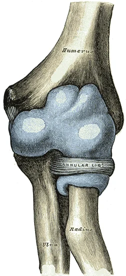
Capsule of elbow-joint (distended). Anterior aspect.

Figure 1: Capsule of elbow-joint (distended) (anterior aspect).
The elbow joint is a synovial joint, characterised by the presence of a joint cavity. The walls of this space are formed by the articular capsule, a fibrous connective tissue structure that is attached to each bone just outside the area of the bone’s articulating surface. The bones of the joint articulate with each other within the joint cavity. Functionally, the ability of the bones to move smoothly against each other within the joint cavity means that the elbow is classified as a diarthrosis. [3]
DefinitionHinge Joint
A hinge joint allows only for bending and straightening motions along a single axis, and is thus functionally classified as a uniaxial joint. [3]
In a hinge joint, the convex end of one bone articulates with the concave end of the adjoining bone. A good example is the elbow joint, with the articulation between the trochlea of the humerus and the trochlear notch of the ulna. Other hinge joints of the body include the knee, ankle, and interphalangeal joints between the phalanx bones of the fingers and toes. [3]
The articular capsule surrounds the joint, attaching to the humerus, radius, and ulna just outside the articular surfaces. The synovial membrane lines the inner surface of the capsule, excluding the articular cartilage. It produces synovial fluid to lubricate the joint. The capsule is reinforced by ligaments, including the radial collateral ligament laterally and the ulnar collateral ligament medially. The synovial membrane extends into the radial notch of the ulna, allowing rotation of the radial head.
NoteSynovial Membrane
The synovial membrane lines the joint cavity and secretes synovial fluid, which nourishes the articular cartilage and reduces friction between moving parts.
Additional support at the elbow is provided by the muscles and their tendons that act across the joint. A tendon is the dense connective tissue structure that attaches a muscle to bone. As forces acting on a joint increase, the body will automatically increase the overall strength of contraction of the muscles crossing that joint, thus allowing the muscle and its tendon to serve as a “dynamic ligament” to resist forces and support the joint. This type of indirect support by muscles is very important at the shoulder joint, for example, where the ligaments are relatively weak. However, at the elbow, the bony congruence and strong collateral ligaments provide primary stability, with muscles providing dynamic reinforcement during movement. [3]
The synovial membrane of the elbow joint is extensive. It lines the capsule and also projects into the coronoid and olecranon fossae of the humerus. This allows for full extension and flexion without impingement of the membrane. The membrane is vascularised and innervated, contributing to joint sensation and homeostasis.
| Structure | Description |
|---|---|
| Articular Capsule | Fibrous layer attached to bones outside articular surfaces. |
| Synovial Membrane | Inner layer secreting synovial fluid, lining capsule and fossae. |
| Radial Collateral Ligament | Reinforces lateral aspect of capsule. |
| Ulnar Collateral Ligament | Reinforces medial aspect of capsule. |
| Annular Ligament | Holds radial head against ulna, part of proximal radioulnar joint. |
The elbow joint is functionally classified as a uniaxial joint. Pivot and hinge joints are functionally classified as uniaxial joints. This classification reflects the single plane of motion permitted by the hinge mechanism.
Figure 2Gray329
Long description
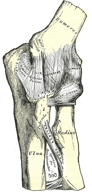
Left elbow joint, showing anterior and internal ligaments

Figure 2: Left elbow joint, showing anterior and internal ligaments.
The elbow joint is a complex hinge formed by the articulation of the distal humerus with the proximal ulna and radius. Understanding its stability requires examining the intrinsic and extrinsic ligaments, as well as the anatomical boundaries of the cubital fossa.
The primary stabilizers of the elbow are the collateral ligaments. The ulnar collateral ligament (UCL) is a triangular band on the medial side. It arises from the medial epicondyle of the humerus and inserts into the medial margin of the coronoid process and the medial side of the olecranon. It is the primary restraint against valgus stress. It resists varus stress. [1]
| Ligament | Origin | Insertion | Function |
|---|---|---|---|
| Ulnar Collateral | Medial epicondyle | Coronoid process, Olecranon | Resists valgus stress |
| Radial Collateral | Lateral epicondyle | Annular ligament, Olecranon | Resists varus stress |
The annular ligament is a strong band that encircles the head of the radius. It attaches to the anterior and posterior margins of the radial notch of the ulna. This ligament holds the radial head against the ulna, allowing rotation during pronation and supination. [1]
The cubital fossa is a triangular depression on the anterior aspect of the elbow. It contains important neurovascular structures. The boundaries are defined by the pronator teres medially, the brachioradialis laterally, and an imaginary line between the epicondyles superiorly. The floor is formed by the brachialis and supinator muscles.
Figure 3915 Elbow Joint
Long description
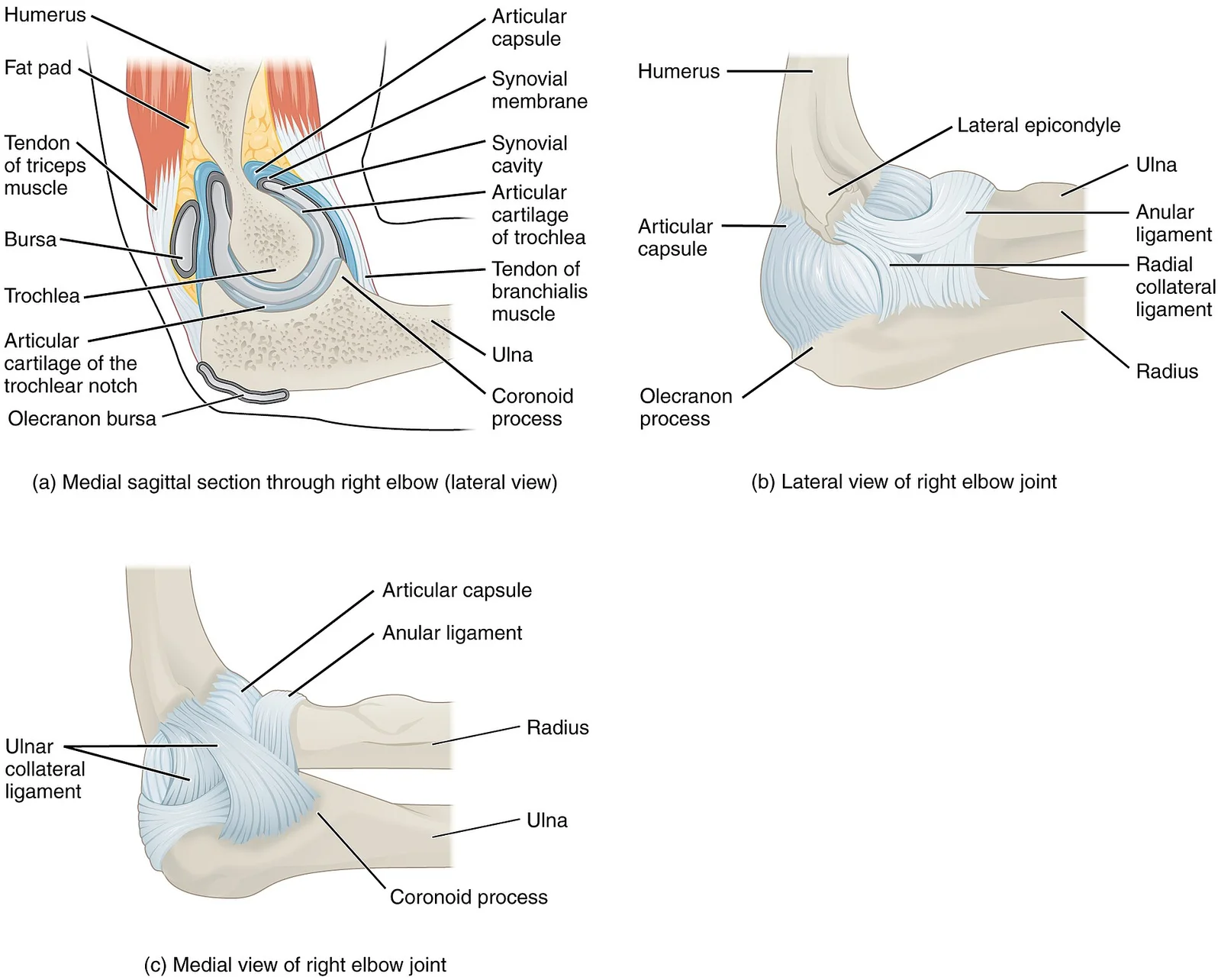
(a) The elbow is a hinge joint that allows only for flexion and extension of the forearm. (b) It is supported by the ulnar and radial collateral ligaments. (c) The annular ligament supports the head of the radius at the proximal radioulnar joint, the pivot joint that allows for rotation of the radiu

Figure 3: (a) The elbow is a hinge joint that allows only for flexion and extension of the forearm.
The articular capsule is thin anteriorly and posteriorly but thickened laterally and medially by the collateral ligaments. The cubital fossa is outlined, showing the relationship of the brachial artery and median nerve deep to the bicipital aponeurosis.
Figure 4Gray330
Long description
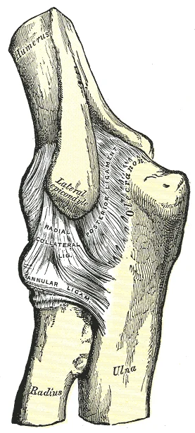
Left elbow-joint, showing posterior and radial collateral ligaments.

Figure 4: Left elbow-joint, showing posterior and radial collateral ligaments.
Clinical pearlClinical Pearl
The ulnar nerve passes posterior to the medial epicondyle, not within the cubital fossa. Injury here causes sensory loss in the medial one and a half digits.
Exam trapCommon Trap
Do not confuse the annular ligament with the radial collateral ligament. The annular ligament stabilizes the radial head against the ulna, while the RCL stabilizes the joint against varus stress.
Its stability relies on a combination of bony congruence, ligamentous support, and protective soft tissue structures. Understanding the spatial relationships of these components is essential for clinical examination and surgical intervention.
Bursae are fluid-filled sacs that reduce friction between moving structures. They are classified by their anatomical position relative to the skin, muscles, and tendons.
DefinitionBursae
Bursae contain a lubricating fluid that serves to reduce friction between structures. [3]
There are three main types of bursae relevant to the upper limb. Subcutaneous bursae prevent friction between the skin and an underlying bone. Submuscular bursae protect muscles from rubbing against a bone or another muscle. A subtendinous bursa prevents friction between bone and a muscle tendon. Tendon sheaths contain a lubricating fluid and surround tendons to allow for smooth movement of the tendon as it crosses a joint. [3]
Figure 5804 Humerus and Elbow
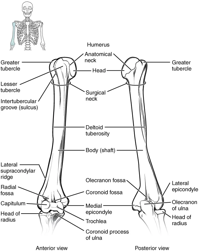
Long description
The humerus is the single bone of the upper arm region. It articulates with the radius and ulna bones of the forearm to form the elbow joint.

Figure 5: The humerus is the single bone of the upper arm region.
The most clinically significant bursa is the olecranon bursa. It lies between the skin and the olecranon process of the ulna. This is a subcutaneous bursa. Other bursae may lie between muscles or tendons, such as the subtendinous bursa under the triceps tendon. These structures ensure that repetitive motion does not damage the underlying bone or tendon.
The articular capsule encloses the joint. It is thin on its anterior and posterior aspects, but is thickened along its outside margins by strong intrinsic ligaments. These ligaments prevent side-to-side movements and hyperextension. [1]
On the medial side is the triangular ulnar collateral ligament. This arises from the medial epicondyle of the humerus and attaches to the medial side of the proximal ulna. Reconstructive surgical repair of this ligament is referred to as Tommy John surgery, named for the former major league pitcher who was the first person to have this treatment. [1]
On the lateral side of the elbow is supported by the radial collateral ligament. This arises from the lateral epicondyle of the humerus and then blends into the lateral side of the annular ligament. The annular ligament encircles the head of the radius. This ligament supports the head of the radius as it articulates with the radial notch of the ulna at the proximal radioulnar joint. This is a pivot joint that allows for rotation of the radius during supination and pronation of the forearm. [1]
The ulnar nerve passes posterior to the medial epicondyle of the humerus. This bony projection is prominent and easily palpable. The nerve lies in a groove behind the epicondyle, protected by the cubital tunnel. It is vulnerable to compression or injury here. The medial epicondyle is larger and more robust than the lateral epicondyle because it serves as the attachment point for the powerful grasping muscles of the anterior forearm. The lateral epicondyle gives rise to the weaker posterior forearm muscles. The proximity of the ulnar nerve to the medial epicondyle means that trauma to this area can directly affect nerve function, leading to sensory loss in the hand and weakness in grip.
The ulna is the medial bone of the forearm, running alongside the radius, which is the lateral bone of the forearm[1]. [2]
The radius is lateral, with its head articulating with the capitulum. The brachialis muscle lies anterior to the distal humerus, inserting onto the ulnar tuberosity. The biceps brachii tendon crosses the joint anteriorly, inserting onto the radial tuberosity. The triceps brachii tendon inserts onto the olecranon process posteriorly. These structures demonstrate the mechanical arrangement for flexion and extension.
The primary movements at the elbow are flexion and extension. Flexion is produced mainly by the biceps brachii and brachialis muscles. The biceps brachii also contributes to supination of the forearm. Extension is produced by the triceps brachii muscle. The triceps brachii has three heads: the lateral, medial, and long heads. The long head originates from the infraglenoid tubercle of the scapula. The lateral and medial heads originate from the posterior surface of the humerus. All three heads converge into a single tendon that inserts onto the olecranon process of the ulna.
| Muscle | Origin | Insertion | Action | Nerve Supply |
|---|---|---|---|---|
| Biceps brachii | Supraglenoid tubercle and supraglenoid ridge | Radial tuberosity | Flexion, supination | Musculocutaneous |
| Brachialis | Distal anterior humerus | Coronoid process and tuberosity of ulna | Flexion | Musculocutaneous |
| Triceps brachii | Infraglenoid tubercle and posterior humerus | Olecranon process | Extension | Radial |
The proximal radioulnar joint allows for rotation of the radius around the ulna. This joint is formed by the articulation between the head of the radius and the radial notch of the ulna. The radial notch is a small, smooth area on the lateral side of the ulna. This articulation permits supination and pronation, which are essential for orienting the hand. The joint is stabilized by the annular ligament, which encircles the head of the radius. [2]
The stability of the elbow is provided by strong ligaments and the congruence of the articular surfaces. The ulnar collateral ligament stabilizes the medial side, while the radial collateral ligament stabilizes the lateral side. These ligaments prevent excessive varus and valgus stress. The joint capsule encloses the articulation and is reinforced by these collateral ligaments.
NoteClinical Relevance
Dislocation of the elbow joint is a serious injury that can damage the ulnar nerve, which runs posterior to the medial epicondyle of the humerus.
The elbow joint is a complex hinge mechanism that permits flexion and extension, while also allowing rotation of the forearm through the proximal radioulnar articulation. Stability is not derived from a single structure but from the interplay of bony congruence, ligamentous constraints, and dynamic muscular support. Understanding these factors requires examining the specific anatomical relationships that prevent dislocation and control movement.
The ulna is positioned medially, with its trochlear notch gripping the trochlea of the humerus. This bony interlock provides significant medial stability. Laterally, the radial head articulates with the capitulum.
Bony stability is inherent in the shape of the articulating surfaces. The trochlea of the humerus fits into the trochlear notch of the ulna, creating a deep hinge. This geometry limits lateral movement and prevents dislocation during flexion and extension. The radial head acts as a secondary stabilizer against valgus stress. When the elbow is extended, the olecranon process of the ulna engages with the olecranon fossa of the humerus, locking the joint in extension. This bony block prevents hyperextension and provides stability when weight-bearing through the upper limb.
Ligamentous structures provide static stability. The ulnar collateral ligament is the primary restraint against valgus stress. It consists of anterior, posterior, and transverse bands. The anterior band is the most important component, resisting valgus forces particularly in flexion. The radial collateral ligament complex resists varus stress. The annular ligament encircles the radial head, securing it within the radial notch of the ulna. This allows the radius to rotate during pronation and supination without dislocating. The quadrate ligament connects the neck of the radius to the ulna, providing additional stability to the proximal radioulnar joint.
Dynamic stability is provided by the muscles crossing the joint. The biceps brachii and brachialis are primary flexors, while the triceps brachii is the main extensor. These muscles not only generate movement but also compress the joint surfaces, enhancing stability. The common flexor and extensor tendons originate from the medial and lateral epicondyles respectively, contributing to the collateral ligamentous support. The integrity of these muscular attachments is essential for maintaining joint congruence during functional activities.
Clinical pearlKey Stability Concept
The elbow relies on bony congruence for hinge stability, ligaments for collateral stability, and muscles for dynamic compression.
The elbow joint is a complex hinge formed by the articulation of the humerus with the radius and ulna. Its stability and function rely on a robust vascular and neural network. Understanding this supply requires applying Hilton’s law, which states that the nerves supplying a joint also supply the muscles that move it and the skin overlying it. This principle ensures coordinated protection and movement.
Centrally, the brachial artery descends along the medial side of the humerus. As it approaches the elbow, it bifurcates into the radial and ulnar arteries, though significant collateral circulation exists before this split.
Regarding nerves, the image highlights the ulnar nerve passing posterior to the medial epicondyle of the humerus. This is a critical landmark. The nerve lies in the cubital tunnel, protected by the cubital tunnel retinaculum. Anterior to the joint, the median nerve is visible, running between the two heads of the pronator teres muscle. Laterally, the radial nerve is seen dividing into its superficial and deep branches. The deep branch, or posterior interosseous nerve, pierces the supinator muscle. The superficial branch continues distally along the radius. The musculocutaneous nerve terminates as the lateral antebrachial cutaneous nerve, supplying the skin of the lateral forearm.
The blood supply is derived from three main arteries: the brachial, radial, and ulnar arteries. The brachial artery gives off the profunda brachii artery, which contributes to the lateral collateral ligament supply. The radial artery arises from the brachial bifurcation and runs laterally. The ulnar artery runs medially and deeply. These vessels anastomose extensively around the joint capsule. This collateral circulation is vital for healing after injury.
The nerve supply follows Hilton’s law closely. The musculocutaneous nerve supplies the anterior compartment muscles, such as the biceps brachii. The radial nerve supplies the posterior compartment, including the triceps brachii. The median and ulnar nerves supply the anterior forearm flexors. The skin over the elbow is supplied by branches from all four nerves, providing redundant sensory innervation. This redundancy is protective, ensuring that damage to one nerve does not result in complete sensory loss.
DefinitionHilton's Law
The nerves supplying a joint also innervate the muscles that move the joint and the skin overlying it.
The clinical significance of this anatomy is profound. Compression of the ulnar nerve at the medial epicondyle can lead to cubital tunnel syndrome. This condition causes numbness and weakness in the hand. The rich blood supply means that fractures of the distal humerus rarely result in avascular necrosis of the joint surfaces, unlike the femoral neck. However, soft tissue injuries can disrupt these vessels, leading to compartment syndrome.
| Nerve | Primary Supply |
|---|---|
| Musculocutaneous | Anterior compartment muscles |
| Radial | Posterior compartment muscles |
| Median | Anterior forearm flexors |
| Ulnar | Medial forearm flexors |
The integration of vascular and neural structures allows for precise motor control and sensory feedback. The anastomotic network ensures resilience against trauma. Medical students must recognise these relationships to diagnose nerve injuries and plan surgical approaches. The elbow is not just a hinge; it is a dynamic interface of supply and movement.
The primary articulation is the humeroulnar joint, formed by the trochlea of the humerus and the trochlear notch of the ulna. This hinge joint allows for flexion and extension. The olecranon process of the ulna is prominent posteriorly, fitting into the olecranon fossa of the humerus during full extension. The coronoid process of the ulna is anterior, stabilizing the joint during flexion. The radial head is circular and articulates with the capitulum. The proximal radioulnar joint is located between the radial head and the radial notch of the ulna. The joint space should be uniform, indicating healthy cartilage. Any narrowing suggests degenerative changes. The alignment of the humerus, radius, and ulna is critical for normal function. The bony contours should be smooth without irregularities. The epiphyseal lines, if present, indicate skeletal immaturity. Understanding these landmarks is crucial for interpreting elbow injuries accurately. The humerus forms the upper part of the joint, while the radius and ulna form the lower part. The articulation allows for a wide range of motion, primarily in the sagittal plane. The annular ligament encircles the radial head, holding it against the ulna. The joint capsule surrounds the entire joint, providing protection and lubrication. The synovial membrane lines the capsule and produces synovial fluid. The radiological assessment focuses on the integrity of the bony structures and the alignment of the joint surfaces. Any disruption in these structures can lead to significant functional impairment.
DefinitionHumeroulnar Joint
The hinge joint between the trochlea of the humerus and the trochlear notch of the ulna, allowing flexion and extension.
The elbow joint is a complex synovial articulation primarily involving the humerus, radius, and ulna. It functions as a modified hinge joint, permitting flexion and extension, while the proximal radioulnar joint allows for pronation and supination. Understanding the bony landmarks and soft tissue relationships is critical for clinical examination and intervention.
Elbow dislocation is the most common large joint dislocation in adults, typically resulting from a fall on an outstretched hand. The mechanism usually involves valgus stress and axial loading, leading to disruption of the lateral collateral ligament complex followed by medial structures. Posterior dislocation is the predominant pattern, where the ulna and radius displace posteriorly relative to the humerus.
Fracture-dislocations are common, particularly in older adults with osteoporotic bone. The coronoid process of the ulna is frequently fractured, compromising anterior stability. The radial head is also susceptible to fracture, which can lead to chronic instability if not addressed. The "terrible triad" of the elbow consists of elbow dislocation, radial head fracture, and coronoid process fracture. This combination results in significant instability and requires careful surgical management.
Clinical examination begins with inspection for swelling, deformity, or open wounds. Palpation should assess the lateral and medial epicondyles, the radial head, and the olecranon. The lateral epicondyle is the origin of the common extensor tendon, while the medial epicondyle is the origin of the common flexor tendon.
Range of motion should be assessed actively and passively. Flexion is limited by contact between the olecranon and the olecranon fossa. Extension is limited by the anterior capsule and bony impingement. The carrying angle is normally valgus, averaging approximately 5 to 15 degrees. Deviation from this norm may indicate malunion or ligamentous laxity.
Special tests include the varus and valgus stress tests to assess collateral ligament integrity. The lateral pivot shift test can detect posterolateral rotatory instability. Neurovascular assessment is mandatory, particularly of the median, ulnar, and radial nerves, as well as the brachial artery.
Joint aspiration may be required for diagnostic analysis of synovial fluid or therapeutic decompression. The lateral approach is often preferred due to the relative safety of the surrounding structures. The needle is inserted just anterior and distal to the radial head, in the soft tissue window between the radial head and the capitulum of the humerus.
The medial approach involves insertion just anterior and distal to the medial epicondyle, avoiding the ulnar nerve which lies posterior to the epicondyle in the cubital tunnel. The posterior approach is generally avoided due to the risk of contaminating the joint with skin flora and the difficulty of maintaining a sterile field near the olecranon.
DefinitionElbow Joint
A synovial hinge joint formed by the humeroulnar, humeroradial, and proximal radioulnar articulations, allowing flexion, extension, pronation, and supination.
| Structure | Location | Clinical Significance |
|---|---|---|
| Lateral Epicondyle | Distal humerus, lateral side | Origin of common extensor tendon; tennis elbow |
| Medial Epicondyle | Distal humerus, medial side | Origin of common flexor tendon; golfers elbow |
| Radial Head | Proximal radius | Common fracture site; pivot for pronation/supination |
| Olecranon | Proximal ulna | Forms the point of the elbow; insertion of triceps |
DefinitionElbow joint overview
The elbow is a hinge joint formed by the humerus, radius and ulna, allowing flexion/extension and limited supination/pronation. The joint capsule is reinforced by the medial and lateral collateral ligaments, which resist varus and valgus stresses respectively. The radial head articulates with the capitellum of the humerus, while the trochlea of the humerus articulates with the ulna’s trochlear notch. The annular ligament encircles the radial head, maintaining its position during forearm rotation. The ulnar collateral ligament (UCL) attaches from the medial epicondyle of the humerus to the sublime tubercle of the ulna and is the primary restraint to valgus stress. The medial and lateral epicondyles are palpable landmarks for assessing ligament integrity. The radial head is the most prominent structure on the lateral side, and the medial epicondyle is the most prominent on the medial side. The joint is surrounded by a synovial membrane that produces lubricating fluid, and the joint capsule is continuous with the surrounding fascia. The elbow is a common site of injury in sports, with valgus stress causing UCL sprains, especially in overhead athletes. The lateral epicondylitis (“tennis elbow”) results from repetitive extension of the wrist and forearm, causing micro‑tears of the extensor carpi radialis brevis tendon at the LCL attachment. The radial head can be fractured in a fall onto an outstretched hand, with the fracture line typically running transversely across the head. The olecranon can fracture in a direct blow to the posterior elbow, often with a displaced fragment that can be palpable. The most common elbow fracture in children is the supracondylar fracture of the humerus, which can compromise the brachial artery and median nerve if displaced. The joint’s stability is maintained by the congruent articular surfaces, the collateral ligaments, the annular ligament and the surrounding musculature, including the biceps brachii, brachialis, triceps brachii, pronator teres and supinator. The biceps brachii tendon attaches to the radial tuberosity and contributes to supination and elbow flexion. The triceps brachii inserts on the olecranon and is the primary extensor of the elbow. The brachialis originates from the humerus and inserts on the ulna, providing powerful flexion independent of the biceps. The joint’s innervation is primarily by the median, ulnar and radial nerves, with proprioceptive fibres from the joint capsule and ligaments. In clinical practice, a thorough inspection, palpation of the epicondyles, assessment of ligament laxity, range of motion testing and neurovascular examination are essential for diagnosing elbow pathology.
The coronoid fossa of the anterior humerus is a larger depression that receives the coronoid process of the ulna when the forearm is flexed[1]. [2]
Another frequent mistake involves the collateral ligaments. Students often assume the medial (ulnar) collateral ligament is the primary stabiliser against varus stress. While it is crucial, the lateral collateral ligament complex is equally vital for rotational stability. Confusing the attachments of these ligaments can result in incorrect diagnosis of ligamentous injuries. The interosseous membrane of the forearm, a sheet of dense connective tissue, unites the ulna and radius bones along the interosseous border of the ulna[1]. This structure is often overlooked when considering forearm stability, yet it transmits forces from the hand to the elbow.
Misidentifying the nerve supply is a third common pitfall. The ulnar nerve passes posterior to the medial epicondyle, a location vulnerable to trauma. Students sometimes confuse this with the radial nerve, which winds around the radial shaft. The radial nerve is not directly related to the medial epicondyle. Correctly mapping nerve courses is essential for predicting sensory and motor deficits in elbow trauma. The supinator muscle, which wraps around the radial neck, can entrap the posterior interosseous nerve, a branch of the radial nerve. This distinction is critical for differentiating between radial nerve palsy and posterior interosseous nerve syndrome.
A fourth error is neglecting the bony landmarks used for clinical examination. The three prominent bony points of the elbow are the medial epicondyle, lateral epicondyle, and the olecranon. In extension, these form a straight line. In flexion, they form an isosceles triangle. Students often forget this geometric relationship, which is key to distinguishing dislocation from fracture. The olecranon process of the ulna is the most posterior structure and is easily palpable. Its position relative to the epicondyles changes with flexion and extension.
Fifthly, students may confuse the movements allowed at the elbow. The humeroulnar joint allows only flexion and extension. It does not permit rotation. Rotation occurs at the proximal radioulnar joint, which is part of the elbow complex but functionally distinct. Assuming the elbow allows rotation can lead to incorrect assessment of forearm pronation and supination. The radius rotates around the ulna during these movements. The interosseous border of the ulna serves as the attachment for the interosseous membrane, which stabilises this rotation[1].
Finally, a common trap is ignoring the relations of the cubital fossa. The biceps tendon inserts on the radial tuberosity. Students often assume it inserts on the ulna. This error affects understanding of elbow flexion strength. The brachialis muscle, lying deep to the biceps, is the primary flexor. It inserts on the ulnar tuberosity. Recognising the deep anatomy is essential for surgical approaches. The median nerve lies medial to the brachial artery in the fossa. Misidentifying this order can lead to iatrogenic injury during venepuncture or arterial line placement.
Knowledge of the elbow joint is indispensable at the bedside for assessing trauma and chronic conditions. When a patient presents with elbow pain, the clinician must first determine if the joint is dislocated or fractured. This relies on the anatomical relationships of the epicondyles and olecranon. If the elbow is flexed, these three points form a triangle. If the elbow is extended, they align in a straight line. Deviation from this pattern suggests a fracture or dislocation[1]. The olecranon fossa receives the olecranon during extension, providing a bony stop. Understanding this helps in reducing dislocations manually.
In the operating theatre, precise knowledge of the collateral ligaments is required for reconstruction. Medial collateral ligament reconstruction is common in throwing athletes. The surgeon must identify the anterior bundle of the ligament, which runs from the medial epicondyle to the sublime tubercle of the ulna. Lateral collateral ligament reconstruction involves the annular ligament and the radial collateral ligament. The annular ligament holds the radial head against the ulna. Damage to this ligament can lead to radial head subluxation. The interosseous membrane also provides stability, attaching to the interosseous border of the ulna[1]. Surgeons must preserve this attachment to maintain forearm integrity.
On imaging, the elbow is viewed in anteroposterior and lateral projections. The lateral view is critical for assessing the anterior and posterior fat pads. A posterior fat pad sign indicates an intra-articular effusion, often due to a fracture. The olecranon fossa is visible on the lateral view. Fractures of the olecranon can extend into this fossa. The radial head is also assessed for fractures. The proximal radioulnar joint must be congruent. Misalignment suggests a fracture-dislocation. The humeroradial joint space must be uniform. Narrowing suggests arthritis.
In examinations, students are often asked to identify structures in cross-section or on diagrams. The cubital fossa contents are a favourite topic. From lateral to medial, the order is: Biceps tendon, Brachial artery, Median nerve. This mnemonic is essential. The brachialis muscle lies deep to the biceps. It is the main flexor. The triceps muscle extends the elbow. It inserts on the olecranon. The anconeus muscle assists in extension. It is located on the lateral aspect of the olecranon.
The elbow is also used in assessing neurological deficits. The ulnar nerve is tested by asking the patient to abduct their fingers. Weakness suggests ulnar nerve pathology. The radial nerve is tested by wrist extension. The median nerve is tested by thumb opposition. These tests rely on the motor innervation of the forearm muscles. The muscles originate from the medial and lateral epicondyles. The common flexor tendon arises from the medial epicondyle. The common extensor tendon arises from the lateral epicondyle.
In daily practice, the elbow is involved in many activities. Lifting objects requires elbow flexion. Pushing objects requires elbow extension. The triceps muscle is key for extension. The biceps and brachialis are key for flexion. The supinator and pronator muscles rotate the forearm. The supinator originates from the lateral epicondyle and the supinator crest of the ulna. The pronator teres originates from the medial epicondyle. Understanding these origins helps in diagnosing tendonitis.
The elbow joint is also important in paediatric orthopaedics. Radial head subluxation, or 'nursemaid's elbow', is common in young children. It occurs when the annular ligament slips over the radial head. Reduction involves supination and flexion of the elbow. This maneuver returns the ligament to its normal position. The anatomy of the annular ligament is crucial for this procedure. It encircles the radial neck. It attaches to the ulna.
Finally, the elbow is used in assessing systemic diseases. Rheumatoid arthritis often affects the elbow joint. Synovial inflammation leads to pain and stiffness. Osteoarthritis is less common but can occur after trauma. The joint space narrows, and osteophytes form. The olecranon fossa may show sclerosis. Imaging helps differentiate these conditions. Clinical examination reveals tenderness and swelling. The range of motion is limited. Understanding the normal anatomy helps in recognising these pathological changes.
The elbow is a compound joint with three articulations within one capsule. The olecranon fossa receives the olecranon during full extension. The medial and lateral epicondyles serve as attachment points for forearm muscles. The cubital fossa contains the biceps tendon, brachial artery, and median nerve. The ulnar nerve passes posterior to the medial epicondyle. The radial head is held in place by the annular ligament. Flexion is primarily performed by the biceps and brachialis muscles. Extension is primarily performed by the triceps muscle.
Answer each one in your head first, then open the answer.
Which bony depression receives the olecranon during extension?
Show answerHide answer
Olecranon fossaWhat is the order of structures in the cubital fossa from lateral to medial?
Show answerHide answer
Biceps tendon, Brachial artery, Median nerveWhich nerve passes posterior to the medial epicondyle?
Show answerHide answer
Ulnar nerveWhat muscle is the primary flexor of the elbow?
Show answerHide answer
BrachialisWhat ligament holds the radial head against the ulna?
Show answerHide answer
Annular ligament
- Betts JG, Young KA, Wise JA, et al. Anatomy of Selected Synovial Joints. OpenStax, Anatomy and Physiology 2e (CC BY-NC-SA 4.0). Accessed .
- Betts JG, Young KA, Wise JA, et al. Bones of the Upper Limb. OpenStax, Anatomy and Physiology 2e (CC BY-NC-SA 4.0). Accessed .
- Betts JG, Young KA, Wise JA, et al. Synovial Joints. OpenStax, Anatomy and Physiology 2e (CC BY-NC-SA 4.0). Accessed .
Figure credits
- Figure 1: Henry Vandyke Carter, via Wikimedia Commons, Public domain, Public domain.
- Figure 2: Henry Vandyke Carter, via Wikimedia Commons, Public domain, Public domain.
- Figure 3: OpenStax College, via Wikimedia Commons, CC BY 3.0, CC BY 3.0. resized.
- Figure 4: Henry Vandyke Carter, via Wikimedia Commons, Public domain, Public domain.
- Figure 5: OpenStax College, via Wikimedia Commons, CC BY 3.0, CC BY 3.0.
Educational content for medical students and doctors. It is not a substitute for clinical judgement or local guidelines.