Arm compartments
Anatomical study of the anterior and posterior compartments of the upper arm.
These septa create separate spaces for muscles with different innervations and vascular supplies. The anterior compartment contains the flexor muscles, while the posterior compartment houses the extensors. Understanding these boundaries is critical for diagnosing compartment syndromes and planning surgical approaches. [1]
It contains three main muscles: the biceps brachii, brachialis, and coracobrachialis. These muscles are primarily responsible for elbow flexion and forearm supination. The compartment is supplied by the brachial artery, which runs centrally within the compartment, accompanied by two venae comitantes. The musculocutaneous nerve pierces the coracobrachialis to supply the anterior muscles. [2]
| Muscle | Origin | Insertion | Action | Nerve Supply |
|---|---|---|---|---|
| Biceps brachii | Supraglenoid tubercle; coracoid process | Radial tuberosity | Flexion of elbow; supination | Musculocutaneous |
| Brachialis | Anterior distal humerus | Coronoid process of ulna | Flexion of elbow | Musculocutaneous |
| Coracobrachialis | Coracoid process | Mid-shaft of humerus | Flexion and adduction of arm | Musculocutaneous |
The posterior compartment is bounded by the same septa and the posterior surface of the humerus. It contains the triceps brachii and the anconeus. These muscles extend the elbow joint. The radial nerve and the profunda brachii artery run through this compartment. The radial nerve is particularly vulnerable as it passes through the radial groove of the humerus. [2]
It descends through the anterior compartment, lying medial to the humerus initially, then crossing anterior to the elbow joint. The radial groove is a shallow depression on the posterior surface of the humerus. It houses the radial nerve and the profunda brachii artery. The radial nerve winds around the posterior aspect of the humerus in this groove. Injury to the radial nerve in the radial groove can lead to wrist drop. The brachial artery and veins are closely associated with the median nerve in the anterior compartment. This relationship is important for venipuncture and arterial line placement.
Clinical pearlClinical Pearl
The radial nerve is at risk in mid-shaft humeral fractures due to its course in the radial groove.
The triceps brachii, the primary extensor of the elbow joint, is innervated by the seventh and eighth cervical nerves via the radial nerve; its deep distal fibers, occasionally termed the subanconeus, insert directly into the posterior ligament and synovial membrane of the elbow. The long head of the biceps brachii originates from the supraglenoid tubercle, its tendon traversing the shoulder joint capsule within a synovial sheath before arching over the humeral head and descending in the intertubercular groove, where it is retained by the transverse humeral ligament and a fibrous prolongation from the pectoralis major tendon. The short head arises from the coracoid process in common with the coracobrachialis. The lateral intermuscular septum extends from the greater tubercle crest along the lateral supracondylar ridge to the lateral epicondyle, blending with the deltoid tendon and providing attachment for the triceps posteriorly and the brachialis, brachioradialis, and extensor carpi radialis longus anteriorly; it is perforated by the radial nerve and the profunda brachii artery. The thicker medial intermuscular septum runs from the lesser tubercle crest along the medial supracondylar ridge to the medial epicondyle, blending with the coracobrachialis tendon and attaching the triceps and brachialis; it transmits the ulnar nerve, the superior ulnar collateral artery, and the posterior branch of the inferior ulnar collateral artery. Accessory heads of the triceps occasionally cross the brachial artery or insert into the medial septum, creating potential sites of neurovascular compression.
The arm is divided into anterior and posterior compartments by the medial and lateral intermuscular septa, which extend from the humeral supracondylar ridges to the respective epicondyles. The lateral septum, blended with the deltoid tendon, gives attachment to the triceps posteriorly and to brachialis, brachioradialis, and extensor carpi radialis longus anteriorly; it is pierced by the radial nerve and the profunda brachii artery. The thicker medial septum, continuous with the coracobrachialis tendon, attaches triceps posteriorly and brachialis anteriorly, transmitting the ulnar nerve, the superior ulnar collateral artery, and the posterior branch of the inferior ulnar collateral artery. The triceps lateral head originates above the radial groove between teres minor and the groove, while the medial head arises below the groove, extending from teres major to within 2.5 cm of the trochlea; both heads also attach to their respective septa. At the elbow, the biceps tendon expands into the lacertus fibrosus (bicipital fascia), which crosses the brachial artery obliquely to blend with the deep fascia of the forearm flexor origins. An accessory third head of biceps occurs in approximately 10% of individuals, typically arising from brachialis and inserting into the lacertus fibrosus, often lying posterior to the brachial artery. [3]
The extracellular fluid (ECF) constitutes the remaining one-third of body water. Approximately 20 percent of the ECF is plasma, which transports blood cells, proteins, electrolytes, nutrients, gases, and wastes. The interstitial fluid (IF) lies between cells and plasma, separated from the ICF by a selectively permeable cell membrane. Water movement between these compartments occurs along osmotic gradients, driven by differences in solute concentration. Water moves from areas of high water concentration (low solute) to low water concentration (high solute). [3]
Hydrostatic pressure also forces fluid movement between compartments. Active transport processes require ATP to move solutes against their concentration gradients. The composition of these compartments differs significantly. Plasma and IF are similar, containing high sodium, chloride, and bicarbonate, but plasma has higher protein levels. In contrast, the ICF has elevated potassium, phosphate, magnesium, and protein. [3]
In neuroanatomy, the striatum contains two distinct compartments: striosomes and the matrix. Both contain medium spiny neurons (MSNs) expressing D1 and D2 dopamine receptors, though not in equal proportions. These compartments receive input from both intratelencephalic (IT) and pyramidal tract (PT) cortical neurons. In Huntington’s disease, there is preferential degeneration of D2-expressing MSNs in the indirect pathway, leading to hyper-kinesia. Experiments on selective vulnerability of striosome or matrix compartments in Huntington’s disease have yielded variable results. [4]
Synthetic compartments have been developed to mimic natural cellular structures, with microfluidic platforms allowing for highly controlled construction. These synthetic systems can exhibit complex behaviors like oscillations, though they require tight control of reagent concentrations.
This tendon is easily palpable and serves as a key landmark for locating the brachial artery. The brachial artery lies medial to the biceps tendon within the fossa. It can be compressed against the medial side of the humerus to control bleeding from the forearm. The median nerve is located medial to the artery, deep to the bicipital aponeurosis. This aponeurosis is a strong fibrous band that extends from the biceps tendon to the deep fascia of the forearm. It protects the underlying neurovascular bundle during venepuncture.
The lateral border of the biceps brachii is defined by the lateral head of the muscle. Lateral to this border lies the brachioradialis muscle. The cephalic vein runs along the lateral aspect of the arm, superficial to the brachioradialis. It is often used for intravenous access. The basilic vein runs along the medial aspect of the arm. It pierces the deep fascia in the mid-arm to join the brachial veins. This location is critical for avoiding accidental arterial puncture.
The posterior compartment is bounded laterally by the lateral head of the triceps brachii. This muscle forms the bulk of the posterior arm. The nerve can be injured in mid-shaft fractures of the humerus. The triceps tendon inserts onto the olecranon process of the ulna. This insertion is palpable at the elbow joint.
The medial border of the posterior compartment is formed by the medial head of the triceps. The ulnar nerve runs posterior to the medial epicondyle of the humerus. This location is known as the cubital tunnel. The nerve is superficial and vulnerable to compression or injury here. The medial epicondyle is a palpable bony prominence on the medial side of the elbow. It serves as an attachment point for the common flexor tendon.
The lateral epicondyle is a palpable bony prominence on the lateral side of the elbow. It serves as an attachment point for the common extensor tendon. The radial nerve divides into superficial and deep branches near this area. The superficial branch runs under the brachioradialis muscle. It provides sensory innervation to the dorsum of the hand. The deep branch innervates the extensor muscles of the forearm.
The skin over the anterior arm is thin and mobile. This allows for easy palpation of the underlying muscles. The skin over the posterior arm is thicker and more adherent. This provides protection for the underlying neurovascular structures. The subcutaneous tissue contains the cutaneous nerves and veins. These structures are important for clinical examination and procedures.
| Structure | Location | Clinical Significance |
|---|---|---|
| Biceps Tendon | Anterior elbow | Palpable; landmark for brachial artery |
| Brachial Artery | Medial to biceps tendon | Pulse point; compression site |
| Median Nerve | Medial to artery | Deep to bicipital aponeurosis |
| Cephalic Vein | Lateral arm | Venepuncture site |
| Basilic Vein | Medial arm | Pierces deep fascia |
| Triceps Tendon | Posterior elbow | Palpable; inserts on olecranon |
| Ulnar Nerve | Posterior medial epicondyle | Vulnerable to compression |
The arm comprises the humerus, the single bone of the upper limb segment between the shoulder and elbow. The humeral shaft forms the central structural axis, providing attachment for the major muscle groups that control forearm and wrist movement. Understanding the bony landmarks is essential for identifying compartmental boundaries and potential sites of injury, particularly in the context of humeral shaft fractures.
At the proximal end, the head articulates with the glenoid cavity, while the anatomical neck lies just distal to it. The surgical neck, a common site for fractures, is located below the tubercles. The shaft exhibits the deltoid tuberosity laterally, where the deltoid muscle inserts. The medial and lateral epicondyles mark the distal expansion, flanking the trochlea and capitulum which form the elbow joint.
The humeral shaft is susceptible to fracture, often resulting from direct trauma or falls. Such fractures can compromise the radial nerve, which runs in the radial groove on the posterior aspect of the shaft. The bony integrity of the shaft is critical for maintaining the alignment of the elbow and wrist joints. The periosteum covering the shaft is rich in sensory nerves, contributing to the pain associated with fractures.
The cortical bone of the shaft provides resistance to bending and torsional forces. The medullary cavity contains bone marrow. The nutrient foramen, typically located on the posterior surface, supplies blood to the inner bone. Disruption of these structures during a fracture can lead to significant bleeding and swelling within the confined fascial compartments.
The distal humerus widens to form the epicondyles. The medial epicondyle serves as the origin for the flexor muscles of the forearm, while the lateral epicondyle anchors the extensor muscles. The intercondylar region contains the trochlea and capitulum, which articulate with the ulna and radius respectively. These landmarks are crucial for surgical approaches and for assessing joint stability after injury.
The proximal humerus includes the greater and lesser tubercles. The lesser tubercle anchors the subscapularis. The intertubercular groove, or bicipital groove, runs between these tubercles and contains the tendon of the long head of the biceps brachii. This groove is a key landmark for identifying the rotator cuff muscles and the biceps tendon.
The deltoid tuberosity is a roughened area on the lateral aspect of the shaft. It is the insertion point for the deltoid muscle, which is the primary abductor of the arm. The radial groove spirals around the posterior shaft, housing the radial nerve and profunda brachii artery. Injury to this area can result in radial nerve palsy, leading to wrist drop.
The medial and lateral supracondylar ridges extend from the epicondyles up the shaft. These ridges serve as attachment points for the medial and lateral intermuscular septa, which divide the arm into anterior and posterior compartments. The anterior compartment contains the biceps brachii, brachialis, and coracobrachialis muscles. The posterior compartment contains the triceps brachii and anconeus muscles.
The humeral shaft’s geometry influences the mechanics of the elbow joint. The carrying angle, formed by the distal humerus, allows the forearm to swing clear of the body. This angle is more pronounced in females. Fractures of the shaft can alter this angle, leading to functional impairment.
The bony landmarks also serve as reference points for neurovascular structures. The median nerve passes anterior to the elbow, while the ulnar nerve passes posterior to the medial epicondyle. The radial nerve winds around the radial groove. Knowledge of these relationships is vital for preventing iatrogenic injury during surgical fixation of humeral shaft fractures.
The arm is divided into two distinct compartments by the deep fascia and intermuscular septa. The deep fascia, known as the brachial fascia, encloses the entire limb. It is thickened medially and laterally to form the medial and lateral intermuscular septa, respectively. These septa extend from the deep fascia to the humerus, effectively separating the anterior and posterior compartments. This anatomical arrangement ensures that muscles within each compartment share a common vascular and nervous supply.
Anterior to the humerus lies the anterior compartment, containing the flexor muscles. Posterior to the humerus lies the posterior compartment, containing the extensor muscles.
The anterior compartment contains the flexor muscles of the arm. These muscles are primarily responsible for flexion at the elbow joint. The compartment is innervated by the musculocutaneous nerve, which provides motor supply to all muscles within this space. The blood supply is derived from the brachial artery, which runs through the compartment alongside the nerve. [2]
| Muscle | Origin | Insertion | Action | Nerve Supply |
|---|---|---|---|---|
| Biceps brachii | Supraglenoid tubercle and coracoid process | Radial tuberosity and bicipital aponeurosis | Flexion of elbow; supination of forearm | Musculocutaneous nerve |
| Brachialis | Anterior surface of distal half of humerus | Coronoid process and tuberosity of ulna | Flexion of elbow | Musculocutaneous nerve and radial nerve |
| Coracobrachialis | Tip of coracoid process | Medial surface of humeral shaft | Flexion and adduction of arm | Musculocutaneous nerve |
The posterior compartment contains the extensor muscles of the arm. These muscles are primarily responsible for extension at the elbow joint. The compartment is innervated by the radial nerve, which provides motor supply to all muscles within this space. The blood supply is derived from the profunda brachii artery, a branch of the brachial artery. [2]
| Muscle | Origin | Insertion | Action | Nerve Supply |
|---|---|---|---|---|
| Triceps brachii | Long head: infraglenoid tubercle; Lateral head: posterior humerus above spiral groove; Medial head: posterior humerus below spiral groove | Olecranon process of ulna | Extension of elbow | Radial nerve |
| Anconeus | Lateral epicondyle of humerus | Lateral surface of olecranon and proximal ulna | Assists in elbow extension | Radial nerve |
The intermuscular septa play a crucial role in compartmentalising the arm. They prevent the spread of infection or haemorrhage between compartments. Clinically, this separation is significant in conditions such as compartment syndrome, where increased pressure within a closed fascial space can compromise circulation. The anterior compartment is more commonly affected due to its larger muscle mass and tighter fascial enclosure. Understanding these boundaries is essential for surgical approaches and diagnostic imaging interpretation.
This anatomical arrangement ensures that muscles within each compartment share a common nerve supply and vascularisation, a principle critical for understanding clinical presentations of nerve injury.
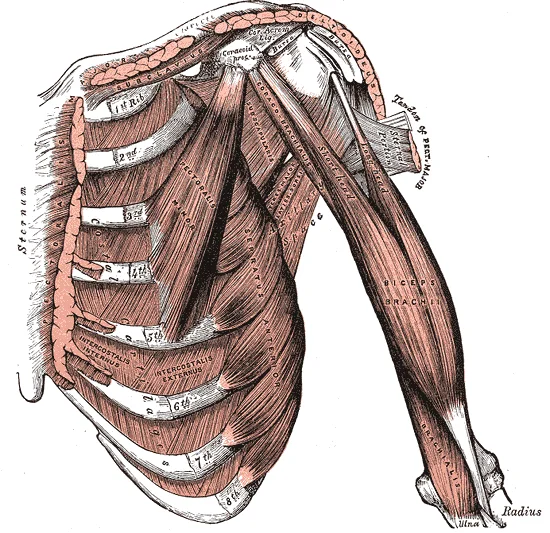
Figure 1Arm muscles front deep
Long description
deep front muscles of arm. Labelled illustration from Wikimedia Commons, used under its open licence.

Figure 1: Deep front muscles of arm.
The anterior compartment contains three muscles: the biceps brachii, brachialis, and coracobrachialis. These muscles primarily act to flex the elbow and supinate the forearm.
DefinitionAnterior Compartment
The anterior compartment of the arm contains the biceps brachii, brachialis, and coracobrachialis, all innervated by the musculocutaneous nerve.
The biceps brachii has two heads. The short head originates from the coracoid process, while the long head originates from the supraglenoid tubercle. Both heads insert onto the radial tuberosity and the bicipital aponeurosis. Its actions are elbow flexion and forearm supination. It is supplied by the musculocutaneous nerve.
The brachialis lies deep to the biceps brachii. It originates from the anterior surface of the distal half of the humeral shaft. It inserts onto the ulnar tuberosity and the coronoid process of the ulna. Its sole action is elbow flexion. It is supplied by the musculocutaneous nerve, with a small contribution from the radial nerve to its lateral part.
The coracobrachialis originates from the tip of the coracoid process. It inserts onto the medial surface of the humeral shaft, midway down. It acts to flex and adduct the arm at the shoulder. It is supplied by the musculocutaneous nerve.
| Muscle | Origin | Insertion | Action | Nerve Supply |
|---|---|---|---|---|
| Biceps Brachii | Short head: Coracoid process; Long head: Supraglenoid tubercle | Radial tuberosity, Bicipital aponeurosis | Elbow flexion, Forearm supination | Musculocutaneous |
| Brachialis | Anterior distal humerus | Ulnar tuberosity, Coronoid process | Elbow flexion | Musculocutaneous (Radial contribution) |
| Coracobrachialis | Coracoid process | Medial humeral shaft | Arm flexion, Adduction | Musculocutaneous |
DefinitionPosterior Compartment
The posterior compartment of the arm contains the triceps brachii, innervated by the radial nerve.
The triceps brachii has three heads. The long head originates from the infraglenoid tubercle of the scapula. The lateral head originates from the posterior humerus, superior to the radial groove. The medial head originates from the posterior humerus, inferior to the radial groove. All three heads converge to insert onto the olecranon process of the ulna. Its action is elbow extension, with the long head also assisting in shoulder extension and adduction. It is supplied by the radial nerve.
The radial nerve travels through the arm in the radial groove, lying against the humerus. It gives off branches to the medial and lateral heads of the triceps before piercing the lateral intermuscular septum to enter the anterior compartment. The musculocutaneous nerve pierces the coracobrachialis muscle and runs between the biceps brachii and brachialis, supplying both.
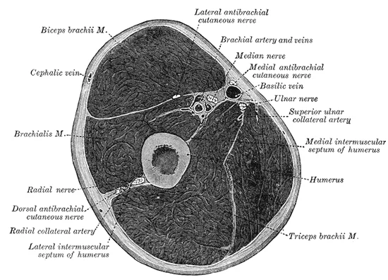
Figure 2Gray413
Long description
Cross-section through the middle of upper arm. (Eycleshymer and Schoemaker. 84 )

Figure 2: Cross-section through the middle of upper arm (Eycleshymer and Schoemaker.
| Muscle | Origin | Insertion | Action | Nerve Supply |
|---|---|---|---|---|
| Triceps Brachii | Long head: Infraglenoid tubercle; Lateral head: Posterior humerus (supr groove); Medial head: Posterior humerus (infr groove) | Olecranon process of ulna | Elbow extension | Radial |
The arterial supply to the arm compartments is derived primarily from the axillary artery and its major branches. It courses through the axilla, passing deep to the pectoralis minor muscle. The third part of the axillary artery gives rise to the brachial artery, which is the principal vessel of the arm. The brachial artery descends in the anterior compartment of the arm, running deep to the biceps brachii and brachialis muscles. It is accompanied by the median nerve and the basilic vein. As it reaches the cubital fossa, the brachial artery bifurcates into the radial and ulnar arteries, which supply the forearm.
The branches of the brachial artery are critical for understanding compartmental blood flow. The inferior ulnar collateral artery and the superior ulnar collateral artery are also branches that contribute to the anastomotic network around the elbow. These vessels ensure collateral circulation if the main arterial flow is compromised. The radial artery continues into the lateral forearm, while the ulnar artery continues into the medial forearm.
| Vessel | Origin | Course | Main Branches | Termination |
|---|---|---|---|---|
| Axillary | Subclavian | Axilla | Thoracoacromial, Lateral thoracic | Brachial |
| Brachial | Axillary | Anterior arm | Deep brachial, Inferior ulnar collateral | Radial and Ulnar |
| Deep Brachial | Brachial | Posterior arm | Nutrient artery | Anastomosis |
Understanding these vascular relationships is essential for assessing compartment syndrome, where increased pressure within a closed fascial space can compromise arterial inflow. The deep brachial artery is particularly vulnerable in posterior arm injuries. Clinical examination of the arm must include palpation of the radial and ulnar pulses to assess distal perfusion. A lack of pulse does not necessarily indicate arterial occlusion, as collateral circulation may maintain flow. However, significant pressure changes can lead to ischemia, necessitating prompt intervention. The anatomical knowledge of these vessels allows clinicians to predict the effects of trauma or surgical procedures on the arm’s blood supply.
The venous system of the upper limb operates on a hierarchical principle, moving deoxygenated blood from the deep tissues towards the central circulation. This process relies on two distinct networks: the deep veins and the superficial veins. Understanding their arrangement is critical for interpreting clinical signs and planning interventions.
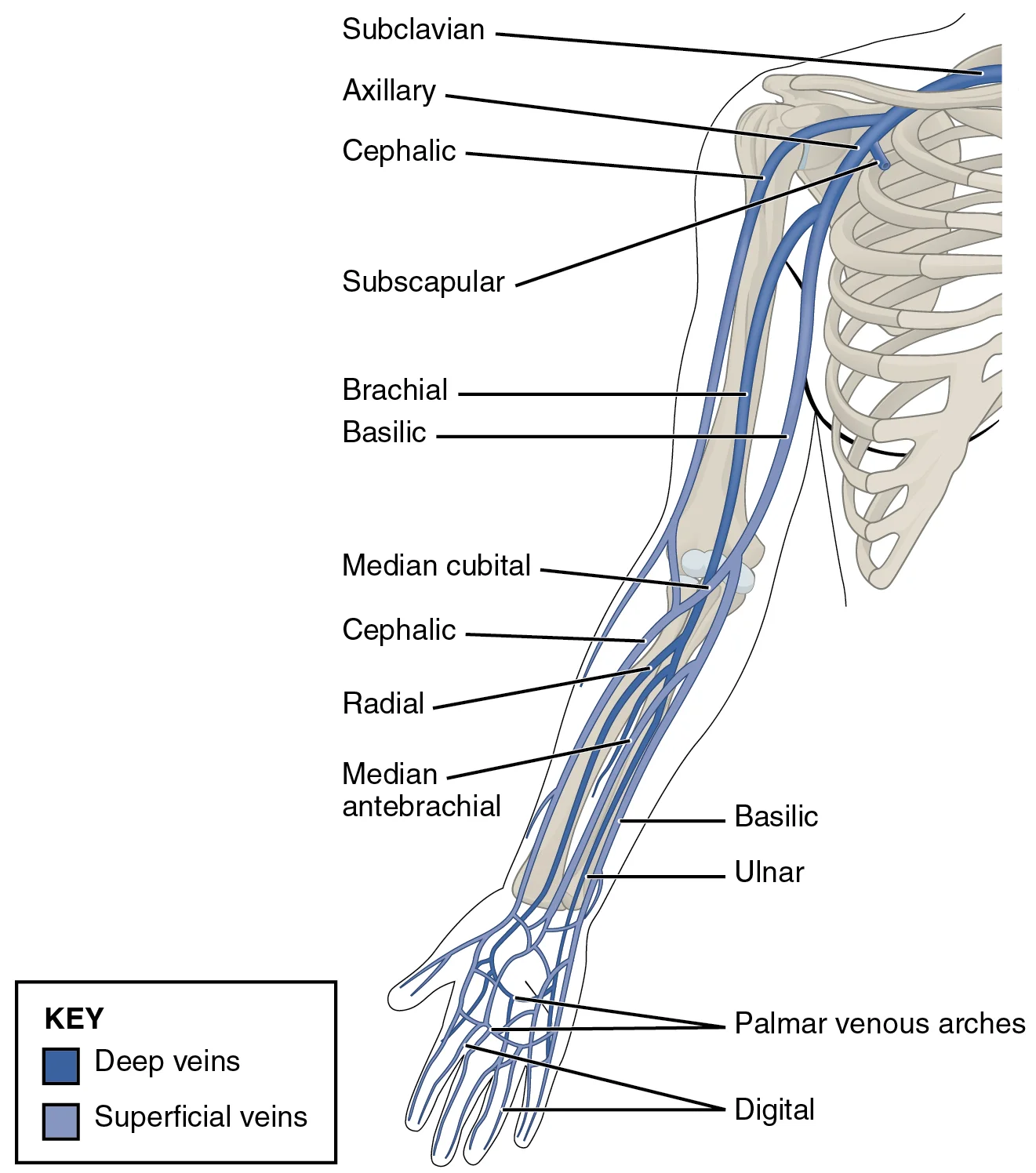
Figure 32134 Thoracic Upper Limb Veins
Long description
Illustration from Anatomy & Physiology, Connexions Web site. http://cnx.org/content/col11496/1.6/, Jun 19, 2013.

Figure 3: Illustration from Anatomy & Physiology, Connexions Web site (http://cnx.org/content/col11496/1.6/, jun 19, 2013).
Deep veins typically accompany arteries of the same name. They are arranged in pairs, known as venae comitantes, which lie on either side of the artery. This arrangement facilitates efficient blood return through the compression of muscles during contraction. The deep veins of the arm include the radial, ulnar, and brachial veins. The radial and ulnar veins arise from the deep palmar venous plexus. They ascend along the respective bones, receiving muscular branches. In the distal arm, they unite to form the brachial veins. At the lower border of the teres major muscle, the brachial veins join the basilic vein to form the axillary vein.
Superficial veins lie within the subcutaneous tissue, superficial to the deep fascia. They do not accompany arteries directly but drain into the deep system via perforating veins. The two major superficial veins are the cephalic and basilic veins. The cephalic vein originates from the dorsal venous network of the hand. It ascends along the lateral aspect of the forearm and arm. In the deltopectoral groove, it pierces the clavipectoral fascia to drain into the axillary vein. This piercing point is clinically significant for venous cutdown procedures.
Disruption of these pathways can lead to venous congestion or thrombosis. Compression of the axillary vein can impair drainage from both superficial and deep systems. Understanding the anatomical relationships allows for accurate localization of pathology. For instance, swelling in the medial arm may indicate basilic vein thrombosis, while lateral swelling suggests cephalic involvement. The deep veins are less accessible but more prone to deep vein thrombosis following trauma or catheterization.
The arm is divided into anterior and posterior compartments, each containing specific nerves that provide motor and cutaneous supply. Understanding these nerves requires examining their origin, course, relations, branches, and termination.
The musculocutaneous nerve arises from the lateral cord of the brachial plexus. It pierces the coracobrachialis muscle and descends between the biceps brachii and brachialis muscles. It terminates as the lateral antebrachial cutaneous nerve, which supplies the skin of the lateral forearm.
It terminates by dividing into superficial and deep branches.
The median nerve arises from the medial and lateral cords of the brachial plexus. It descends anterior to the elbow joint, medial to the brachial artery. It passes between the two heads of the pronator teres. It terminates by dividing into superficial and deep branches.
| Nerve | Motor Supply | Cutaneous Supply |
|---|---|---|
| Musculocutaneous | Coracobrachialis, Biceps brachii, Brachialis | Lateral forearm |
| Radial | Triceps brachii, Brachioradialis, Extensor carpi radialis longus | Posterior arm, posterior forearm, dorsal hand |
| Median | Flexor carpi radialis, Palmaris longus, Flexor digitorum superficialis | Lateral palm, digits 1-3 and half of digit 4 |
The lymphatic drainage of the upper limb follows a predictable pattern that mirrors the venous return, ensuring efficient removal of interstitial fluid and immune surveillance. Understanding this pathway is critical for assessing the spread of infection or malignancy from the arm compartments. The system is divided into superficial and deep components, which generally drain in parallel before converging at specific nodal stations.
From there, the vessels continue up the arm, accompanying the basilic vein towards the axilla.
The axillary lymph nodes are grouped into five categories: pectoral, subscapular, humeral, central, and apical. The subscapular nodes drain the posterior chest wall and the back of the arm. The central nodes collect lymph from all the other axillary groups, except the apical nodes. Finally, the apical nodes receive drainage from the central nodes and the upper part of the breast. Efferent vessels from the apical nodes form the subclavian trunk, which drains into the venous angle.
Clinical significance arises when these pathways are obstructed or invaded. Lymphoedema of the arm can result from surgical removal of axillary nodes, leading to swelling in the arm compartments. Similarly, metastatic spread from skin cancers on the hand often presents first in the epitrochlear nodes before reaching the axilla. The clear distinction between superficial and deep drainage helps in staging diseases and determining the extent of surgical intervention required for the arm compartments.
The arm is divided into two main compartments by the medial and lateral intermuscular septa, which extend from the deep fascia to the humerus. These septa create distinct anatomical spaces that house specific structures. The anterior compartment, also known as the flexor compartment, contains the biceps brachii, brachialis, and coracobrachialis muscles. The posterior compartment, or extensor compartment, contains the triceps brachii and anconeus muscles.
Laterally, the radial nerve runs in the radial groove on the posterior aspect of the humerus. The medial and lateral epicondyles of the humerus serve as attachment points for the intermuscular septa and provide origin for forearm muscles.
The neurovascular structures follow specific courses. The ulnar nerve passes posterior to the medial epicondyle, entering the forearm through the cubital tunnel.
The spaces between the compartments are potential sites for fluid accumulation or infection spread. The anterior compartment is bounded by the deep fascia, the intermuscular septa, and the humerus. The posterior compartment shares similar boundaries. The intermuscular septa themselves are thickened areas of deep fascia that provide attachment for muscle origins.
The relations of the humerus are critical for understanding clinical presentations. Fractures of the humeral shaft can injure the radial nerve due to its close proximity in the radial groove. Supracondylar fractures may damage the brachial artery or median nerve. The anatomical arrangement ensures that the neurovascular bundles are somewhat protected by the surrounding muscle mass, but they remain vulnerable to direct trauma or compression.
The contents of the arm compartments are organised to facilitate movement. The anterior compartment muscles are primarily responsible for flexion of the elbow and supination of the forearm. The posterior compartment muscles extend the elbow and stabilise the joint. The nerves supplying these muscles arise from the brachial plexus, with the musculocutaneous nerve supplying the anterior compartment and the radial nerve supplying the posterior compartment.
The deep fascia of the arm is continuous with the fascia of the forearm and axilla. It encloses the muscles and neurovascular structures, providing a compartmentalised environment. This arrangement limits the spread of pathology but also predisposes to compartment syndrome if pressure increases significantly within a closed space.
The learning of these relations is essential for clinical examination and surgical procedures. Understanding the spatial relationships allows for accurate localisation of nerve injuries and vascular compromise. The humerus acts as a landmark for identifying the course of the radial nerve and the position of the brachial artery.
The humerus and its relations are fundamental to upper limb anatomy. The bone provides attachment for muscles and protection for neurovascular structures. The intermuscular septa define the compartments and guide the course of nerves and vessels. The specific relations of the humerus determine the clinical significance of fractures and injuries in this region.
The humerus lies centrally, surrounded by the brachial fascia. Anteriorly, the anterior compartment contains the biceps brachii and brachialis muscles. Posteriorly, the posterior compartment houses the triceps brachii. The medial and lateral intermuscular septa attach the fascia to the humerus, creating these closed spaces. Understanding this layout is essential for interpreting imaging in trauma or compartment syndrome.
DefinitionCompartment Syndrome
A clinical condition where increased pressure within a closed fascial space compromises circulation and tissue function.
On plain radiographs, the arm compartments are not directly visualised as soft tissue structures unless there is significant swelling or gas. However, the bony landmarks are critical. The humeral shaft provides the rigid boundary. In cases of fracture, displacement of bone fragments can impinge on the neurovascular bundles within these compartments. Radiographs are primarily used to assess the integrity of the humerus rather than the soft tissue compartments themselves.
| Compartment | Primary Muscles | Key Neurovascular Structures |
|---|---|---|
| Anterior | Biceps brachii, Brachialis, Coracobrachialis | Musculocutaneous nerve, Brachial artery |
| Posterior | Triceps brachii, Anconeus | Radial nerve, Profunda brachii artery |
Computed tomography (CT) provides superior detail of the bony cortex and can detect subtle cortical destruction or mineralization. CT is particularly useful in assessing the extent of soft tissue swelling and identifying foreign bodies or gas within the compartments. It offers a clear depiction of cortical destruction, which is vital in evaluating pathological fractures or tumours affecting the arm.
Exam trapCT Limitation
CT is excellent for bone but less sensitive than MRI for early soft tissue oedema or muscle ischaemia.
Magnetic resonance imaging (MR) is the gold standard for evaluating soft tissue compartments. It demonstrates bone marrow involvement and accompanying soft tissue masses with high sensitivity. MR images often show heterogeneous signal intensity on all pulse sequences. In conditions like compartment syndrome, T2-weighted images reveal hyperintense signal within the muscles due to oedema. Gadolinium-enhanced T1-weighted images show marked heterogeneous enhancement in areas of inflammation or necrosis. MRI is well suited for demonstrating the full extent of muscle injury or tumour infiltration.
Ultrasound is a dynamic tool for assessing the arm compartments. It can visualise muscle tears, haematomas, and fluid collections in real-time. It is particularly useful for guiding aspiration or biopsy of soft tissue masses. Ultrasound can also assess blood flow in the brachial artery using Doppler techniques, helping to rule out vascular compromise in compartment syndrome.
The arm is divided into two distinct compartments by the intermuscular septa, each housing specific muscles and neurovascular structures. Understanding this compartmental organisation is vital for interpreting injuries and planning surgical interventions. The anterior compartment contains the flexor muscles, while the posterior compartment houses the extensors. This anatomical arrangement dictates the pattern of nerve palsies and the spread of pathology.
The radial nerve originates from the posterior cord of the brachial plexus and descends posterior to the humeral shaft. The radial nerve lies directly against the bone in the mid-shaft region, making it vulnerable to injury during fractures.
The radial nerve is the most commonly injured nerve in humeral shaft fractures. It supplies the triceps brachii, anconeus, brachioradialis, and extensor muscles of the forearm. Injury results in wrist drop and sensory loss over the dorsal aspect of the hand. The clinical examination involves assessing extension of the wrist and fingers. Understanding these relations helps in localizing the level of injury.
Surface landmarks are crucial for clinical assessment. The medial and lateral epicondyles of the humerus are palpable at the elbow. The radial groove is not directly palpable but is located midway down the posterior humerus. Clinical procedures such as intramuscular injections must avoid the radial nerve. The deltoid muscle is a safe site, but the triceps region requires caution.
DefinitionRadial Nerve
A major terminal branch of the posterior cord, supplying the posterior compartment of the arm and forearm.
| Compartment | Muscle | Action | Nerve Supply |
|---|---|---|---|
| Anterior | Biceps Brachii | Flexion of elbow | Musculocutaneous |
| Anterior | Brachialis | Flexion of elbow | Musculocutaneous |
| Posterior | Triceps Brachii | Extension of elbow | Radial |
| Posterior | Anconeus | Extension of elbow | Radial |
Fractures of the humeral shaft are classified by location. Mid-shaft fractures are particularly associated with radial nerve palsy. The mechanism of injury often involves direct trauma or indirect forces. Clinical evaluation includes neurovascular assessment distal to the injury. Imaging confirms the fracture pattern and assesses nerve involvement.
The arm is divided into two distinct fascial compartments, each with specific contents and clinical significance. Understanding the boundaries and contents is fundamental for interpreting injuries and pathologies.
The anterior compartment, also known as the flexor compartment, is enclosed by the brachial fascia and the intermuscular septa. It contains the biceps brachii, brachialis, and coracobrachialis muscles. These muscles are primarily responsible for flexion of the elbow joint.
DefinitionAnterior Compartment
The anterior compartment of the arm contains the flexor muscles and is innervated by the musculocutaneous nerve.
| Muscle | Origin | Insertion | Action | Nerve Supply |
|---|---|---|---|---|
| Biceps brachii | Supraglenoid tubercle and coracoid process | Radial tuberosity and bicipital aponeurosis | Flexion of elbow; supination of forearm | Musculocutaneous nerve |
| Brachialis | Anterior surface of distal humerus | Coronoid process and tuberosity of ulna | Flexion of elbow | Musculocutaneous nerve and radial nerve |
| Coracobrachialis | Tip of coracoid process | Medial surface of mid-shaft of humerus | Flexion and adduction of arm | Musculocutaneous nerve |
A common trap is confusing the innervation of the brachialis. While primarily supplied by the musculocutaneous nerve, it also receives a branch from the radial nerve. This dual supply is a frequent exam question. Injury to the musculocutaneous nerve results in weakness of elbow flexion and loss of sensation over the lateral forearm.
The posterior compartment, or extensor compartment, lies behind the humerus and the intermuscular septa. It contains the triceps brachii and the anconeus muscle. These muscles extend the elbow joint.
DefinitionPosterior Compartment
The posterior compartment contains the extensor muscles and is innervated by the radial nerve.
| Muscle | Origin | Insertion | Action | Nerve Supply |
|---|---|---|---|---|
| Triceps brachii | Infraglenoid tubercle and posterior humerus | Olecranon process of ulna | Extension of elbow | Radial nerve |
| Anconeus | Lateral epicondyle of humerus | Lateral surface of olecranon | Assists in elbow extension | Radial nerve |
Fractures of the mid-shaft of the humerus are particularly dangerous because the radial nerve is tightly adherent to the bone in the radial groove. Such fractures can lead to radial nerve palsy, characterized by wrist drop and sensory loss over the dorsal aspect of the hand. This is a critical clinical correlation for medical students to master.
Students frequently confuse the natural history and malignant potential of fibro-osseous lesions, leading to significant errors in clinical reasoning. A primary error is assuming that all fibrous dysplasia (FD) carries a high risk of malignant transformation. In reality, the monostotic form of the disease occurs most frequently and represents approximately 75% of all FD cases. Malignancies in monostotic FD are very rare, whereas the frequency of malignant change is increased with the polyostotic form of FD. Students must also recognize that while individuals with polyostotic FD are at an increased risk of malignant change compared with monostotic FD cases, the absolute number of malignancies in monostotic FD may appear higher simply due to the significantly higher number of monostotic than polyostotic FD cases. [1]
Another critical pitfall involves the aetiology of secondary sarcomas. Many students incorrectly attribute sarcoma development in FD primarily to prior radiation therapy. Although many authors have emphasized the importance of radiation therapy in the development of sarcoma in FD cases, radiotherapy is no longer recommended for the treatment of FD. In recent cohorts, no patient had received previous radiation treatment, suggesting that radiotherapy is not the main trigger of sarcoma in FD. Instead, surgical irritation may play a role; half of the patients in recent studies had a history of surgery, including curettage and internal fixation or curettage and bone grafting. Areas of surgical irritation, including irritation resulting from bone grafts and internal fixation, could be the niches in which mesenchymal stem cells develop tumors. However, evidence is not conclusive, as there was not enough evidence to prove that surgical irritation caused the development of secondary sarcomas in the 5 patients investigated. [1]
Diagnostic confusion between Osteofibrous Dysplasia (OFD) and Adamantinoma (AD) is another frequent error. Students often rely solely on lesion size to differentiate these entities. However, Bethapudi et al. reported that even though small lesions tend to support the diagnosis of OFD and differentiated AD more than AD, the size of lesions is less crucial in differentiating larger lesions. To date, no distinguishing imaging characteristics to differentiate OFD from differentiated AD or AD have been established. Furthermore, biopsy sampling errors can lead to misdiagnosis. OFD may still be misdiagnosed as AD on pathological examination if insufficient tissue is taken. Papagelopoulos et al. reported that OFD was initially diagnosed on a small biopsy of the lesions, but AD was ultimately diagnosed following sufficient tissue was taken. Thus, percutaneous or other limited biopsy specimens might lead to sampling errors that should be considered, and a large tissue specimen is recommended for differentiation. [2]
Finally, students often misunderstand the prognostic implications of OFD. While OFD always has an excellent prognosis because it is a benign lesion in nature, the lesion generally disappears and does not induce other harmful complications in most adulthood. However, there is an association between the benign lesion of OFD and the malignant lesion of AD. Although most OFD patients with excellent follow-up do not progress to AD, the minority of OFD cases have described the progression of OFD to AD, but this can be interpreted as an initial misdiagnosis or biopsy sampling error. Some scholars believe that OFD was a precursor lesion that can progress to typical full-blown AD. As the presence of an intermediate lesion, differentiated AD seemed to favor that possibility. Consequently, Most et al. recommended that all OFD be treated aggressively due to the risk that OFD might progress to AD and sampling error. Additionally, Oka et al. reported the first case of secondary osteosarcoma associated with OFD, and alerted oncologists that OFD may develop into secondary osteosarcoma during long-term follow-up. [2]
This knowledge is applied at the bedside to determine the urgency of intervention and the nature of follow-up. This age distribution is consistent with the data previously reported in the literature. Clinicians also note that sarcoma in FD seemed to predominately occur in males, although other literature on malignancy in FD reported no obvious gender predominance. Understanding the total medullary cavity involvement helps in prognosis; the total medullary cavity involvement is less frequently found in OFD and differentiated AD and differentiated AD. However, the complete involvement of the medullary cavity and cortex could be detected in most AD patients. Besides, soft-tissue involvement and moth-eaten margins have also been described in AD patients. [1] [2]
In the operating theatre, the decision to biopsy or resect is guided by the risk of sampling error and malignant potential. Because accurate diagnosis directly affects treatment decision-making and prognosis in patients with OFD, surgeons must ensure adequate tissue sampling. The recommendation for a large tissue specimen is based on the risk that limited specimens may miss the malignant component if the lesion is actually differentiated AD or AD. Surgeons must also be aware that unilocular OFD in images needs to be differentiated with osteoid osteoma, intra-cortical abscess, and intra-cortical hemangioma. This differential diagnosis prevents unnecessary aggressive resection for benign conditions that mimic malignancy. [2]
On imaging, MRI features are interpreted with caution. MRI can provide some evidence, but it is not the gold standard for diagnosing OFD. Students must understand why imaging is heterogeneous. Firstly, cystic, hemorrhagic, and even cartilaginous differentiation might influence the signal intensity and lead to heterogeneous signal intensity. Secondly, collagen density, the cells, and the degree of mineralization in the osteoid matrix could also affect the signal intensity. Moreover, the imaging features of OFD are similar to other fibroblastic stromal tumors that do not always show such different signal intensity patterns and can present a comparatively well-enhanced pattern that is likely to reflect rich fibrovascular stroma. Therefore, imaging alone cannot confirm the diagnosis, necessitating histological correlation. [2]
In examinations, students are tested on the distinction between precursor lesions and primary malignancies. The possibility of progression of differentiated AD to an AD should be kept in mind, especially when the destructive changes are observed on imaging. This concept is crucial for answering questions about the natural history of fibro-osseous lesions. Students must also recall that the exact pathogenesis of transformation of sarcoma in FD cases remains largely elusive. Current knowledge about the pathogenesis of transformation of FD remains limited, and further investigation is warranted. This highlights the importance of long-term monitoring rather than assuming a static benign course for all fibro-osseous lesions. [2] [1]
Clinical pearlBiopsy Depth
Always obtain a large tissue specimen for fibro-osseous lesions to avoid sampling errors that misdiagnose Adamantinoma as Osteofibrous Dysplasia.
Diagnostic Approach to Fibro-osseous Lesions
- Identify LesionFibro-osseous mass on imaging
- Question: Monostotic or Polyostotic FD?
Polyostotic
- High RiskIncreased risk of malignant change
Monostotic
- Low RiskMalignancies are very rare
Monostotic FD represents approximately 75% of all FD cases. Malignancies in monostotic FD are very rare. The frequency of malignant change is increased with the polyostotic form of FD. Sarcoma in FD seemed to predominately occur in males. The mean age at the time of sarcoma occurrence was 46.5 ± 15.9 years. [1] 80% of patients were past the fourth decade of life. [1] Radiotherapy is no longer recommended for the treatment of FD. A large tissue specimen is recommended for differentiation of OFD and AD.
Answer each one in your head first, then open the answer.
What percentage of FD cases are monostotic?
Show answerHide answer
75%What is the mean age at sarcoma occurrence in FD?
Show answerHide answer
46.5 ± 15.9 yearsIs radiotherapy recommended for FD?
Show answerHide answer
NoWhat type of biopsy is recommended for OFD?
Show answerHide answer
Large tissue specimenWhat is the prognosis of OFD?
Show answerHide answer
Excellent
- Qu N, Yao W, Cui X, Zhang H. Malignant transformation in monostotic fibrous dysplasia: clinical features, imaging features, outcomes in 10 patients, and review. Medicine 2015. Accessed .
- Liu R, Tong L, Wu H, Guo Q, Xu L, Sun Z, Yan H. Osteofibrous dysplasia: a narrative review. Journal of orthopaedic surgery and research 2024. Accessed .
- Betts JG, Young KA, Wise JA, et al. Body Fluids and Fluid Compartments. OpenStax, Anatomy and Physiology 2e (CC BY-NC-SA 4.0). Accessed .
- Crittenden JR, Graybiel AM. Basal Ganglia disorders associated with imbalances in the striatal striosome and matrix compartments. Frontiers in neuroanatomy 2011;5:59. doi:10.3389/fnana.2011.00059. Accessed .
Figure credits
Educational content for medical students and doctors. It is not a substitute for clinical judgement or local guidelines.