Shoulder joint
The shoulder complex comprises three joints enabling wide mobility, stabilised primarily by muscular rather than ligamentous structures.
The shoulder complex is not a single entity but a functional unit comprising three distinct synovial joints: the glenohumeral, acromioclavicular, and sternoclavicular joints. Synovial joints are characterised by a fluid-filled joint cavity that allows articulating bone surfaces to move smoothly against each other without direct fibrous or cartilaginous connection. This structural arrangement provides increased joint mobility compared to fibrous or cartilaginous joints. [2]
The glenohumeral joint is a multiaxial ball-and-socket joint, the same structural type as the hip joint. It is the only ball-and-socket joint in the upper limb, allowing the head of the humerus to articulate with the glenoid cavity of the scapula. This configuration permits flexion, extension, abduction, adduction, circumduction, and medial or lateral rotation of the humerus. As a ball-and-socket joint, it possesses the greatest range of motion of any joint in the body. [1] [2]
Despite this mobility, the glenohumeral joint is inherently unstable. Intrinsic ligaments, such as the coracohumeral and glenohumeral ligaments, provide only limited support. The primary support is provided by muscles crossing the joint, specifically the four rotator cuff muscles. These muscles are the supraspinatus, infraspinatus, teres minor, and subscapularis. They arise from the scapula and attach to the greater or lesser tubercles of the humerus. Their tendons encircle the head of the humerus and fuse with the anterior, superior, and posterior walls of the articular capsule. This fusion c [1]
The acromioclavicular joint is a plane (gliding) joint formed between the acromion of the scapula and the clavicle. Plane joints have flat or slightly curved articulating surfaces of approximately the same size. This allows the bones to slide against each other, although the motion is usually small and tightly constrained by surrounding ligaments. Functionally, plane joints are classified as multiaxial, though specific movement is limited by ligaments. [2]
The ligaments surrounding the acromioclavicular joint are relatively weak. Consequently, the primary support comes from the coracoclavicular ligament. This strong connective tissue band anchors the coracoid process of the scapula to the inferior surface of the acromial end of the clavicle. This ligament provides important indirect support for the joint. It also transmits forces from the upper limb to the clavicle. Injury to this joint often results from a hard fall onto the elbow or outstretched [3]
The sternoclavicular joint connects the medial end of the clavicle to the manubrium of the sternum. It is a synovial joint that allows for elevation, depression, protraction, and retraction of the clavicle. This movement is essential for the full range of motion of the upper limb. The joint is stabilised by strong ligaments, including the costoclavicular ligament, which limits excessive movement.
DefinitionRotator Cuff
The rotator cuff is the thickening of the articular capsule formed by the fusion of the tendons of the supraspinatus, infraspinatus, teres minor, and subscapularis muscles.
| Muscle | Origin | Insertion | Action | Nerve Supply |
|---|---|---|---|---|
| Supraspinatus | Supraspinous fossa | Greater tubercle | Abduction | Suprascapular nerve |
| Infraspinatus | Infraspinous fossa | Greater tubercle | Lateral rotation | Suprascapular nerve |
| Teres minor | Lateral border | Greater tubercle | Lateral rotation | Axillary nerve |
| Subscapularis | Subscapular fossa | Lesser tubercle | Medial rotation | Upper and lower subscapular nerves |
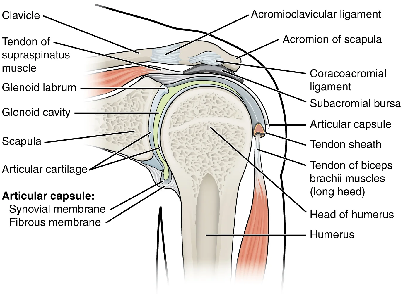
Figure 1914 Shoulder Joint
Long description
The glenohumeral (shoulder) joint is a ball-and-socket joint that provides the widest range of motions. It has a loose articular capsule and is supported by ligaments and the rotator cuff muscles.

Figure 1: The glenohumeral (shoulder) joint is a ball-and-socket joint that provides the widest range of motions.
The shoulder joint is a multiaxial ball‑and‑socket synovial joint that permits a very large range of motion. The large, rounded humeral head articulates with the small, shallow glenoid cavity of the scapula, which is only about one third the size of the humeral head. The socket is slightly deepened by a small lip of fibrocartilage called the glenoid labrum, which extends around the outer margin of the cavity. The articular capsule that surrounds the glenohumeral joint is relatively thin and loos [1][2]
The stability of the shoulder is largely dependent on the balance between the large range of motion and the relatively weak ligamentous support. The glenoid cavity is shallow, so the joint relies on muscular control and the labrum to maintain congruence. The labrum increases the depth of the socket by about 1 mm, improving stability. The intrinsic ligaments, although weak, help prevent excessive anterior or posterior translation of the humeral head. The capsule is lax in most positions but tightens in extension, preventing dislocation when the arm is moved behind the body.
The shoulder joint’s mobility is a direct consequence of its anatomical configuration. The large, rounded humeral head and shallow glenoid cavity, combined with a thin, loose capsule and minimal intrinsic ligamentous support, give the joint a very large range of motion. However, this mobility comes at the cost of stability, which is compensated by the labrum, intrinsic ligaments, and muscular control. Understanding the interplay between these structures is essential for recognising the mechanism
Figure 2Gray327
Long description
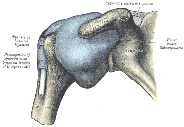
Capsule of shoulder-joint (distended). Anterior aspect.

Figure 2: Capsule of shoulder-joint (distended) (anterior aspect).
The shoulder joint, clinically termed the glenohumeral joint, is a multiaxial ball-and-socket articulation formed by the head of the humerus and the glenoid cavity of the scapula. It possesses the largest range of motion of any joint in the body. This extensive mobility is achieved at the expense of structural stability, as the joint lacks robust inherent bony support. [1]
Figure 3Shoulder Capsule
Long description
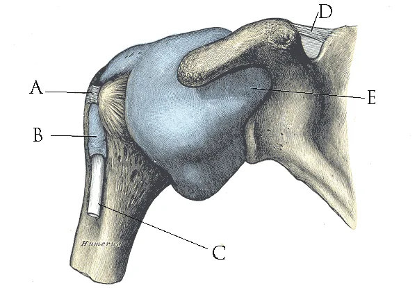
Shoulder Capsule.png. Labelled illustration from Wikimedia Commons, used under its open licence.

Figure 3: Shoulder Capsule.png.
png. The capsule is attached to the margin of the glenoid cavity superiorly and inferiorly, and to the anatomical neck of the humerus medially and laterally. The capsule is relatively thin and lax, particularly inferiorly, allowing for wide movement. The coracohumeral ligament extends from the base of the coracoid process to the greater tubercle of the humerus. The transverse humeral ligament bridges the intertubercular groove, holding the tendon of the long head of the biceps brachii in place. The synovial membrane lines the inner surface of the fibrous capsule. It covers the neck of the humerus and the margins of the glenoid cavity but does not cover the articular cartilage of the humeral head or the glenoid labrum. A notable feature is the synovial sheath surrounding the tendon of the long head of the biceps brachii as it passes through the intertubercular groove. This sheath allows the tendon to glide smoothly within the joint cavity. The synovial membrane secretes synovial fluid, a thick, slimy substance that lubricates the joint and nourishes the avascular articular cartilage.
The stability of the shoulder relies heavily on dynamic support from the rotator cuff muscles and their tendons. These muscles act as "dynamic ligaments," contracting to resist forces and maintain the humeral head within the glenoid cavity. This is crucial because the static ligaments of the shoulder are relatively weak compared to those of other joints. The articular cartilage, composed of hyaline cartilage, covers the articulating surfaces. It acts like a low-friction coating, allowing smooth movement without damaging the underlying bone. Unlike cartilaginous joints, the articular cartilages
DefinitionGlenohumeral Joint
A ball-and-socket joint formed by the articulation between the head of the humerus and the glenoid cavity of the scapula.
Clinical pearlStability vs Mobility
Freedom of movement is due to the lack of structural support, offset by a loss of stability.
| Ligament | Location | Function |
|---|---|---|
| Glenohumeral ligaments | Anterior capsule | Reinforces anterior capsule |
| Coracohumeral ligament | Coracoid to humerus | Reinforces superior capsule |
| Transverse humeral ligament | Intertubercular groove | Holds biceps tendon |
Figure 4Gray326
Long description
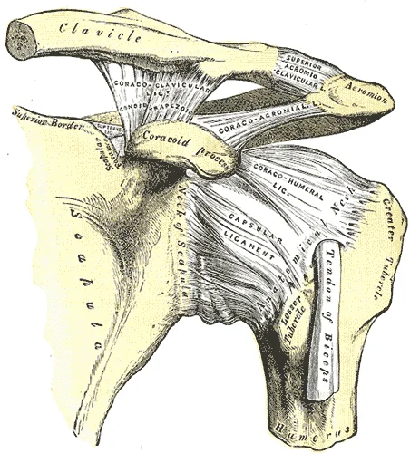
The left shoulder and acromioclavicular joints, and the proper ligaments of the scapula.

Figure 4: The left shoulder and acromioclavicular joints, and the proper ligaments of the scapula.
The coracoclavicular ligament is prominent, connecting the coracoid process of the scapula to the clavicle. It consists of two parts: the trapezoid ligament laterally and the conoid ligament medially. These are crucial for suspending the scapula from the clavicle. The coracoacromial ligament arches over the joint, forming the coracoacromial arch with the acromion and coracoid process. This arch prevents superior displacement of the humeral head. The acromioclavicular ligament connects the acromion to the clavicle, stabilising the acromioclavicular joint. The costoclavicular ligament anchors the medial clavicle to the first rib, providing stability to the sternoclavicular joint.
This reinforcement is vital because the glenoid cavity is shallow. The ball-and-socket nature of the joint allows multiaxial movement. The humerus can move in anterior-posterior and medial-lateral directions. It can also rotate around its long axis. The shallow socket formed by the glenoid cavity allows the shoulder joint an extensive range of motion. This mobility comes at the cost of stability. Ligaments must compensate for the lack of bony constraint. [1]
Injuries to the shoulder joint are common. Repetitive use of the upper limb, particularly in abduction such as during throwing, swimming, or racquet sports, may lead to acute or chronic inflammation of the bursae or muscle tendons, a tear of the glenoid labrum, or degeneration or tears of the rotator cuff. This can occur when force is applied to the humerus when the upper limb is fully abducted, as when diving to catch a baseball and landing on your hand or elbow. Inflammatory responses to any shoulder injury can lead to the formation of scar tissue between the articular capsule and surrounding structures, thus reducing shoulder mobility, a condition called adhesive capsulitis (“frozen shoulder”). [2]
The acromioclavicular joint is supported by the acromioclavicular and coracoclavicular ligaments. The scapula then separates from the clavicle, with the weight of the upper limb pulling the shoulder downward. This dislocation injury of the acromioclavicular joint is known as a “shoulder separation” and is common in contact sports such as hockey, football, or martial arts.
| Ligament | Attachments | Function |
|---|---|---|
| Coracoclavicular | Coracoid process to clavicle | Suspends scapula; resists superior displacement |
| Coracoacromial | Coracoid process to acromion | Forms arch; prevents superior humeral head displacement |
| Glenohumeral | Glenoid rim to humeral neck | Strengthens superior and anterior capsule walls |
| Acromioclavicular | Acromion to clavicle | Stabilises acromioclavicular joint |
| Costoclavicular | First rib to clavicle | Stabilises sternoclavicular joint |
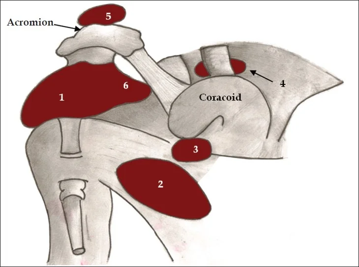
The shoulder joint is a complex synovial articulation that requires extensive support structures to maintain stability while allowing a wide range of motion. Central to this function are the bursae, which are thin connective tissue sacs filled with lubricating liquid. These structures are located in regions where skin, ligaments, muscles, or muscle tendons can rub against each other, usually near a body joint. Bursae reduce friction by separating the adjacent structures, preventing them from rub [2] [1]
Figure 5Bursae shoulder joint normal
Long description
Diagram of normal bursae surrounding the shoulder joint: (1) subacromial-subdeltoid bursa, (2) subscapular recess, (3) subcoracoid bursa, (4) coracoclavicular bursa, (5) supra-acromial bursa and (6) medial extension of s

Figure 5: Diagram of normal bursae surrounding the shoulder joint: (1) subacromial-subdeltoid bursa, (2) subscapular recess, (3) subcoracoid bursa, (4) coracoclavicular bursa, (5) supra-acromial bursa and (6) medial extension of s.
The subacromial bursa lies between the acromion process of the scapula and the head of the humerus, specifically beneath the supraspinatus tendon.
DefinitionBursa
A thin connective tissue sac filled with lubricating liquid that reduces friction between adjacent structures near a joint.
The glenoid labrum is a fibrocartilaginous rim attached to the margin of the glenoid cavity of the scapula. It deepens the socket, providing stability for the head of the humerus. The glenohumeral ligaments are thickenings of the joint capsule that reinforce the anterior aspect of the joint. They limit excessive abduction and external rotation of the arm.
| Type | Location | Function |
|---|---|---|
| Subcutaneous | Between skin and bone | Allows skin to move smoothly over the bone |
| Submuscular | Between muscle and bone | Protects muscles from rubbing against a bone [2] |
| Subtendinous | Between tendon and bone | Prevents friction between bone and a muscle tendon |
The clavicle is an anterior bone whose sternal end articulates with the manubrium of the sternum at the sternoclavicular joint. The acromial end of the clavicle articulates with the acromion of the scapula at the acromioclavicular joint. This arrangement provides a stable yet mobile foundation for the shoulder joint. The bursae and intra-articular structures work together to minimise wear and tear, ensuring efficient movement of the upper limb. [2] [3]
The shoulder joint, or glenohumeral joint, is a synovial ball-and-socket joint that provides the upper limb with an extensive range of motion. This mobility allows for complex actions such as throwing a ball or typing, distinguishing it from the more robust, stability-focused joints of the lower limb. The joint consists of the head of the humerus articulating with the glenoid cavity of the scapula. Stability is maintained by both static structures, such as the glenoid labrum and ligaments, and d [1]
The humeral head is centrally located, seated within the shallow glenoid cavity. Surrounding the humeral head are the four rotator cuff muscles, which are critical for joint integrity.
DefinitionRotator Cuff
The rotator cuff muscles serve to hold the head of the humerus in position within the glenoid cavity. By constantly adjusting their strength of contraction to resist forces acting on the shoulder, these muscles serve as “dynamic ligaments” and thus provide the primary structural support for the glenohumeral joint. [1]
The muscles acting on the shoulder can be categorised by their primary action. Abduction is initiated by the supraspinatus, with further assistance from the deltoid. Adduction is primarily performed by the pectoralis major, latissimus dorsi, and teres major. Flexion involves the anterior deltoid and pectoralis major, while extension is driven by the posterior deltoid and latissimus dorsi. Medial rotation is produced by the subscapularis, pectoralis major, latissimus dorsi, and teres major. Lateral rotation is performed by the infraspinatus and teres minor.
| Muscle | Origin | Insertion | Action | Nerve Supply |
|---|---|---|---|---|
| Supraspinatus | Supraspinous fossa | Greater tubercle of humerus | Abduction (initiation) | Suprascapular nerve |
| Infraspinatus | Infraspinous fossa | Greater tubercle of humerus | Lateral rotation | Suprascapular nerve |
| Teres minor | Lateral border of scapula | Greater tubercle of humerus | Lateral rotation | Axillary nerve |
| Subscapularis | Subscapular fossa | Lesser tubercle of humerus | Medial rotation | Upper and lower subscapular nerves |
| Deltoid | Clavicle, acromion, spine | Deltoid tuberosity | Abduction (middle), Flexion/Extension (anterior/posterior) | Axillary nerve |
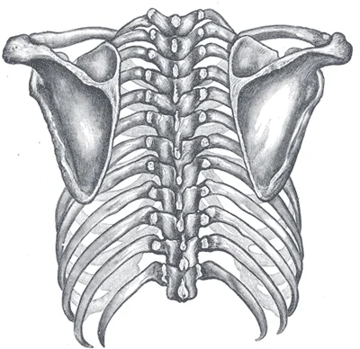
The scapula plays a vital role in these movements. It is located on the posterior side of the shoulder and is surrounded by muscles on both its anterior and posterior sides. The scapula has three margins: superior, medial, and lateral. The spine of the scapula runs across its upper portion, ending laterally in the acromion. The acromion forms the bony tip of the superior shoulder region and articulates with the lateral end of the clavicle. The coracoid process projects anteriorly and serves as an attachment site for muscles of the anterior chest and arm. The fossae of the scapula, including the supraspinous, infraspinous, and subscapular fossae, provide large surface areas for muscle attachment. These muscles cross the shoulder joint to act on the humerus, enabling the wide array of movements possible at this joint. The clavicle acts as a strut that extends laterally to support the scapula, holding the shoulder joint superiorly and laterally from the body trunk, allowing for maximal freedom of motion. [3]
Figure 6Gray412
Long description
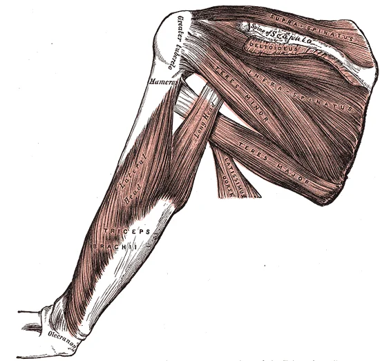
—Muscles on the dorsum of the scapula, and the Triceps brachii.

Figure 6: —Muscles on the dorsum of the scapula, and the Triceps brachii.
The shoulder joint, or glenohumeral joint, is the most mobile joint in the body, a feature that necessitates a complex system of static and dynamic stabilizers to maintain congruence during movement. Unlike the hip, which relies on a deep bony socket, the shoulder relies on soft tissue structures and muscular coordination. The suprascapular notch is located on the superior border. The scapula also has three corners, two of which are the superior and inferior angles. The third corner is occupied by the glenoid cavity. Posteriorly, the spine separates the supraspinous and infraspinous fossae, and then extends laterally as the acromion. The subscapular fossa is located on the anterior surface of the scapula. The coracoid process projects anteriorly, passing inferior to the lateral end of the clavicle. [3]
Figure 7Gray204
Long description
Posterior view of the thorax and shoulder girdle. (Morris.)

Figure 7: Posterior view of the thorax and shoulder girdle (Morris.).
The humeral head is centrally located, articulating with the shallow glenoid cavity of the scapula. The joint capsule surrounds the articulation, attaching to the glenoid rim and the anatomical neck of the humerus.
Figure 8Gray328
Long description
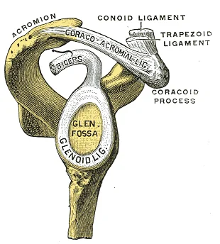
Glenoid fossa of right side. Labelled illustration from Wikimedia Commons, used under its open licence.

Figure 8: Glenoid fossa of right side.
Static stability is primarily provided by the glenohumeral ligaments and the glenoid labrum. The glenoid labrum is a ring of fibrocartilage that deepens the shallow glenoid fossa, increasing the surface area for articulation and enhancing suction effects. The glenohumeral ligaments are thickenings of the anterior joint capsule. The superior glenohumeral ligament runs from the supraglenoid tubercle to the lesser tubercle. The middle glenohumeral ligament extends from the glenoid rim to the lesser tubercle. The inferior glenohumeral ligament complex is the primary restraint to anterior dislocation in abduction. The joint capsule is lax inferiorly to allow for wide range of motion, but reinforced superiorly by the coracoacromial ligament, which forms a protective arch over the joint.
Dynamic stability is provided by the rotator cuff muscles, which compress the humeral head into the glenoid fossa during movement. The supraspinatus initiates abduction, while the infraspinatus and teres minor externally rotate the arm. The subscapularis internally rotates and adducts the arm. These muscles work in concert to center the humeral head, preventing superior migration and anterior translation. The long head of the biceps brachii also contributes to stability by anchoring the humeral head superiorly.
Clinical pearlKey Concept
The shoulder's mobility comes at the cost of stability, relying heavily on muscular control rather than bony congruence.
| Muscle | Origin | Insertion | Action | Nerve Supply |
|---|---|---|---|---|
| Supraspinatus | Supraspinous fossa | Greater tubercle | Abduction | Suprascapular nerve |
| Infraspinatus | Infraspinous fossa | Greater tubercle | External rotation | Suprascapular nerve |
| Teres minor | Lateral border | Greater tubercle | External rotation | Axillary nerve |
| Subscapularis | Subscapular fossa | Lesser tubercle | Internal rotation | Upper and lower subscapular nerves |
The shoulder joint, or glenohumeral joint, is a synovial ball-and-socket joint that permits a wide range of motion. Its stability relies on both static structures, such as the glenoid labrum and joint capsule, and dynamic structures, primarily the rotator cuff muscles. Understanding the vascular and neural architecture is critical for surgical approaches and interpreting clinical deficits.
The thoracoacromial artery branches are noted supplying the deltoid and pectoral muscles.
The blood supply to the shoulder joint is derived from several arterial sources. The anterior circumflex humeral artery and the posterior circumflex humeral artery form an anastomosis around the surgical neck of the humerus, supplying the joint capsule and the head of the humerus. The suprascapular artery, a branch of the thyrocervical trunk, supplies the supraspinatus and infraspinatus muscles and contributes to the joint capsule. The thoracoacromial artery, specifically its deltoid branch, supplies the deltoid muscle and contributes to the periarticular anastomosis. These vessels ensure adequate perfusion to the joint structures and surrounding musculature.
Nerve supply follows Hilton's law, which states that the nerves supplying a joint also supply the muscles moving the joint and the skin overlying it. The axillary nerve, derived from the posterior cord of the brachial plexus, is the primary nerve supplying the shoulder joint. It passes through the quadrangular space, accompanied by the posterior circumflex humeral artery, and winds around the surgical neck of the humerus. It provides articular branches to the glenohumeral joint and motor innervation to the deltoid and teres minor muscles. Injury to the axillary nerve can result in weak [1]
The suprascapular nerve, also from the upper trunk of the brachial plexus (C5-C6), supplies the supraspinatus and infraspinatus muscles. It passes through the suprascapular notch, under the superior transverse scapular ligament, and provides articular branches to the joint. The suprascapular nerve is vulnerable to compression at the suprascapular notch, which can lead to atrophy of the supraspinatus and infraspinatus muscles.
Bursae reduce friction between moving parts. The subacromial-subdeltoid bursa lies between the supraspinatus tendon and the acromion, allowing smooth movement of the deltoid muscle over the rotator cuff. Inflammation of this bursa, known as subacromial bursitis, is a common cause of shoulder pain and restricted movement. [1][2]
DefinitionHilton's law
The nerves supplying a joint also innervate the muscles moving the joint and the skin overlying it.
| Structure | Origin | Course | Relations |
|---|---|---|---|
| Axillary Nerve | Posterior cord (C5-C6) | Through quadrangular space | Wraps around surgical neck of humerus |
| Suprascapular Nerve | Upper trunk (C5-C6) | Under superior transverse scapular ligament | Supplies supraspinatus and infraspinatus |
| Posterior Circumflex Humeral Artery | Axillary artery | Around surgical neck of humerus | Accompanies axillary nerve |
Understanding the shoulder joint on imaging requires a systematic approach to identifying bony landmarks, joint spaces, and soft tissue structures. The glenohumeral joint is the primary articulation, formed by the head of the humerus and the glenoid cavity of the scapula. On an anteroposterior (AP) view, the humeral head should appear congruent with the glenoid fossa. Any asymmetry in the joint space may indicate pathology such as arthritis or instability. The acromioclavicular (AC) joint lies superiorly, connecting the acromion process of the scapula to the distal clavicle. This joint is often assessed for separation injuries, which are common in contact sports.
The rotator cuff muscles, particularly the supraspinatus, insert onto the greater tuberosity of the humerus. The subacromial space, located between the acromion and the humeral head, is critical for assessing impingement. A narrowed space suggests tendinopathy or bursitis. The coracoid process projects anteriorly and serves as an attachment point for the short head of the biceps brachii and the coracobrachialis muscle. The clavicle extends laterally to meet the acromion, forming the AC joint. The distal clavicle should align with the acromion; any step-off indicates ligamentous injury.
DefinitionGlenohumeral Joint
The articulation between the head of the humerus and the glenoid cavity of the scapula, stabilized by the rotator cuff and glenohumeral ligaments.
Magnetic resonance imaging (MRI) provides superior soft tissue contrast, allowing detailed evaluation of the rotator cuff tendons, labrum, and muscles. T1-weighted images show anatomy clearly, while T2-weighted images highlight fluid and edema, useful for detecting tears or inflammation. The supraspinatus tendon is the most commonly injured rotator cuff tendon, passing under the acromion. On MRI, a full-thickness tear appears as a gap in the tendon with fluid signal intensity. Partial-thickness tears may show increased signal within the tendon substance without a complete break. The subscapularis tendon inserts on the lesser tuberosity and is assessed for integrity in cases of anterior instability.
| Muscle | Origin | Insertion | Action | Nerve Supply |
|---|---|---|---|---|
| Supraspinatus | Supraspinous fossa | Greater tuberosity | Abduction | Suprascapular nerve |
| Infraspinatus | Infraspinous fossa | Greater tuberosity | External rotation | Suprascapular nerve |
| Teres minor | Lateral border of scapula | Greater tuberosity | External rotation | Axillary nerve |
| Subscapularis | Subscapular fossa | Lesser tuberosity | Internal rotation | Upper and lower subscapular nerves |
Ultrasound is a dynamic, real-time modality ideal for evaluating rotator cuff pathology. It allows comparison with the asymptomatic side and assessment of tendon movement during abduction. The long head of the biceps tendon runs in the bicipital groove, anterior to the humeral head. Dislocation or tendinopathy of this tendon can be visualized easily. The subacromial-subdeltoid bursa lies between the rotator cuff and the deltoid muscle; effusion here indicates bursitis.
Exam trapImpingement Sign
A narrowed subacromial space on imaging suggests impingement, but clinical correlation is essential as asymptomatic narrowing can occur.
Anterior dislocation is the most common type. This usually occurs when the arm is abducted and externally rotated, forcing the humeral head against the anterior inferior capsule. The humeral head typically displaces subcoracoidly. A key complication is injury to the axillary nerve, which wraps around the surgical neck of the humerus. Patients may present with loss of sensation over the lateral aspect of the shoulder, known as the regimental badge [1]
The shoulder joint, or glenohumeral joint, is a synovial ball-and-socket joint formed by the head of the humerus and the glenoid cavity of the scapula. It is the most mobile joint in the body, prioritising range of motion over stability. This inherent instability makes it susceptible to dislocation, particularly anteriorly.
Anterior dislocation is the most common type. This usually occurs when the arm is abducted and externally rotated, forcing the humeral head against the anterior inferior capsule. The humeral head typically displaces subcoracoidly. Patients may present with loss of sensation over the lateral aspect of the shoulder, known as the regimental badge
Clinical examination begins with inspection for swelling or deformity. The patient may hold the arm in slight abduction and external rotation. To assess for anterior instability, the apprehension test is performed. For posterior instability, the jerk test is useful. The examiner applies an axial load to the humerus while adducting and
Shoulder joint aspiration is indicated for suspected septic arthritis or haemarthrosis. The patient sits upright with the arm adducted. It is directed medially and slightly inferiorly towards the glenohumeral joint space. Ultrasound guidance improves accuracy and reduces complications. The needle should penetrate the capsule to enter the joint cavity. Aspiration of cloudy fluid suggests infection, while blood indicates trauma or coagulopathy.
Recurrent dislocation is the most frequent long-term complication, especially in young athletes. Osteoarthritis may develop secondary to repeated instability or labral damage. Nerve injuries, particularly to the axillary nerve, can lead to deltoid weakness and sensory loss. Vascular injury to the axillary artery is rare but serious, requiring immediate surgical exploration. Frozen shoulder, or adhesive capsulitis, can occur after immobilisa
The shoulder joint, or glenohumeral joint, is a synovial ball-and-socket joint that provides the body with the greatest range of motion. This mobility comes at the cost of stability, relying heavily on soft tissue structures rather than bony congruence. Understanding the anatomy requires a systematic approach to the bones, ligaments, muscles, and neurovascular supply.
The joint is formed by the articulation between the head of the humerus and the glenoid cavity of the scapula. The humeral head is large and spherical, while the glenoid cavity is shallow and small. This disparity in size means that the bony architecture alone provides minimal stability. The glenoid cavity is deepened by the glenoid labrum, a fibrocartilaginous rim attached to the margin of the glenoid. This labrum increases the surface area for articulation and serves as an attachment point for the long head of the biceps tendon and the glenohumeral ligaments.
Stability is primarily maintained by the joint capsule and the rotator cuff muscles. The articular capsule is thin and lax superiorly but reinforced by the glenohumeral ligaments anteriorly and inferiorly. These ligaments prevent excessive abduction and lateral rotation. The coracohumeral ligament strengthens the superior aspect of the capsule. The transverse humeral ligament bridges the interval between the greater and lesser tubercles of the humerus, holding the tendon of the long head of the biceps brachii in the intertubercular groove.
The rotator cuff is a critical stabilizer of the shoulder joint. It consists of four muscles that surround the joint and attach the humerus to the scapula. These muscles are often remembered by the mnemonic SITS: Supraspinatus, Infraspinatus, Teres minor, and Subscapularis.
| Muscle | Origin | Insertion | Action | Nerve Supply |
|---|---|---|---|---|
| Supraspinatus | Supraspinous fossa | Superior facet of greater tubercle | Initiates abduction | Suprascapular nerve |
| Infraspinatus | Infraspinous fossa | Middle facet of greater tubercle | Lateral rotation | Suprascapular nerve |
| Teres minor | Lateral border of scapula | Inferior facet of greater tubercle | Lateral rotation | Axillary nerve |
| Subscapularis | Subscapular fossa | Lesser tubercle | Medial rotation | Upper and lower subscapular nerves |
The shoulder joint receives its blood supply from the anterior and posterior circumflex humeral arteries, which are branches of the axillary artery. The axillary nerve and the suprascapular nerve provide the primary innervation. The axillary nerve wraps around the surgical neck of the humerus and supplies the deltoid and teres minor muscles. It also provides sensory innervation to the skin over the lower deltoid. Injury to this nerve can result in weakness of abduction and loss of sensation over the lateral shoulder.
Shoulder dislocation is the most common major joint dislocation. Anterior dislocations are far more common than posterior ones due to the weakness of the anterior capsule. The head of the humerus typically displaces anteriorly and inferiorly, often lodging beneath the coracoid process. This injury can damage the axillary nerve, leading to deltoid paralysis. Rotator cuff tears are common in older adults and those involved in overhead activities. The supraspinatus tendon is the most frequently affected due to its vulnerable position under the acromion.
Students frequently confuse the articulations of the shoulder girdle with the true glenohumeral joint. A primary error is assuming the sternoclavicular joint is a synovial hinge joint similar to the elbow. It is, in fact, a saddle joint that allows for complex movements including elevation, depression, protraction, retraction, and rotation. Another common mistake is overlooking the role of the intervertebral discs and zygapophysial joints in overall spinal stability. While these are not part of [2]
A significant trap involves the ligaments of the elbow, which students often mistakenly attribute to shoulder stability. The ulnar collateral ligament arises from the medial epicondyle of the humerus and attaches to the medial side of the proximal ulna. Injury to this ligament, particularly its strongest anterior portion which resists hyperextension, is associated with forceful extensions of the forearm, such as in baseball pitchers. This is distinct from shoulder pathology. Similarly, the cruciate ligaments of the knee are often confused with shoulder ligaments. The anterior cruciate ligament becomes tight when the knee is extended, resisting hyperextension. The posterior cruciate ligament prevents the femur from sliding anteriorly off the tibia. These structures are irrelevant to shoulder anatomy but are frequent distractors in examinations. [1]
Students also misinterpret the locking mechanism of the knee as applicable to the shoulder. In the knee, the medial rotation of the femur serves to lock it into full extension. This is initiated by the popliteus muscle laterally rotating the femur to unlock the knee. The shoulder does not have an equivalent bony locking mechanism; its stability relies on the rotator cuff and labrum. Furthermore, morphological differences in the clavicle are often ignored. In females, the clavicle is shorter, thinner, and less curved, whereas in males, it is heavier, longer, and has greater curvature and rougher attachment surfaces[1]. Failing to account for these sex differences can lead to errors in radiological interpretation or forensic identification. [3]
Exam trapElbow vs Shoulder Ligaments
The ulnar collateral ligament resists hyperextension of the elbow, not the shoulder. Injury is linked to forceful forearm extension.
DefinitionZygapophysial Joints
These are plane joints between articular processes, providing limited motion and determining regional spinal mobility.
At the bedside, understanding the shoulder girdle is critical for assessing posture and referred pain. Clinicians must distinguish between primary shoulder pathology and pain originating from the cervical spine. The extensive range of motion in the cervical region allows for significant flexion, extension, lateral flexion, and rotation. Pathology here may mimic shoulder pain. Knowledge of clavicular morphology aids in physical examination; a thicker, more curved clavicle suggests male anatomy or manual labour history. This information can be relevant in trauma cases where fracture [1]
In the operating theatre, precise anatomical knowledge is vital for procedures involving the shoulder girdle. While the elbow and knee ligaments are not directly involved, understanding the principles of ligamentous stability is transferable. For instance, the thickening of the articular capsule by intrinsic ligaments in the elbow prevents side-to-side movements and hyperextension. Similar principles apply to the shoulder, where the glenohumeral ligaments and rotator cuff provide stability. Surgeons must be aware of the sex differences in clavicular structure, as the heavier, rougher surfaces in males may affect screw purchase or plate fixation in fractures[1].
On imaging, radiologists must identify the correct anatomical landmarks. The distinction between the sternoclavicular joint and the acromioclavicular joint is fundamental. Misidentifying the zygapophysial joints of the spine as shoulder structures can lead to diagnostic errors. The orientation of articular processes in the cervical region allows for extensive motion, which can be assessed on dynamic X-rays. In contrast, the thoracic region's limited motion due to overlapping spinous processes and the thoracic cage is a key differentiator. Recognizing these differences helps in diagnosing spina
In examinations, students are tested on the fixed anatomical relationships. The origin, course, relations, branches, and termination of nerves and vessels are standard questions. For example, the axillary nerve's course around the surgical neck of the humerus is a frequent topic. Understanding the locking mechanism of the knee, while not shoulder-specific, tests the student's ability to distinguish between joint types. The femur's medial rotation locking the knee is a specific mechanical detail. The popliteus muscle's role in unlocking the knee is another key fact. These details, while from different joints, test the same anatomical reasoning skills required for the shoulder.
Shoulder Pain Assessment
- Patient presents with shoulder painInitial assessment
- Question: Is pain referred from neck?
Yes
- Assess cervical spine mobilityCheck for flexion, extension, lateral flexion, rotation
No
- Assess shoulder jointCheck for instability, rotator cuff integrity
Clinical pearlClavicle Morphology
Female clavicles are shorter, thinner, and less curved; male clavicles are heavier, longer, and rougher[1].
The zygapophysial joints are plane joints providing limited motion between vertebrae. [1] Cervical articular processes are flattened and face upward or downward, allowing extensive motion. [1] The ulnar collateral ligament arises from the medial epicondyle and attaches to the proximal ulna. The anterior cruciate ligament resists hyperextension when the knee is extended. The posterior cruciate ligament prevents anterior sliding of the femur on the tibia. Female clavicles are shorter, thinner, and less curved than male clavicles[1]. The popliteus muscle laterally rotates the femur to unlock the knee. Thoracic motion is limited by overlapping spinous processes and the thoracic cage.
Answer each one in your head first, then open the answer.
What type of joint are the zygapophysial joints?
Show answerHide answer
Plane jointsWhich ligament arises from the medial epicondyle of the humerus?
Show answerHide answer
Ulnar collateral ligamentWhat motion does the popliteus muscle produce to unlock the knee?
Show answerHide answer
Lateral rotation of the femurHow does the female clavicle differ from the male clavicle?
Show answerHide answer
Shorter, thinner, and less curvedWhich ligament resists hyperextension of the knee?
Show answerHide answer
Anterior cruciate ligament
- Betts JG, Young KA, Wise JA, et al. Anatomy of Selected Synovial Joints. OpenStax, Anatomy and Physiology 2e (CC BY-NC-SA 4.0). Accessed .
- Betts JG, Young KA, Wise JA, et al. Synovial Joints. OpenStax, Anatomy and Physiology 2e (CC BY-NC-SA 4.0). Accessed .
- Betts JG, Young KA, Wise JA, et al. The Pectoral Girdle. OpenStax, Anatomy and Physiology 2e (CC BY-NC-SA 4.0). Accessed .
Figure credits
- Figure 1: OpenStax College, via Wikimedia Commons, CC BY 3.0, CC BY 3.0.
- Figure 2: Henry Vandyke Carter, via Wikimedia Commons, Public domain, Public domain.
- Figure 3: Gray327.png: Henry Vandyke Carter derivative work: Raf db (talk), via Wikimedia Commons, Public domain, Public domain.
- Figure 4: Henry Vandyke Carter, via Wikimedia Commons, Public domain, Public domain.
- Figure 5: Zameer Hirji, via Wikimedia Commons, CC BY 3.0, CC BY 3.0.
- Figure 6: Henry Vandyke Carter, via Wikimedia Commons, Public domain, Public domain.
- Figure 7: Henry Vandyke Carter, via Wikimedia Commons, Public domain, Public domain.
- Figure 8: Henry Vandyke Carter, via Wikimedia Commons, Public domain, Public domain.
Educational content for medical students and doctors. It is not a substitute for clinical judgement or local guidelines.