Brachial plexus
Network of cervical nerves supplying the upper limb
The brachial plexus is formed by the ventral rami of spinal nerves, which reorganise into trunks, divisions, cords and terminal branches that innervate the shoulder, arm and hand. Clinically it is remembered by three classic patterns of dysfunction. “Waiter’s tip” describes weakness of shoulder muscles producing a drooping arm that cannot be raised – a sign of upper‑plexus injury. “Claw hand” reflects loss of intrinsic hand muscles supplied by th [1]
Thoracic outlet syndrome (TOS) illustrates how external compression of the plexus produces neurogenic symptoms. The thoracic outlet is the anatomical corridor between the clavicle and the first rib, comprising three potential spaces through which the brachial plexus, subclavian artery and vein pass. Compression within these spaces may generate pain, paraesthesia, weakness and muscle atrophy of the upper limb. Neurogenic TOS accounts for the majority of cases, with 85–90 % of patients showing com [5][6]
Injury rates after surgical decompression highlight the importance of careful technique. Transaxillary first‑rib resection for neurogenic TOS has been associated with brachial plexus injury in 0.6 % of patients in one multi‑institutional series, whereas another series reported a 9 % incidence, rising to 4 % after a supraclavicular approach. These figures underscore that even routine procedures can jeopardise the plexus. [5]
DefinitionBrachial Plexus
A network of nerves arising from C4‑T1 that supplies motor and sensory innervation to the upper limb.
| Pattern | Typical lesion level | Main muscles affected | Clinical sign |
|---|---|---|---|
| Waiter’s tip | Upper (C5‑C6) | Deltoid, supraspinatus, infraspinatus | Drooping arm, inability to abduct above shoulder height [1] |
| Claw hand | Lower (C8‑T1) | Interossei, lumbricals (ulnar) | Hyperextension of MCP joints, flexion of IP joints |
| Mixed NTOS | Upper + lower (C5‑T1) | Variable | Combined shoulder weakness and hand intrinsic atrophy |
Approach to a patient with suspected brachial plexus pathology
- HistoryMechanism of injury, onset, distribution of symptoms
- Question: Upper‑plexus signs?
Yes
- Assess C5‑C6 myotomesDeltoid, biceps, wrist extensors
No
- Assess lower‑plexus myotomesHand intrinsics, finger flexors
- Question: Evidence of compression?
Yes
- Evaluate for TOSPhysical exam, imaging of thoracic outlet
No
- Consider traumatic or avulsion injuryImaging, EMG
- ManagementConservative therapy, surgical referral if refractory
The brachial plexus is a complex network of nerve fibres formed by the reorganisation of axons from the spinal nerves C4 through T1. Unlike cranial nerves, which connect directly to the brain, or isolated spinal nerves, these fibres intermingle to form systemic nerves that innervate the upper limb. The structure is built in a strict sequence: roots, trunks, divisions, cords, and finally branches. [1]
The plexus originates from the ventral primary rami of spinal nerves C4 through T1. Specifically, the roots are C5, C6, C7, C8, and T1. These roots emerge from the spinal cord and pass through the intervertebral foramina. The sensory axons enter the spinal cord via the dorsal root, while motor fibres emerge via the ventral root. Each spinal nerve is a mixed nerve containing both sensory and motor axons. [1]
The roots unite to form three trunks. The superior trunk is formed by the union of specific cervical nerves. The middle trunk is a continuation of another cervical nerve. The inferior trunk is formed by the union of lower cervical and first thoracic nerves. These trunks lie in the posterior triangle of the neck, deep to the sternocleidomastoid muscle. [1]
Each trunk divides into an anterior and a posterior division. These divisions are located behind the clavicle. The anterior divisions generally contribute to flexor compartments, while posterior divisions contribute to extensor compartments.
The divisions reorganise to form three cords named for their relationship to the axillary artery. The lateral cord is formed by the anterior divisions of the superior and middle trunks. The posterior cord is formed by the posterior divisions of all three trunks. The medial cord is formed by the anterior division of the inferior trunk. [1]
The cords give rise to the terminal branches of the plexus. The lateral cord gives rise to the lateral pectoral nerve and contributes to the musculocutaneous nerve. The medial cord gives rise to the medial pectoral nerve, medial brachial cutaneous nerve, medial antebrachial cutaneous nerve, and the ulnar nerve. The posterior cord gives rise to the upper and lower subscapular nerves, thoracodorsal nerve, axillary nerve, and radial nerve. [1]
Figure 1Gray808
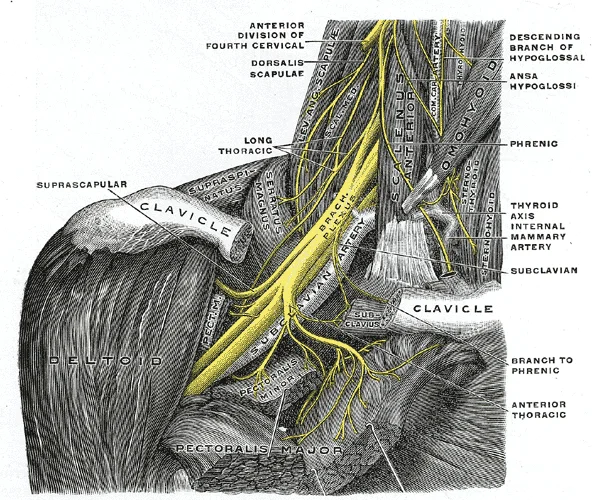
Long description
The right brachial plexus with its short branches, viewed from in front. The sternomastoid and trapezius muscles have been completely removed, the omohyoid and subclavius have been partially removed; a piece has been sawed out of the clavicle; the pectoralis muscles have been incised and reflected.

Figure 1: The right brachial plexus with its short branches, viewed from in front.
Lesions of the plexus are classified by level. Erb’s palsy involves injury to the upper trunk (C5-C6), often due to traction during birth. Klumpke’s palsy involves complete injury of all elements of the brachial plexus, potentially causing Horner’s syndrome if the sympathetic fibres are affected. Thoracic outlet syndrome can compress these structures as they pass through the thoracic outlet. [3]
The brachial plexus is a complex network of nerve fibres that conveys motor and sensory information to the upper limb. It originates from the ventral rami of spinal nerves C4 to T1. These roots emerge from the spinal cord and pass between the anterior and middle scalene muscles in the neck. The plexus then courses laterally and inferiorly, passing behind the clavicle to enter the axilla. In the axilla, the plexus is intimately related to the axillary artery and vein. The cords of the plexus are [1]
Figure 2Gray809
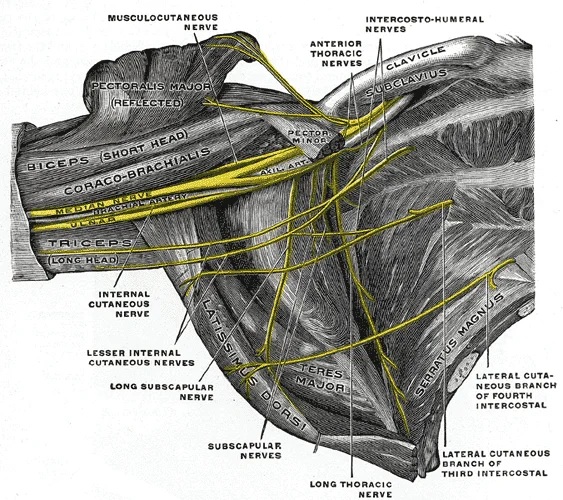
Long description
The right brachial plexus (infraclavicular portion) in the axillary fossa; viewed from below and in front. The Pectoralis major and minor muscles have been in large part removed; their attachments have been reflected. (Spalteholz.)

Figure 2: The right brachial plexus (infraclavicular portion) in the axillary fossa; viewed from below and in front.
The trunks are formed above the clavicle. The divisions are located behind the clavicle. In the axilla, the cords surround the axillary artery. The lateral cord is anterior and lateral to the artery. The medial cord is medial and posterior to the artery. The posterior cord is posterior to the artery. This spatial arrangement is crucial for understanding clinical procedures and potential sites of injury.
DefinitionBrachial Plexus
A network of nerve fibres formed by the ventral rami of spinal nerves C5 to T1, supplying the upper limb.
The relations in the neck are defined by the scalene muscles. The anterior scalene muscle lies anterior to the plexus. The middle scalene muscle lies posterior to the plexus. The phrenic nerve runs on the anterior surface of the anterior scalene muscle. This relationship is important during surgical approaches to the plexus.
Figure 3Gray806
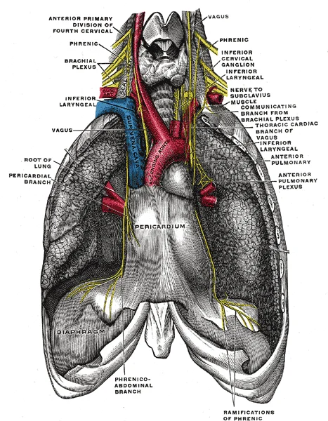
Long description
The phrenic nerve and its relations with the vagus nerve.

Figure 3: The phrenic nerve and its relations with the vagus nerve.
In the axilla, the plexus is related to the axillary vessels. The axillary artery is the central landmark. The cords of the plexus are arranged around the artery. The lateral cord is formed by the anterior divisions of the upper and middle trunks. The medial cord is formed by the anterior division of the lower trunk. The posterior cord is formed by the posterior divisions of all three trunks. The axillary vein lies medial to the artery and the medial cord.
| Structure | Relation to Axillary Artery |
|---|---|
| Lateral Cord | Anterior and lateral |
| Medial Cord | Medial and posterior |
| Posterior Cord | Posterior |
| Axillary Vein | Medial |
The brachial plexus gives rise to several major nerves. The musculocutaneous nerve arises from the lateral cord. The median nerve is formed by branches from the lateral and medial cords. The ulnar nerve arises from the medial cord. The radial nerve arises from the posterior cord. The axillary nerve also arises from the posterior cord. These nerves supply the muscles and skin of the upper limb.
NoteClinical Relevance
Understanding the course and relations of the brachial plexus is essential for diagnosing nerve injuries and planning surgical interventions.
Injuries to the brachial plexus can occur at various levels. Avulsion injuries involve the tearing of nerve roots from the spinal cord. These injuries are often caused by traction forces. Compression injuries can also occur, particularly in the thoracic outlet. Understanding the anatomy helps in localising the site of injury.
Figure 4The Brachial Plexus
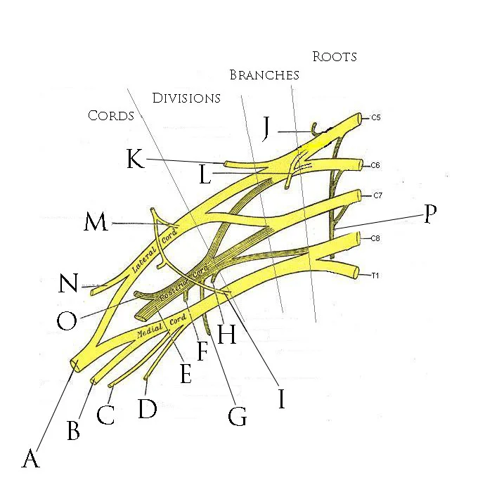
Long description
Anterior view of right brachial plexus. Illustration. Modified by Mattopaedia on 02-Jan-2006 from the 1918 Edition of Gray's Anatomy. Original unmodified image sourced from http://www.bartleby.com/107

Figure 4: Anterior view of right brachial plexus (illustration).
Figure 5Gray807
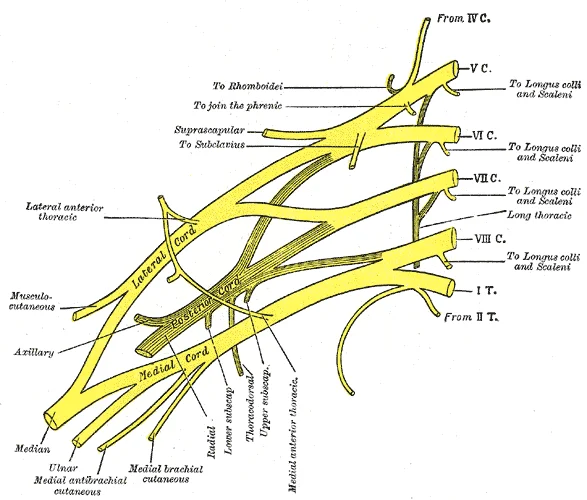
Long description
Plan of brachial plexus. Labelled illustration from Wikimedia Commons, used under its open licence.

Figure 5: Plan of brachial plexus.
DefinitionBrachial plexus
A network of nerves formed by the ventral rami of C5–T1 that supplies the upper limb.
The brachial plexus is organised into roots, trunks, divisions, cords and terminal branches.
The roots (C5–T1) unite to form the superior, middle and inferior trunks. Each trunk splits into an anterior and a posterior division. The divisions recombine to form the lateral, posterior and medial cords, which lie in relation to the axillary artery. The cords give rise to the terminal branches that supply the shoulder, arm, forearm and hand.
| Branch | Target muscles | Function |
|---|---|---|
| Musculocutaneous | Biceps brachii, brachialis, coracobrachialis | Flexion of elbow, supination of forearm |
| Axillary | Deltoid, teres minor | Abduction of arm, external rotation |
| Median | Flexors of wrist and fingers, thenar muscles | Flexion of wrist and fingers, opposition of thumb |
| Ulnar | Flexors of fingers, interossei, hypothenar muscles | Flexion of fingers, abduction/adduction of fingers |
| Radial | Extensors of wrist and fingers, triceps brachii | Extension of elbow, wrist and fingers |
| Branch | Skin territory |
|---|---|
| Musculocutaneous (sensory component) | Lateral forearm |
| Axillary (sensory component) | Lateral shoulder |
| Median (sensory component) | Palmar aspect of thumb, index, middle and half of ring finger |
| Ulnar (sensory component) | Palmar aspect of little finger and half of ring finger, dorsal aspect of little finger |
| Radial (sensory component) | Dorsal aspect of hand and thumb |
The radial nerve also carries proprioceptive fibres from the elbow and wrist joints. The axillary nerve supplies the skin over the deltoid region.
Each trunk divides into anterior and posterior divisions; the anterior divisions of the superior and middle trunks form the lateral cord, the posterior divisions of all three trunks form the posterior cord, and the anterior division of the inferior trunk forms the medial cord.
The caption: “Branches and what they supply of brachial plexus – lateral view of neck and shoulder, showing roots, trunks, divisions, cords and terminal nerves with their muscular and cutaneous territories.”
Figure 61321 Spinal Nerve Plexuses
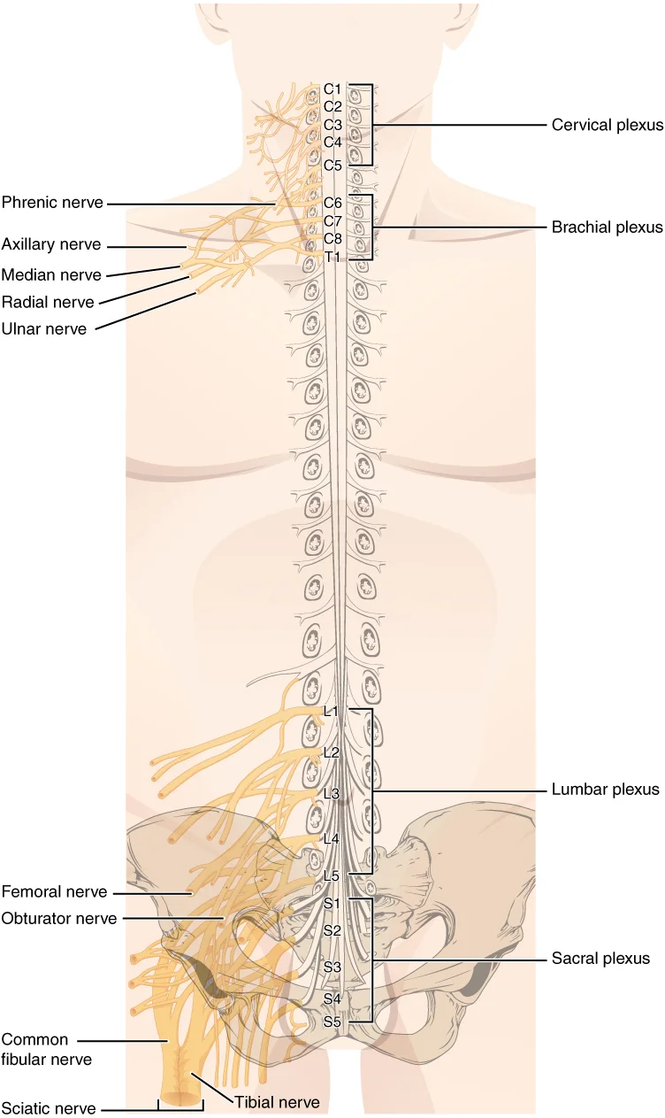
Long description
There are four main nerve plexuses in the human body. The cervical plexus supplies nerves to the posterior head and neck, as well as to the diaphragm. The brachial plexus supplies nerves to the arm. The lumbar plexus supplies nerves to the anterior leg. The sacral plexus supplies nerves to the poste

Figure 6: There are four main nerve plexuses in the human body.
Figure 7Gray817
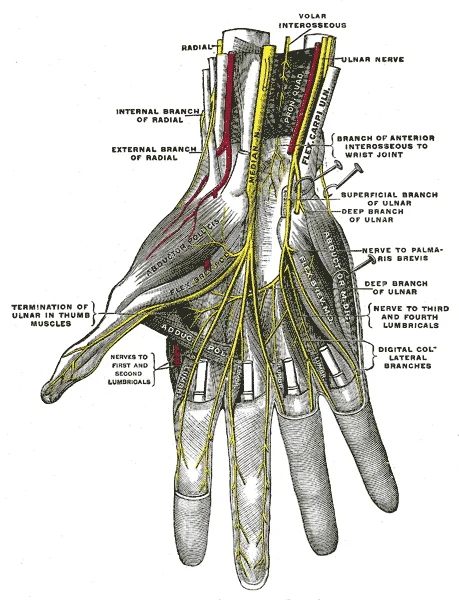
Long description
Deep palmar nerves. (Testut.). Labelled illustration from Wikimedia Commons, used under its open licence.

Figure 7: Deep palmar nerves (Testut.).
Figure 8Gray815
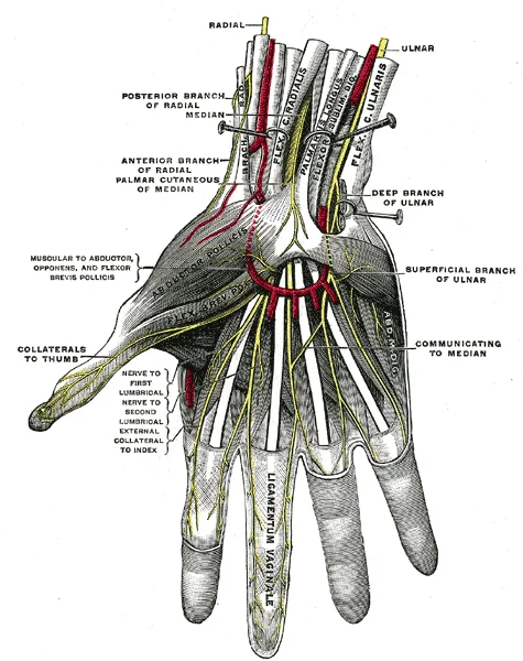
Long description
Superficial palmar nerves. (Testut.). Labelled illustration from Wikimedia Commons, used under its open licence.

Figure 8: Superficial palmar nerves (Testut.).
The brachial plexus is a complex network of nerves that can be injured at various levels, leading to distinct clinical syndromes. Understanding the anatomy and the specific level of injury is crucial for accurate diagnosis and management. The image displays the brachial plexus in an anterior view, with the cervical spine at the top and the shoulder girdle at the bottom. Key labelled structures include the roots (C5-T1), trunks, divisions, cords, and terminal branches. The axillary nerve, a branch of the posterior cord, is highlighted as it wraps around the surgical neck of the humerus. This figure helps visualise how different forces can affect specific parts of the plexus.
Brachial plexus injury (BPI) after shoulder dislocation is most commonly observed in two patient groups. The first comprises patients in whom dislocation is a result of high-energy forces, such as motor vehicle accidents or falls from a height. In these cases, multiple other injuries coexist, including fractures of the shoulder girdle, proximal humerus, and the first rib. In four studies analysing the largest patient groups, high-energy injury was responsible for 36–43% of the cases. [4]
Shoulder dislocation most commonly causes injury to the axillary nerve. Axillary nerve was found to be affected in all patients with neurologic deficit after shoulder dislocation (100%) in several studies. It occupied the first place among injuries to the long nerves of the brachial plexus in all analysed studies but two. The incidence of neurological complications varies from 5.4 to 55%, being more common in primary than in recurrent dislocations (10 and 2%, respectively, according to McLaughlin and McLellan). In the largest prospective study conducted on 3633 patients who suffered shoulder dislocation, the incidence of neurological deficit was 13.5%. [4]
| Lesion | Level | Key Deficits |
|---|---|---|
| Erb’s Palsy | Upper trunk (C5-C6) | Waiter’s tip posture; loss of shoulder abduction and external rotation |
| Klumpke’s Palsy | Lower trunk (C8-T1) | Claw hand; loss of hand intrinsic muscles |
| Axillary Nerve Injury | Posterior Cord | Loss of deltoid and teres minor function; sensory loss over lateral shoulder |
In another study on 240 patients, only 3.3% of them suffered isolated neurological deficit, while in 6.25% of the cases, it was associated with RCT or GTF.
In another study on multitrauma victims, 17% of the patients with infraclavicular lesions as opposed to 52% with supraclavicular lesions required operative intervention. [4]
The type of surgical procedure depends on severity of injury to neural structures, time elapsed from the trauma to operation and response to electrophysiological and nerve action potentials testing. Careful pre- and intraoperative assessment of the severity and type of nerve lesion is extremely important, because unnecessary resection of a regenerating brachial plexus element or performing solely neurolysis of elements for which there is no chance for regeneration does the patient a great harm. External neurolysis is sufficient in cases with nerve continuity and present regenerative nerve action potentials (NAPs). [4]
Surgical candidates should have failed conservative management. Most surgical candidates exhibit nTOS with uncontrolled pain or progressively worsening upper extremity weakness. The surgery of choice is a first rib resection aimed at brachial plexus decompression, typically performed by vascular surgeons. The operation can also be performed by thoracic surgeons, neurosurgeons, orthopedic surgeons, and plastic surgeons. In nTOS, the first rib is removed in addition to a scalenectomy or scalenotomy. [5]
More recently, the introduction of minimally invasive techniques has achieved superior outcomes in first rib removal, as both robotic and thoracoscopically assisted approaches minimize brachial plexus manipulation. Additional training, equipment, and expertise is required but may limit the overall surgical risk. [5]
The distribution of pain can be very wide in NTOS, but commonly occurs in the neck, trapezius, shoulder, arm, and in some cases also manifests as chest pain and occipital headache. The pain should be non-radicular in nature and be present during activities, where it may limit function, as well as at rest. Paraesthesias can be widespread in the upper extremity and fingers. Non-specific descriptions include heaviness with movements above the shoulder, referring to the weakness of the affected muscle groups. Once this is suspected, history taking may enable one to separate whether the upper or lower parts of the brachial plexus are more involved, with lower plexus (C8-T1) resulting in symptoms in the ulnar forearm and hand, as well as the axillary and anterior shoulder region. [6]
The clinical examination of the brachial plexus requires a systematic assessment of motor strength, sensory distribution, and reflexes to localise lesions. Differentiating between upper trunk, lower trunk, and isolated nerve injuries is critical for prognosis and management.
Erb palsy results from injury to the upper trunk (C5–C6). It typically presents with the arm hanging by the side, internally rotated, and extended, with the forearm pronated. This posture is often described as the 'waiter’s tip' position. Klumpke palsy involves the lower trunk (C8–T1). It manifests as a 'claw hand' deformity due to paralysis of the intrinsic hand muscles and flexors of the digits. Sensory loss in Klumpke palsy affects the medial aspect of the forearm and hand. [3]
Neurogenic thoracic outlet syndrome (nTOS) is caused by compression of the C5 through T1 brachial plexus nerve roots and comprises up to 90% of total TOS cases. Congenital anomalies such as cervical ribs or aberrant scalene musculature may entrap these roots. nTOS is often seen in young, active individuals who participate in athletic activities that involve repetitive overhead upper extremity motion and heavy lifting. Diagnosis is based on the history of symptom presentation and clinical exam findings. [5]
Brachial plexus injury (BPI) manifested as mononeuropathy in 18.2% to 90.5% of cases, depending on the study population. Multiple nerve injuries were found more often than injury to a single nerve. In young individuals, high-energy trauma may cause isolated axillary nerve injury, while complex neurological deficits are associated with older age, female sex, and low-energy falls. Single nerve injury is more likely a result of high-energy trauma in younger patients, whereas multiple nerve injury r [4]
Brachial plexus injury (BPI) manifested as mononeuropathy in 18.2% to 90.5% of cases, depending on the study population. Multiple nerve injuries were found more often than injury to a single nerve. In young individuals, high-energy trauma may cause isolated axillary nerve injury, while complex neurological deficits are associated with older age, female sex, and low-energy falls. Single nerve injury is more likely a result of high-energy trauma in younger patients, whereas multiple nerve injury r
Evidence of plexopathy or isolated neuropathy after shoulder dislocation should always raise a suspicion of coexisting arterial injury. The presence of vascular injury may provide information about the site and severity of nerve lesions. Due to rich collateral circulation, the presence of palpable distal pulses does not preclude axillary artery injury. When performed by an experienced examiner, imaging techniques can depict nerve disruption manifested by abrupt termination of the nerve and oedema of the surrounding tissues. Large haematomas in the infraclavicular region should be evacuated and the brachial plexus inspected concurrently before resulting fibrosis causes compression of neural elements. It is difficult to distinguish between nerve disruption and closed nerve injury that has chances for recovery on the basis of clinical examination and electromyography alone. In such cases, magnetic resonance neurography helps to determine whether surgery would be beneficial. [4]
The brachial plexus is a network of nerves formed by the ventral rami of spinal nerves C5 to T1. It provides motor and sensory innervation to the upper limb. Understanding its structure is fundamental for diagnosing neurological deficits.
The roots emerge from the spinal cord and pass between the anterior and middle scalene muscles. They unite to form three trunks: upper, middle, and lower. The upper trunk is formed by C5 and C6. The middle trunk is a continuation of C7. The lower trunk is formed by C8 and T1.
Each trunk divides into an anterior and posterior division. The anterior divisions supply the flexor compartments of the limb. The posterior divisions supply the extensor compartments. The posterior divisions unite to form the posterior cord. The anterior divisions of the upper and middle trunks form the lateral cord. The anterior division of the lower trunk continues as the medial cord.
The cords give rise to five major terminal branches. These are often remembered by the mnemonic "MUSCL": Musculocutaneous, Axillary, Radial, Median, and Ulnar. The musculocutaneous nerve arises from the lateral cord. It innervates the anterior compartment of the arm. The axillary nerve arises from the posterior cord. It supplies the deltoid and teres minor muscles. The radial nerve arises from the posterior cord. It innervates the posterior compartment of the arm and forearm. The median nerve arises from both lateral and medial cords. It supplies most flexors in the forearm and thenar muscles. The ulnar nerve arises from the medial cord. It supplies the medial flexors and intrinsic hand muscles.
The brachial plexus passes through the neck into the axilla. It is closely related to the subclavian and axillary arteries. The artery changes name as it passes anatomical landmarks. For example, the left subclavian artery becomes the axillary artery as it passes through the body wall and into the axillary region, and then becomes the brachial artery as it flows from the axillary region into the upper arm (or brachium). At the level of the axilla, brachial plexus and axillary artery are invested by a common connective tissue sheath. Thus, even minimal swelling within the sheath can cause compression on plexus elements. [2] [4]
Lesions often result from trauma or compression. Erb’s palsy affects the upper trunk (C5-C6). It causes the "waiter’s tip" posture. Klumpke’s palsy affects the lower trunk (C8-T1). It causes claw hand deformity. Thoracic outlet syndrome involves compression of the plexus. Neurogenic TOS is generally more difficult to diagnose as nerve and tissue inflammation lack consistent radiographic evidence[1]. However, as imaging studies evolve, newer modalities with higher quality allow for impr [5]
A common trap is confusing the origin of the median nerve. It is not from a single cord but from both lateral and medial cords. Another trap is assuming sensory loss always matches motor loss. In root lesions, dermatomal patterns are key. In plexus lesions, the distribution follows the cords or trunks. Pain after brachial plexus avulsion involves both peripheral and central components; thereby it is characterized as a mixed (central and peripheral) neuropathic pain syndrome. Timing of brachial plexus surgery after shoulder dislocation remains a controversial issue, which needs balancing between allowing time for spontaneous nerve regeneration and undertaking operative intervention before denervation atrophy occurs that would render the muscles refractory to reinnervation. It is generally accepted that more than 12–18 months’ interval between denervation and reinnervation causes the return of muscle function unlikely to be successful. [3] [4]
Exam trapNerve Origins
Remember the median nerve is dual-origin (lateral and medial cords), unlike the others which are single-cord origins.
Clinical pearlVascular Relation
The brachial plexus and axillary artery share a common connective tissue sheath in the axilla, meaning swelling affects both.
| Nerve | Cord | Main Motor Supply |
|---|---|---|
| Musculocutaneous | Lateral | Anterior arm (biceps) |
| Axillary | Posterior | Deltoid, Teres minor |
| Radial | Posterior | Posterior arm/forearm |
| Median | Lateral/Medial | Forearm flexors, Thenar |
| Ulnar | Medial | Medial forearm flexors, Intrinsic hand |
Diagnostic Approach to Upper Limb Weakness
- Patient presents with weaknessAssess distribution
- Question: Is it isolated to one nerve?
Yes
- Test specific nerve conduction
No
- Question: Does it follow a dermatomal pattern?
Yes
- Suspect root lesion (Radiculopathy)
No
- Suspect Plexus lesion
Students frequently confuse the anatomical boundaries of the thoracic outlet with the vascular structures passing through it, leading to errors in diagnosing Thoracic Outlet Syndrome (TOS). A primary error is assuming that all TOS cases present with identical neurological deficits. The branching anatomy of the brachial plexus is highly variable, meaning that compression at different sites produces distinct constellations of pain, sensory disturbance, and weakness. Consequently, relying on a single symptom profile to diagnose Neurogenic TOS (NTOS) is unreliable. Many classification systems historically included a category of “disputed TOS” for cases where symptomatology suggested NTOS but nerve conduction studies were negative. However, this distinction is increasingly being phased out because conduction defects may not be apparent in earlier stages of compression. Instead, clinicians must recognise that electrodiagnostic studies are changing; recent evidence suggests that conduction deficits are present in a much larger group of patients with NTOS than previously thought. Specifically, testing the medial antebrachial cutaneous nerve and median motor nerve supplying the abductor pollicis brevis often reveals that T1 and C8 derived fibres commonly show conduction deficits. [6] [5]
Another significant pitfall involves the interpretation of imaging. Students often overlook the utility of advanced imaging in visualising the brachial plexus directly. While traditional methods relied on indirect inference, such as assuming a fibrous band caused compression based on symptoms, modern techniques allow for direct visualisation. High-resolution MRI scanners with a 3.0 T magnetic field strength enable MR neurography (MRN), which suppresses signal from surrounding soft tissue structu
A third common mistake is misattributing the severity and nature of pain following Brachial Plexus Injury (BPI). In most of these cases (67%) the pain is predominantly neuropathic. However, when patients with only Brachial Plexus Avulsion (BPA) are analysed, pain is ominous and highly refractory to the usual treatments. This is probable because BPA affects mainly the CNS structures that can suffer influences from the PNS, giving rise to a mixed neuropathic pain syndrome with major central components. Students often underestimate the central component of avulsion pain, treating it solely as peripheral neuropathy. Additionally, while nerve injury complicating shoulder dislocation most often is neurapraxia or axonotmesis, complete disruption or avulsion is very rare. This rarity is due to the fact that traction is exerted at a point relatively distant to the point of exit of nerve roots from the spinal cord and the plexus is mobile and extensible at this level, as well as thanks to the relatively low energy of injury being in most cases simple fall. Assuming avulsion is common after minor trauma is a critical diagnostic error. [3] [4]
Exam trapDiagnostic Trap
Do not rule out NTOS solely on normal nerve conduction studies, as deficits may be absent in early stages or specific to T1/C8 fibres not always captured in standard screens.
Knowledge of the brachial plexus is applied at the bedside primarily through clinical examination to localise lesions. In the operating theatre, understanding the mobile and extensible nature of the plexus is crucial during shoulder procedures, as complete disruption is rare due to the distance between the traction point and the spinal cord exit. Surgeons must be aware that the plexus is embedded in adipose tissue and protected by its mobility, which influences surgical approaches to avoid iatrogenic injury. On imaging, the brachial plexus is evaluated using MR neurography to visualise nerve morphology and signal non-invasively. This technique is essential for identifying the exact site of compression in cases of suspected TOS, moving beyond indirect inference to direct visualisation of the compressed nerve and the compressing structure. Dynamic CT angiography and ultrasound are also used to stratify patients according to pathophysiology, such as identifying branching variants that increase susceptibility to scalene muscle compression. [6]
In examinations, students are tested on the specific nerve roots involved in common presentations. For instance, the identification of conduction deficits in T1 and C8 derived fibres helps differentiate NTOS from other neuropathies. The distinction between neurapraxia, axonotmesis, and complete avulsion is frequently tested, with the understanding that avulsion is rare in simple falls due to the plexus's extensibility. Furthermore, the high prevalence of neuropathic pain (67%) in BPI patients highlights the need for appropriate pain management strategies that address central components, particularly in avulsion injuries.
Diagnostic Approach to NTOS
- Suspect NTOSPain, sensory disturbance, weakness
- Question: Nerve Conduction Studies Normal?
Yes
- Consider Early NTOS or VariantDeficits may be absent early
No
- Confirm T1/C8 DeficitCheck medial antebrachial cutaneous and median motor nerves
- Advanced ImagingUse MR Neurography (3.0 T) to visualise compression directly
| Lesion Type | Key Feature | Clinical Implication |
|---|---|---|
| Neurapraxia/Axonotmesis | Most common after shoulder dislocation | Usually resolves; traction is distant from cord exit |
| Avulsion (BPA) | Rare; high energy or specific traction | Ominous pain; refractory; affects CNS structures |
| NTOS Compression | Variable symptoms due to branching anatomy | Diagnosis challenging; may require MR Neurography |
Clinical pearlImaging Pearl
MR Neurography suppresses fat signal and removes blood pulsation artefacts, allowing direct identification of the compression site and cause. [6]
- The brachial plexus is mobile and extensible, making complete avulsion rare in simple falls.
- Pain affects 71% to 78% of BPI patients, with 67% being predominantly neuropathic.
- BPA causes mixed neuropathic pain with major central components, which is highly refractory.
- NTOS diagnosis is difficult due to branching anatomy leading to variable symptom constellations.
- Conduction deficits in T1 and C8 fibres are common in NTOS, even if standard studies are normal.
- MR Neurography (3.0 T) allows direct visualisation of nerve compression and morphology.
- Ultrasound can identify branching variants that increase susceptibility to scalene muscle compression.
- The "disputed TOS" category is being phased out as diagnostic criteria evolve.
Answer each one in your head first, then open the answer.
What percentage of BPI patients experience predominantly neuropathic pain?
Show answerHide answer
67%Which nerve roots commonly show conduction deficits in NTOS?
Show answerHide answer
T1 and C8Why is complete nerve avulsion rare in simple shoulder dislocations?
Show answerHide answer
The plexus is mobile and extensible, and traction is distant from the cord exitWhat magnetic field strength is typically used for MR Neurography?
Show answerHide answer
3.0 TWhat does MR Neurography suppress to improve visualisation?
Show answerHide answer
Signal from surrounding soft tissue structures, including fat
- Betts JG, Young KA, Wise JA, et al. The Peripheral Nervous System. OpenStax, Anatomy and Physiology 2e (CC BY-NC-SA 4.0). Accessed .
- Betts JG, Young KA, Wise JA, et al. Circulatory Pathways. OpenStax, Anatomy and Physiology 2e (CC BY-NC-SA 4.0). Accessed .
- Teixeira MJ, da Paz MG, Bina MT, et al. Neuropathic pain after brachial plexus avulsion--central and peripheral mechanisms. BMC neurology 2015;15:73. doi:10.1186/s12883-015-0329-x. Accessed .
- Gutkowska O, Martynkiewicz J, Urban M, Gosk J. Brachial plexus injury after shoulder dislocation: a literature review. Neurosurgical review 2020;43(2):407-423. doi:10.1007/s10143-018-1001-x. Accessed .
- Jones MR, Prabhakar A, Viswanath O, et al. Thoracic Outlet Syndrome: A Comprehensive Review of Pathophysiology, Diagnosis, and Treatment. Pain and therapy 2019;8(1):5-18. doi:10.1007/s40122-019-0124-2. Accessed .
- Povlsen S, Povlsen B. Diagnosing Thoracic Outlet Syndrome: Current Approaches and Future Directions. Diagnostics (Basel, Switzerland) 2018;8(1):E21. doi:10.3390/diagnostics8010021. Accessed .
Figure credits
- Figure 1: Henry Vandyke Carter, via Wikimedia Commons, Public domain, Public domain.
- Figure 2: Henry Vandyke Carter, via Wikimedia Commons, Public domain, Public domain.
- Figure 3: Henry Vandyke Carter, via Wikimedia Commons, Public domain, Public domain.
- Figure 4: Brachial_plexus.jpg: Original uploader was Mattopaedia at en.wikipedia derivative work: Rafael Di Marco Barros, via Wikimedia Commons, Public domain, Public domain.
- Figure 5: Henry Vandyke Carter, via Wikimedia Commons, Public domain, Public domain.
- Figure 6: OpenStax, via Wikimedia Commons, CC BY 4.0, CC BY 4.0.
- Figure 7: Henry Vandyke Carter, via Wikimedia Commons, Public domain, Public domain.
- Figure 8: Henry Vandyke Carter, via Wikimedia Commons, Public domain, Public domain.
Educational content for medical students and doctors. It is not a substitute for clinical judgement or local guidelines.