Axilla
Anatomical region of the axilla including vessels, nerves and lymph nodes.
The axilla is a pyramidal space situated between the upper limb and the thoracic wall. It serves as a conduit for neurovascular structures passing from the neck and trunk to the arm. The region contains the axillary artery and vein, the brachial plexus, lymph nodes, fat, accessory breast tissue, skin and subcutaneous glands. Understanding its boundaries is essential for surgical approaches and imaging interpretation. [2]
The superior boundary is formed by the clavicle, scapula and first rib. This aperture connects the axilla to the neck. The posterior wall is defined by the subscapularis, teres major and latissimus dorsi muscles. These muscles provide attachment points for the arm and contribute to the posterior contour. The anterior wall consists of the pectoralis major and minor muscles. The pectoralis minor is a key landmark for dividing the axillary artery into three parts. The medial wall is composed of the serratus anterior and first four ribs. This wall separates the axilla from the thoracic cavity. The lateral wall is formed by the coracobrachialis and short head of the biceps muscle. [2]
The axillary artery is a continuation of the subclavian artery. As the subclavian artery exits the thorax into the axillary region, it is renamed the axillary artery. It branches to supply the region near the head of the humerus via the humeral circumflex arteries. The majority of the vessel continues into the upper arm, or brachium, and becomes the brachial artery. As the brachial artery approaches the coronoid fossa, it bifurcates into the radial and ulnar arteries, which continue into the forearm, or antebrachium. [1]
The axillary vein is a large calibre vessel. By anatomical definition, the axillary vein is a continuation of the brachial vein, originating at the lower margin of the teres major muscle and terminating at the lateral margin of the first rib. The brachial vein is a deep vein that flows into the axillary vein in the brachium. As it passes through the body wall and enters the thorax, the axillary vein becomes the subclavian vein. On both the left and right sides, the subclavian vein forms when the axillary vein passes through the body wall from the axillary region. It fuses with the external and internal jugular veins from the head and neck to form the brachiocephalic vein. [4] [1]
DefinitionAxillary Vein
The axillary vein is a continuation of the brachial vein, originating at the lower margin of the teres major muscle and terminating at the lateral margin of the first rib.
| Wall | Muscle |
|---|---|
| Posterior | Subscapularis, teres major and latissimus dorsi |
| Anterior | Pectoralis major and minor |
| Medial | Serratus anterior |
| Lateral | Coracobrachialis and short head of biceps |
The axilla is not a synovial joint cavity but a complex anatomical space bounded by the shoulder joint superiorly and the chest wall inferiorly. While it does not contain a joint in the traditional sense, it houses the neurovascular bundle that facilitates movement of the upper limb. Understanding the structural integrity of this space is critical for interpreting imaging findings, particularly when assessing for pathology that may mimic joint or soft-tissue disorders. [5]
Imaging of the axilla has evolved significantly with the introduction of ultrasound, allowing for detailed evaluation of the soft tissues and lymph nodes within this region. Radiologists increasingly utilise ultrasound to image the axilla in females presenting with symptoms or newly diagnosed breast cancer. This modality is essential for guiding percutaneous interventions, which are now standard for diagnosing axillary findings and managing patient care. [5]
When initial imaging reveals a suspicious finding, percutaneous procedures such as ultrasound-guided fine-needle aspiration (FNA) or core biopsy are indicated. At most institutions, FNA is the preferred initial approach because it is usually diagnostic and carries minimal complications such as bleeding, infection, or non-diagnostic samples. The procedure typically employs 22–25-gauge needles, with aspirates sent for cytological analysis. In most cases, at least three passes are made to ensure adequate sampling, particularly if the cytologist is not present on site. If lymphoma is suspected, additional aspirates must be obtained for flow cytometry. However, FNA has limitations; it is operator-dependent, requires reliable cytology support, and carries a false-negative rate of 12–23 %. [5]
Axillary core biopsy offers an alternative with different risk profiles. The risks are similar to other image-guided procedures, primarily bleeding and infection. Due to the presence of axillary vessels and nerves, a “no-throw” technique is preferred to minimise the risk of haematoma or unintended nerve damage. If a spring-loaded device is used, it is typically performed on masses that exceed the throw of the device to prevent the needle from passing into unaffected tissue. In a study of 39 patients with suspicious lymphadenopathy, axillary core biopsy demonstrated 90 % sensitivity, 100 % specificity, and 92 % accuracy without clinically significant complications. Recognising the course of nerves alongside axillary vessels allows clinicians to avoid bleeding and nerve injury in almost all cases. [5]
Clinical pearlImaging Pitfall
High-density particles in the axilla on mammography may represent antiperspirant residues rather than lymph node calcification.
Non-malignant findings are also common. The most frequent skin lesion is seborrheic keratosis, a benign raised lesion with a verrucoid appearance. Epidermal inclusion cysts and sebaceous cysts are located at the dermis-subcutaneous fat junction and appear as circumscribed masses, often with a visible tract to the skin surface. Antiperspirant can appear as radiopaque densities on mammography. If high-density particles are seen, the first step is to confirm they are within a lymph node and not on the skin surface. This is done by recalling the patient for a repeat mediolateral oblique (MLO) view after cleaning the axilla to ensure the densities are no longer present. [5]
Following axillary surgery, common findings include skin and trabecular thickening, post-operative fluid collections or lymphoceles, fat necrosis, and recurrence. Rarely, benign tumours such as schwannomas may occur. Antoni A tissue in schwannomas consists of monomorphic spindle-shaped Schwann cells with nuclear palisading and Verocay bodies. Diagnosis on imaging alone is not possible; core needle biopsy suggests the diagnosis if there is a cytologically bland spindle cell lesion with palisading and S-100 protein immunostaining. [5]
DefinitionAxillary Odour
Axillary odour is formed when odourless apocrine secretions are contacted by skin bacteria, specifically Staphylococci and Corynebacteria.
The axilla’s dense hair coverage creates a moist environment with higher bacterial population density and diversity than other skin surfaces. Fresh apocrine secretions are odourless but develop a pungent odour upon contact with skin bacteria. Two main bacterial genera colonise the axilla: Staphylococci and Corynebacteria, with odour formation associated with Corynebacteria population density. [6]
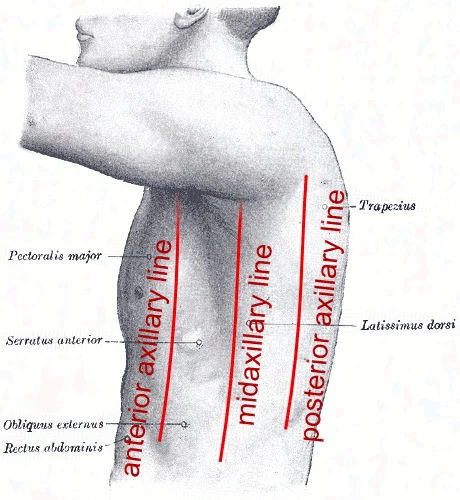
Figure 1Axillary lines
Long description
Surface anatomy of Axillary lines. Labelled illustration from Wikimedia Commons, used under its open licence.

Figure 1: Surface anatomy of Axillary lines.
On the skin surface the most reliable palpable landmark is the cephalic vein, which runs superficially along the lateral aspect of the biceps brachii and can be followed distally into the axillary region where it empties into the axillary vein. In individuals with good muscle tone and little subcutaneous fat the vein is easily visualised as a bluish cord, providing a quick guide to the lateral border of the axilla.
Directly inferior to the lateral border, the axillary tail of Spence (an extension of breast tissue) may be felt as a soft, slightly mobile mound that tapers into the deeper axillary fat pad. Accessory breast tissue, present in 2–6 % of women, often appears as a small, well‑defined lump in the same region and can be mistaken for a lymph node or cyst. Palpation of this tissue should be gentle; it is usually non‑tender unless inflamed. [2]
The axillary lymph nodes lie deep to the subcutaneous tissue, arranged in three levels. The most superficial group (Level I) is situated lateral to the lateral border of the pectoralis minor and can be approximated by pressing the thumb against the lateral chest wall while the fingers rest on the humeral head. A firm, mobile node in this zone is often a normal reactive node; however, a hard, fixed node warrants further imaging.
The skin over the axilla is thin and prone to frictional lesions. Epidermal inclusion cysts are frequently encountered at the dermis‑subcutis junction and are especially common in the axilla. These cysts feel like a smooth, mobile nodule and may have a central punctum. [2]
The labelled structures include: the cephalic vein coursing superficially along the biceps, the axillary tail of Spence extending from the breast, a typical accessory breast tissue nodule, the superficial lymph node group, and a representative epidermal inclusion cyst at the skin-fat interface. The image is oriented with the patient’s right side on the left of the picture, allowing the viewer to relate each superficial landmark to its deeper counterpart. [1]
DefinitionAxillary tail of Spence
A continuation of breast tissue that projects into the axilla, often palpable as a soft mound lateral to the pectoralis major.
NoteClinical tip
When a palpable axillary mass is encountered, first assess the overlying skin for cystic lesions, then locate the cephalic vein to confirm lateral orientation before proceeding to deeper examination.
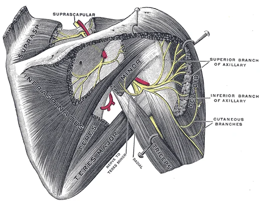
Figure 2Gray810
Long description
Suprascapular and axillary nerves of right side, seen from behind. (Testut.)

Figure 2: Suprascapular and axillary nerves of right side, seen from behind (Testut.).
The axilla is a pyramidal space bounded by the thoracic cage and the shoulder girdle. Understanding its osseous boundaries is fundamental to locating neurovascular structures and interpreting imaging. The anterior wall is formed by the pectoralis major and minor muscles, but the underlying bony framework includes the clavicle superiorly and the upper ribs laterally. The posterior wall is defined by the subscapularis, teres major, and latissimus dorsi muscles, resting against the scapula and the upper ribs. The medial wall consists of the upper four ribs and their intercostal muscles. The lateral wall is the intertubercular groove of the humerus.
The humeral head articulates with the glenoid fossa of the scapula. The coracoid process projects anteriorly from the scapula, serving as an attachment for the short head of the biceps brachii and the coracobrachialis muscle. The clavicle forms the superior boundary, articulating with the acromion process of the scapula. The upper ribs, particularly the first and second, form the medial floor of the axilla. These bones provide the rigid framework within which the axillary vessels and brachial plexus travel.
The radial artery and ulnar artery parallel their namesake bones, giving off smaller branches until they reach the wrist, or carpal region. At this level, they fuse to form the superficial and deep palmar arches that supply blood to the hand, as well as the digital arteries that supply blood to the digits. This vascular pattern demonstrates how arteries follow bony landmarks distally. In the axilla, the axillary artery continues the path of the subclavian artery, running parallel to the humerus within the axillary sheath. [1]
The scapula provides critical landmarks for surgical access. The lateral border of the scapula is adjacent to the long head of the triceps brachii. The inferior angle of the scapula is a palpable landmark at the level of the seventh rib. Recognising these bony relationships allows clinicians to localise pathology. For instance, a mass deep to the pectoralis minor may originate from the ribs or the scapula. The clavicle protects the subclavian vessels as they enter the axilla. Fractures of the clavicle or upper ribs can compromise the axillary contents. Therefore, precise anatomical knowledge of these bones is essential for safe surgical intervention and accurate diagnostic imaging. The axilla also includes muscles and bones of the shoulder and upper rib cage. Therefore, recognising that an axillary mass is within or beneath the muscle is essential in forming an appropriate differential diagnosis. [2]
The fascia is continuous with the clavipectoral fascia superiorly and the latissimus dorsi fascia inferiorly. It encloses the axillary contents, separating them from the surrounding chest wall and upper limb structures. The axillary sheath, a continuation of the prevertebral fascia, surrounds the neurovascular bundle, providing a protective conduit for the brachial plexus and axillary vessels as they pass from the neck into the arm.
The axilla is a pyramidal space bounded by the clavicle superiorly, the scapula posteriorly, the lateral chest wall laterally, and the medial chest wall medially. The walls are formed by muscles and bones. The anterior wall is formed by the pectoralis major and minor muscles, covered by the clavipectoral fascia. The posterior wall is formed by the subscapularis, teres major, and latissimus dorsi muscles. The medial wall is formed by the upper ribs and intercostal muscles, covered by the serratus anterior muscle.
DefinitionAxillary Sheath
The axillary sheath is a tubular fascial covering that extends from the root of the neck to the axilla, enclosing the brachial plexus and axillary vessels.
These nodes drain the upper limb, breast, and superficial thoracic wall. The long thoracic nerve runs along the lateral chest wall, innervating the serratus anterior muscle. The thoracodorsal nerve runs along the posterior wall, innervating the latissimus dorsi muscle.
| Structure | Location | Relation |
|---|---|---|
| Axillary Fascia | Roof of axilla | Continuous with clavipectoral fascia |
| Axillary Sheath | Surrounds neurovascular bundle | Continuation of prevertebral fascia |
| Pectoralis Minor | Anterior wall | Divides axillary nodes into levels |
| Serratus Anterior | Medial wall | Covered by axillary fascia |
When evaluating the axilla, imaging may reveal suspicious findings. If initial imaging shows a suspicious lesion, percutaneous procedures such as ultrasound-guided fine-needle aspiration (FNA) or ultrasound-guided core biopsy may be performed. FNA is typically performed using needles with aspirates sent to cytology. In most cases, multiple passes are made, especially if the cytologist is not on site during the procedure. If lymphoma is of clinical concern, additional aspirates [2]
The risks of axillary core biopsy are similar to other image-guided procedures, namely bleeding and infection. However, given the presence of axillary vessels and nerves coursing through the axilla, a “no-throw” technique is preferred as this minimises the risk of haematoma or unintended nerve damage. In one study of 39 patients with suspicious lymphadenopathy, axillary core biopsy revealed 90 % sensitivity, 100 % specificity and 92 % accuracy without any clinically significant complications. By recognising that the nerves course alongside the axillary vessels, the risk of bleeding and untoward nerve injury can easily be avoided in almost all cases. [2]
Additionally abnormal appearing lymph nodes may be seen related to both benign and neoplastic processes. In general, the differential diagnosis for an enlarged axillary lymph node includes reactive hyperplasia, HIV/immunocompromised state, sarcoidosis or other granulomatous diseases, lymphoma/leukaemia and metastatic disease (most commonly breast cancer, lung cancer, ovarian cancer, gastric cancer or melanoma). Calcifications involving lymph nodes within the axilla can be seen because of either benign or malignant causes. The differential diagnosis for calcified lymph nodes includes prior granulomatous infection, gold therapy in patients with rheumatoid arthritis (Fig. ), collagen vascular disease, and metastasis from breast, thyroid or ovarian cancer. [2]
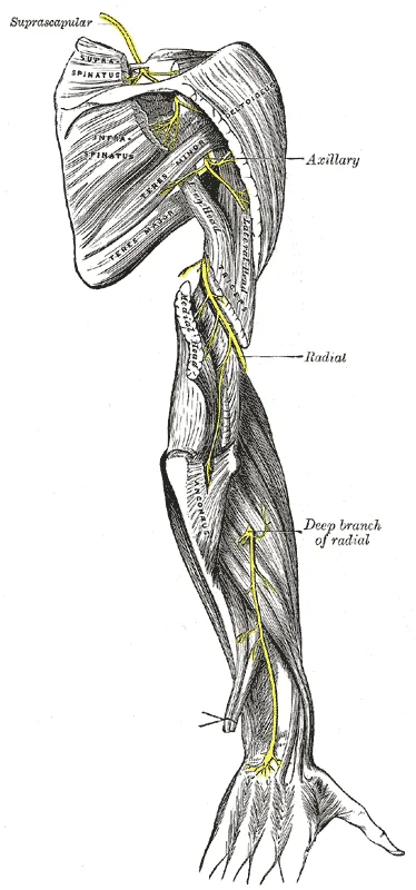
Figure 3Gray818
Long description
The suprascapular, axillary, and radial nerves.

Figure 3: The suprascapular, axillary, and radial nerves.
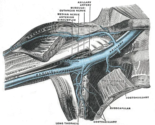
Figure 4Gray576
Long description
Anterior view of right upper limb and thorax - axillary vein and the distal part of the basilic vein and cephalic vein.

Figure 4: Anterior view of right upper limb and thorax - axillary vein and the distal part of the basilic vein and cephalic vein.
Understanding these structures requires visualising the space in relation to the thoracic cage and the arm. [1]
Figure 5Gray809
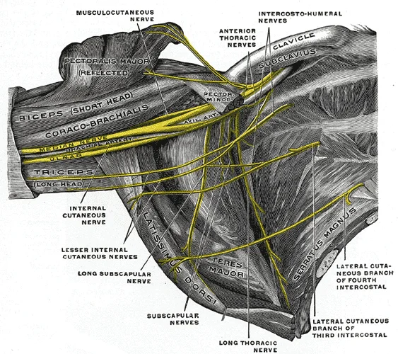
Long description
The right brachial plexus (infraclavicular portion) in the axillary fossa; viewed from below and in front. The Pectoralis major and minor muscles have been in large part removed; their attachments have been reflected. (Spalteholz.)

Figure 5: The right brachial plexus (infraclavicular portion) in the axillary fossa; viewed from below and in front.
The apex of the axilla is the cervico-axillary canal, bounded by the clavicle, first rib, and scapula.
| Muscle | Origin | Insertion | Action | Nerve Supply |
|---|---|---|---|---|
| Pectoralis Major | Clavicle, sternum, ribs | Lateral lip of intertubercular groove | Adduction, medial rotation of arm | Lateral and medial pectoral nerves |
| Pectoralis Minor | Ribs 3-5 | Coracoid process | Stabilises scapula, elevates ribs | Medial pectoral nerve |
| Serratus Anterior | Ribs 1-9 | Medial border of scapula | Protracts scapula | Long thoracic nerve |
| Subscapularis | Subscapular fossa | Lesser tubercle of humerus | Medial rotation of arm | Upper and lower subscapular nerves |
| Teres Major | Inferior angle of scapula | Medial lip of intertubercular groove | Adduction, medial rotation of arm | Lower subscapular nerve |
| Latissimus Dorsi | Spinous processes, iliac crest | Floor of intertubercular groove | Extension, adduction, medial rotation of arm | Thoracodorsal nerve |
The brachial plexus emerges from the neck and passes through the axilla to supply the upper limb. It is arranged in roots, trunks, divisions, cords, and branches. The cords are named in relation to the axillary artery: lateral, medial, and posterior. The lateral cord gives rise to the lateral pectoral nerve and contributes to the musculocutaneous nerve. The medial cord gives rise to the medial pectoral nerve, medial cutaneous nerves of the arm and forearm, and contributes to the median and ulnar nerves. The posterior cord gives rise to the upper and lower subscapular nerves, thoracodorsal nerve, axillary nerve, and radial nerve.
The axillary artery is the continuation of the subclavian artery.
It accompanies the axillary artery and becomes the subclavian vein at the lateral border of the first rib. The cephalic vein drains into the axillary vein.
DefinitionAxillary Space
A pyramidal space between the upper arm and thoracic wall, containing neurovascular structures and lymph nodes.
Figure 6Gray523
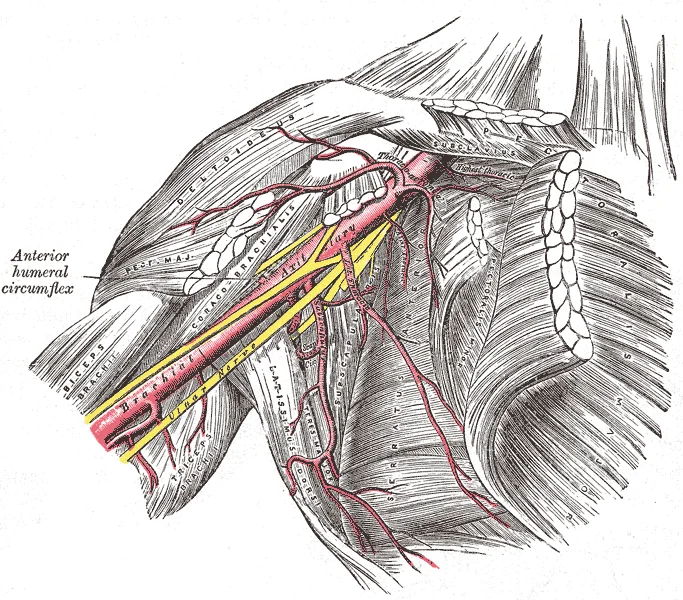
Long description
The axillary artery and its branches. Labelled illustration from Wikimedia Commons, used under its open licence.

Figure 6: The axillary artery and its branches.
Figure 7Axillary limits
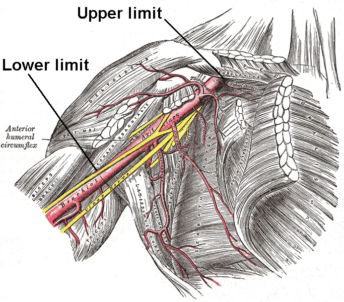
Long description
Limits of the axillary artery. Labelled illustration from Wikimedia Commons, used under its open licence.

Figure 7: Limits of the axillary artery.
The arterial supply to the axilla is derived from the axillary artery, a major vessel that undergoes a series of name changes as it traverses anatomical landmarks, much like a street changing names at an intersection. Specifically, the left subclavian artery becomes the axillary artery as it passes through the body wall and into the axillary region, and subsequently becomes the brachial artery as it flows from the axillary region into the upper arm. The origin of the axillary artery depends on the side of the body. On the right side, the brachiocephalic artery branches into the right subclavian artery and the right common carotid artery. The left subclavian artery arises independently from the aortic arch but follows a similar pattern to the right. Each subclavian artery supplies blood to the arms, chest, shoulders, back, and central nervous system. [1]
The axillary artery is divided into parts by the pectoralis minor muscle, a key anatomical landmark. This division determines the branching pattern of the artery. The first part, proximal to the pectoralis minor, gives rise to the superior thoracic artery. The second part, posterior to the pectoralis minor, gives rise to the thoracoacromial artery and the lateral thoracic artery. The third part, distal to the pectoralis minor, gives rise to the subscapular artery, the anterior circumflex humeral artery, and the posterior circumflex humeral artery. These branches supply the muscles and sk
Figure 8Axillary branches
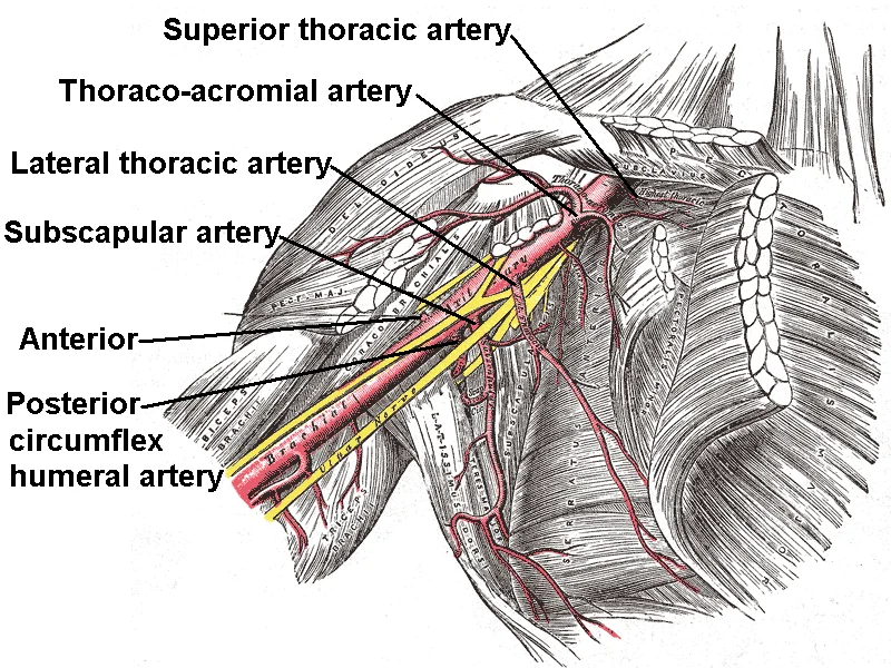
Long description
Branches of axillary artery. Labelled illustration from Wikimedia Commons, used under its open licence.

Figure 8: Branches of axillary artery.
The first part is located medial to the pectoralis minor. The second part is posterior to the pectoralis minor. The third part is lateral to the pectoralis minor. The superior thoracic artery arises from the first part. The thoracoacromial artery arises from the second part. The lateral thoracic artery arises from the second part. The subscapular artery arises from the third part. The anterior circumflex humeral artery arises from the third part. The posterior circumflex humeral artery arises from the third part.
DefinitionAxillary Artery Parts
The axillary artery is divided into three parts by the pectoralis minor muscle, which determines its branching pattern. [1]
The axillary artery continues the supply from the subclavian artery, which also gives rise to the internal thoracic artery, the vertebral artery, and the thyrocervical artery. The internal thoracic artery supplies blood to the thymus, the pericardium of the heart, and the anterior chest wall. The paired vertebral arteries join together to form the large basilar artery at the base of the medulla oblongata. This is an example of an anastomosis. The subclavian artery also gives rise to the thyrocervical artery that provides blood to the thyroid, the cervical region of the neck, and the upper back and shoulder. [1]
| Part | Branch |
|---|---|
| First | Superior thoracic artery |
| Second | Thoracoacromial artery |
| Second | Lateral thoracic artery |
| Third | Subscapular artery |
| Third | Anterior circumflex humeral artery |
| Third | Posterior circumflex humeral artery |
Anastomoses are especially common in veins, where they help maintain blood flow even when one vessel is blocked or narrowed, although there are some important ones in the arteries supplying the brain. The arterial circle in the brain is formed by the anterior cerebral arteries and the posterior cerebral arteries. The anterior cerebral arteries join together to form an anastomosis called the anterior communicating artery. The posterior portion of the arterial circle is formed by a left and a right posterior communicating artery that branches from the posterior cerebral artery. The basilar artery is an anastomosis that begins at the junction of the two vertebral arteries and sends branches to the cerebellum and brain stem. [1]
The venous system is responsible for returning deoxygenated blood to the right atrium of the heart. This blood has already passed through systemic capillaries and is therefore relatively low in oxygen concentration. While arteries and veins often run in parallel, the venous circulation exhibits greater variability than the arterial system. Blood from the head, neck, chest, shoulders, and upper limbs generally flows into the superior vena cava. [1]
The central structure is the axillary vein, which lies medial to the axillary artery. Superiorly, the axillary vein continues as the subclavian vein.
The axillary vein is the continuation of the brachial veins. It begins at the lower border of the teres major muscle. The vein ascends through the axilla, lying medial to the axillary artery. It receives several tributaries that correspond to the branches of the axillary artery. The basilic vein joins the brachial veins to form the axillary vein. The cephalic vein drains into the axillary vein near the lateral border of the pectoralis minor muscle. The thoracoacromial vein and lateral thoracic v [1]
The axillary vein terminates at the lateral border of the first rib, where it becomes the subclavian vein. The subclavian vein then joins the internal jugular vein to form the brachiocephalic vein. This arrangement ensures efficient drainage of the upper limb and shoulder region. [1]
Clinical procedures often involve access to the axillary vein. Ultrasound-guided puncture of the axillary vein is a common technique for central venous access. This method is associated with a short time to obtain access and low complication rates. The axillary vein is superficial and easily visualised with ultrasound. The procedure avoids the risks associated with subclavian puncture, such as pneumothorax. [4]
The axillary vein is closely related to the axillary artery and the brachial plexus. The artery lies lateral to the vein. The medial cord of the brachial plexus lies medial to the vein. The lateral cord lies lateral to the artery. These relationships are important for surgical and interventional procedures.
In summary, the axillary vein drains the upper limb and shoulder region. It is formed by the union of the brachial and basilic veins. The vein terminates at the first rib. Understanding its anatomy is essential for safe vascular access.
The axilla contains a complex network of nerves that provide both motor innervation to the shoulder girdle and upper limb, as well as cutaneous sensation to the skin of the axilla and medial arm. Understanding these structures requires tracing their origins from the brachial plexus and following their courses through the axillary space.
The lateral cord gives rise to the lateral pectoral nerve and contributes to the median nerve. The medial cord gives rise to the medial pectoral nerve, the medial cutaneous nerve of the arm, the medial cutaneous nerve of the forearm, and contributes to the median nerve. The posterior cord gives rise to the upper and lower subscapular nerves, the thoracodorsal nerve, and the axillary and radial nerves. The axillary nerve winds around the surgical neck of the humerus. The radial nerve descends posteriorly. The median nerve lies anterior to the axillary artery. The ulnar nerve lies medial to the axillary artery. The musculocutaneous nerve pierces the coracobrachialis muscle.
The motor supply includes muscles of the upper limb and chest wall. The cutaneous supply includes the skin of the axilla and medial arm.
| Nerve | Motor Supply | Cutaneous Supply |
|---|---|---|
| Axillary | Deltoid, Teres minor | Lateral shoulder skin |
| Radial | Triceps, Brachioradialis, Extensors | Posterior arm, forearm, hand |
| Musculocutaneous | Biceps, Brachialis, Coracobrachialis | Lateral forearm |
| Median | Flexors of forearm, Thenar muscles | Lateral palm, digits 1-3.5 |
| Ulnar | Intrinsic hand muscles, Flexor carpi ulnaris | Medial hand, digit 5 |
| Thoracodorsal | Latissimus dorsi | None |
| Long thoracic | Serratus anterior | None |
| Medial pectoral | Pectoralis major, Pectoralis minor | None |
| Lateral pectoral | Pectoralis major | None |
The axillary nerve arises from the posterior cord. It supplies the deltoid and teres minor muscles. It provides sensation to the skin over the lower deltoid. Injury to this nerve causes weakness in shoulder abduction and loss of sensation over the lateral shoulder. [1]
The radial nerve arises from the posterior cord. It supplies the triceps brachii and forearm extensors. It provides sensation to the posterior arm and forearm. Injury causes wrist drop and sensory loss on the dorsal hand.
The musculocutaneous nerve arises from the lateral cord. It supplies the biceps brachii, brachialis, and coracobrachialis. It provides sensation to the lateral forearm. Injury causes weakness in elbow flexion and sensory loss on the lateral forearm.
The median nerve is formed by contributions from the lateral and medial cords. It supplies most flexors of the forearm and thenar muscles. It provides sensation to the lateral palm and digits. Injury causes median nerve palsy with sensory loss and motor deficits in the hand.
The ulnar nerve arises from the medial cord. It supplies intrinsic hand muscles and flexor carpi ulnaris. It provides sensation to the medial hand and fifth digit. Injury causes claw hand deformity and sensory loss on the medial hand.
The thoracodorsal nerve arises from the posterior cord. It supplies the latissimus dorsi muscle. It has no cutaneous supply. Injury causes weakness in shoulder extension and adduction.
The long thoracic nerve arises from the ventral rami of C5-C7. It supplies the serratus anterior muscle. It has no cutaneous supply. Injury causes winged scapula.
The medial pectoral nerve arises from the medial cord. It supplies the pectoralis minor and major. It has no cutaneous supply.
The lateral pectoral nerve arises from the lateral cord. It supplies the pectoralis major. It has no cutaneous supply.
The axilla contains a complex network of lymphatic vessels and nodes that drain the upper limb, breast, and thoracic wall. Understanding this anatomy is critical for staging breast cancer and managing axillary masses. The lymph nodes are grouped into five levels based on their anatomical relations to the pectoralis minor muscle.
DefinitionAxillary Node Levels
Level I nodes are lateral to the pectoralis minor. Level II nodes lie deep to the pectoralis minor. Level III nodes are medial to the pectoralis minor.
Examination of level I lymph nodes necessitates visualising the axillary vessels as well as scanning the entire fatty contents from the margin of the pectoralis muscles anteromedially to the latissimus dorsi and teres major margin posterolaterally. The lateral thoracic and thoracodorsal arteries and smaller branches will be seen variably along each margin, respectively. Although node groups typically follow these vessels, lymph nodes are frequently found in isolation within the axillary fat. It [2]
Axillary ultrasound should be performed using a high-frequency (7.5–17-MHz) linear-array transducer. A lower frequency (5–7.5-MHz) setting may be needed for larger patients or for patients with a large axillary fat pad; however, the spatial resolution will be compromised. The patient should lie in a supine oblique position, with her hand above her head with the arm abducted and externally rotated (“bathing beauty” position). All findings should be documented in orthogonal planes with and without calipers and a lesion’s largest dimension should also be recorded. Some facilities employ grey-scale and colour Doppler US or power Doppler US during the study, especially when assessing lymph nodes. When colour Doppler US is used, it is important to use low wall filter settings and low velocity settings (higher pulse repetition frequency) to detect abnormal blood flow. [2]
NoteClinical Relevance
Most commonly performed for suspicious lymphadenopathy, the confirmation of metastatic involvement in a lymph node precludes sentinel lymph node biopsy in females with newly diagnosed breast cancer leading to full axillary node dissection. [2]
In one study of 224 patients, the sensitivity of ultrasound-guided FNA for suspicious, indeterminate and normal-appearing axillary nodes was 93, 44 and 11 %, respectively, with an overall sensitivity of 59 % and overall specificity of 100 %. The likelihood of a positive FNA correlated directly with the size of the primary breast malignancy, being 29 % for tumours <1 cm, 50 % for tumours 1–2 cm, 69 % for tumours 2–5 cm and 100 % for tumours >5 cm. Alternatively, percutaneous core needle biopsy [2]
Ultrasound and axillary core biopsy enable adequate pretreatment staging in patients with breast cancer and has a positive impact on their management. The benefit of pre-operative identification of axillary metastases is that it allows the surgeon to proceed directly to full axillary lymph node dissection and to avoid an unnecessary sentinel lymph node biopsy, thereby avoiding a second surgical procedure involving the axillary nodes. Houssami et al. in their meta-analysis found that axillary ultrasound-guided needle biopsy (UNB) had better utility in females with average or higher underlying risk of nodal metastases [2]
Recently, the field of breast cancer surgery has seen a dramatic shift with the results of the American College of Surgeons Oncology Group Z0011 trial. This was despite the fact that 27.3 % of the patients who received ALND had additional positive non-sentinel lymph nodes. [2]
Knowledge of the normal axillary anatomy aids in determining the underlying aetiology of an axillary mass. The differential diagnosis of an axillary mass is broad, including skin lesions, infections, haematoma, lymphadenopathy (hyperplastic, inflammatory, neoplastic or metastatic), accessory breast tissue, fibroadenoma, fibrocystic change, post-operative fluid collections, primary breast cancer and intramuscular neoplasms. For most cases, imaging evaluation entails diagnostic mammography and targeted ultrasound. If intervention is warranted, FNA or core needle biopsies are safe and accurate methods for diagnosis and for guiding the management of an axillary mass. [2]
The nodes are arranged in levels I, II, and III. Level I nodes are lateral to the pectoralis minor. Level II nodes are deep to the pectoralis minor. Level III nodes are medial to the pectoralis minor.
The axilla is a pyramidal space situated between the upper medial aspect of the arm and the lateral chest wall. It serves as a critical conduit for neurovascular structures passing from the neck and thorax to the upper limb. Understanding the spatial relationships within this region is fundamental for clinical procedures and surgical approaches.
The axillary artery lies centrally, acting as the primary landmark. Medial to the artery runs the axillary vein, which is typically more superficial and compressible. Lateral to the artery and vein, the cords of the brachial plexus are arranged in a specific pattern relative to the artery. The axillary sheath encloses these neurovascular bundles, extending from the neck into the axilla. This sheath provides a protective fascial covering. The contents are organised into three parts relative to the pectoralis minor muscle. The first part lies medial to the muscle, the second part posterior to it, and the third part lateral to it. This organisation dictates the branching pattern of the axillary artery. The axillary vein receives tributaries corresponding to the arterial branches. The nerves of the brachial plexus surround the artery, providing motor and sensory innervation to the upper limb. The lymph nodes are clustered along the vessels, particularly in the lateral and central groups. These nodes drain the upper limb, breast, and superficial thoracic wall. The boundaries of the axilla include the apex, base, and four walls. The apex is formed by the outer border of the first rib, the superior border of the scapula, and the clavicle. The base is formed by the skin and fascia of the axillary fold. The anterior wall is formed by the pectoralis major and minor muscles. The posterior wall is formed by the subscapularis, teres major, and latissimus dorsi muscles. The medial wall is formed by the upper ribs and intercostal muscles, covered by the serratus anterior muscle.
DefinitionAxillary Sheath
The axillary sheath is a continuation of the prevertebral fascia that encloses the axillary artery, axillary vein, and brachial plexus within the axilla. [4]
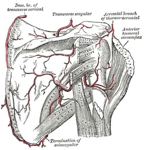
The axillary artery is a direct continuation of the subclavian artery. It begins at the outer border of the first rib and ends at the inferior border of the teres major muscle, where it becomes the brachial artery. The artery is divided into three parts by the pectoralis minor muscle. The first part gives off the superior thoracic artery. The second part gives off the thoracoacromial artery and the lateral thoracic artery. The third part gives off the subscapular artery, the anterior circumflex humeral artery, and the posterior circumflex humeral artery. The subscapular artery is the largest branch and divides into the circumflex scapular artery and the thoracodorsal artery.
Figure 9Gray524
Long description
The scapular and circumflex arteries. Labelled illustration from Wikimedia Commons, used under its open licence.

Figure 9: The scapular and circumflex arteries.
| Muscle | Location | Action | Nerve Supply |
|---|---|---|---|
| Pectoralis Major | Anterior wall | Adducts and medially rotates arm | Lateral and medial pectoral nerves |
| Pectoralis Minor | Anterior wall | Depresses shoulder | Medial pectoral nerve |
| Subscapularis | Posterior wall | Medially rotates arm | Upper and lower subscapular nerves |
| Teres Major | Posterior wall | Adducts and medially rotates arm | Lower subscapular nerve |
| Latissimus Dorsi | Posterior wall | Extends, adducts and medially rotates arm | Thoracodorsal nerve |
| Serratus Anterior | Medial wall | Protracts scapula | Long thoracic nerve |
The axillary vein is formed by the union of the brachial veins and the basilic vein. It accompanies the axillary artery throughout its course. The vein lies medial to the artery in the first part, posterior to it in the second part, and medial to it in the third part. The vein receives the cephalic vein at its lateral border. It continues as the subclavian vein at the outer border of the first rib. The axillary vein is a low-pressure system, making it suitable for venous access. However, careful technique is required to avoid arterial puncture.
Exam trapVenous Access
The axillary vein is more medial and superficial than the artery, and lacks pulsation, making it distinguishable by ultrasound. [4]
The brachial plexus provides the neural supply. It is formed by the ventral rami of C5 to T1. The cords are named for their relationship to the axillary artery. The lateral cord gives rise to the lateral root of the median nerve and the musculocutaneous nerve. The medial cord gives rise to the medial root of the median nerve, the ulnar nerve, and the medial brachial and antebrachial cutaneous nerves. The posterior cord gives rise to the radial nerve and the axillary nerve. These nerves pass through the axilla to innervate the upper limb. The lymph nodes are arranged in five groups: pectoral, subscapular, humeral, central, and apical. The apical nodes drain into the subclavian lymphatic trunk. These nodes are clinically significant in breast cancer staging.
The axilla is a pyramidal space containing vital neurovascular structures and lymphatic tissue. Its assessment relies on multiple imaging modalities, each offering distinct advantages for visualising anatomy and pathology.
Ultrasound is the primary modality for initial evaluation of axillary lymph nodes and soft tissue structures. It provides real-time imaging of vessel patency and node morphology. When evaluating lymph nodes, ultrasound is 94 % sensitive and 72 % specific in characterising nodes as suspicious or benign based on size and morphologic features. Normal nodes appear oval with a hyperechoic fatty hilum. Suspicious nodes often lose this hilum, becoming rounder and hypoechoic. The colour gain should be increased high enough to detect subtle flow without causing a colour noise artefact. This allows differentiation between benign reactive nodes and malignant infiltration. In cases of cat-scratch disease, lymph nodes are enlarged on ultrasound, appearing as hypoechoic lobular or oval masses with central hyperaemia and occasional adjacent fluid. The presence of asymmetry and a hyperechoic hilum may differentiate cat-scratch disease from other aetiologies. Ultrasound is also useful for evaluation of fistula or sinus tracts. It can detect complications such as pneumothorax earlier than radiological control during procedures. [2] [3]
Computed tomography (CT) is beneficial for assessing for chest wall involvement or invasion. It provides a broader anatomical context than ultrasound. Dual-energy CT can perform material decomposition, identifying intracapsular and extracapsular rupture and extracapsular silicone. This is particularly relevant when evaluating silicone implants or granulomatous reactions. CT findings of fat necrosis are not well described but tend to correspond with MRI findings with central fat and peripheral enhancement if contrast is given. The fibrosis has attenuation values similar to fibroglandular tissue or linear densities resembling fibrous bands. Calcifications occasionally are seen on CT. Lymph nodes are enlarged on mammography, which is often correlated with CT findings in staging. [3]
Magnetic resonance imaging (MRI) offers superior soft-tissue contrast. On MRI, the most frequent finding in inflammatory conditions was focal or diffuse asymmetrical signal intensity changes without significant mass effect. Nodular lesions can also be seen with hypointense T1 regions and hyperintense T2 regions. Dynamic contrast-enhanced MRI can show mass-like enhancement, ring-like enhancement and/or nodular enhancement, while time-intensity curves can vary. MRI shows a wide spectrum of appeara The involved parenchyma demonstrates restricted diffusion with ADC valves consistently lower than seen in normal breast parenchyma [3]
The axillary artery and vein lie anterior to the subscapularis muscle. The brachial plexus cords surround the axillary artery. Lymph nodes are clustered around the axillary vein. The long thoracic nerve runs along the lateral chest wall. The thoracodorsal nerve accompanies the thoracodorsal artery.
The axilla is a pyramidal space connecting the neck to the upper limb, serving as a critical conduit for neurovascular structures. Understanding its boundaries is essential for surgical approaches and interpreting imaging. The apex, or cervico-axillary canal, is bounded by the outer border of the first rib, the superior border of the scapula, and the posterior border of the clavicle. This narrow passage transmits the neurovascular bundle into the arm. The base of the axilla is formed by the axillary fascia and skin, while the walls are defined by surrounding muscles and bones. The anterior wall is formed by the pectoralis major and minor muscles. The posterior wall consists of the subscapularis, teres major, and latissimus dorsi muscles. The medial wall is formed by the upper four ribs and the serratus anterior muscle. The lateral wall is narrow, formed by the intertubercular groove of the humerus. [1]
The axillary artery is centrally located, coursing from the apex to the inferior border of the teres major. It is accompanied by the axillary vein, which lies anteromedial to the artery. The three cords of the brachial plexus are named for their relationship to the second part of the axillary artery: lateral, medial, and posterior. In the context of node dissection, the axillary lymph nodes are grouped into levels. Level I nodes are lateral to the pectoralis minor. Level II nodes are posterior to the pectoralis minor. Level III nodes are medial to the pectoralis minor, extending to the apex. Injury to the axillary artery during surgery can lead to significant haemorrhage or ischaemia of the upper limb. Nerve palsies, particularly of the long thoracic nerve, can result in winging of the scapula due to serratus anterior paralysis. [2]
Clinical pearlSurgical Landmark
The pectoralis minor muscle is the key landmark for dividing axillary nodes into three levels, guiding the extent of dissection in breast cancer surgery.
The axillary vein is formed by the union of the brachial and basilic veins. It becomes the subclavian vein at the outer border of the first rib. Venous thrombosis in this region, known as Paget-Schroetter syndrome, can occur after strenuous activity. Lymphatic drainage from the breast primarily flows to the axillary nodes, making them crucial in staging breast cancer. Ultrasound imaging is often used to assess nodal morphology. Eccentric cortical thickening is more suspicious than diffuse thickening, but both are non-specific and concerning enough to warrant intervention in most cases (Fig. ). Findings seen in cases with more advanced nodal involvement, such as effacement of the fatty hilum or a rounded hypoechoic mass, have a higher positive predictive value in patients with invasive breast cancer. Replacement of the entire node or a portion of the node by an ill-defined mass is highly suspicious for malignant involvement. Occasionally, nodal microcalcifications can be seen on imaging (Fig. ). [2] [3]
| Level | Anatomical Boundary | Clinical Significance |
|---|---|---|
| Level I | Lateral to pectoralis minor | First echelon drainage from breast |
| Level II | Posterior to pectoralis minor | Intermediate drainage |
| Level III | Medial to pectoralis minor | Apical drainage |
The axilla is a pyramidal space bounded by the chest wall, humerus, and scapula. Its contents are organised into three levels relative to the pectoralis minor muscle. Level I lies lateral to the muscle, Level II posterior, and Level III medial. This anatomical division is critical for staging breast cancer and planning surgical clearance.
DefinitionAxillary Lymph Nodes
Groups of lymph nodes that drain the upper limb, breast, and superficial thoracic wall, classified into levels based on their position relative to the pectoralis minor muscle.
The axillary vein is formed by the union of the brachial veins and the basilic vein. It runs medial to the axillary artery initially, then crosses anterior to become the subclavian vein at the lateral border of the first rib. Understanding this relationship is vital for central line insertion.
Exam trapVein vs Artery
Do not confuse the axillary vein with the subclavian vein. The axillary vein becomes the subclavian vein at the outer border of the first rib. Puncture errors here can lead to pneumothorax.
The brachial plexus cords are named for their relationship to the axillary artery. The lateral cord is lateral, the medial cord is medial, and the posterior cord is posterior. This mnemonic helps recall the origin of major nerves like the musculocutaneous and median nerves.
Clinical pearlPectoralis Minor Landmark
The pectoralis minor muscle is the key landmark. It divides the axillary artery into three parts. The first part is proximal, the second posterior, and the third distal to the muscle.
Lymphatic drainage from the breast predominantly flows to the axillary nodes. Approximately 75% of lymph from the breast drains into the axillary nodes, while the remainder drains to the internal thoracic nodes. This explains why axillary node status is a prognostic indicator.
These features range from benign to indeterminate, requiring biopsy for definitive diagnosis. [3]
The nodes are tender and swollen, often resolving within a month. [3]
Exam trapGranulomatous Causes
Granulomatous lymphadenitis can be secondary to breast conditions like silicone granuloma or idiopathic causes. Cat-scratch disease is another infectious cause to consider in differential diagnosis. [3]
The axillary vein is a useful bail-out alternative for CIED lead implantation. If the cephalic vein is absent or unsuitable, the axillary vein can be used to avoid intrathoracic puncture. [4]
Students frequently confuse the anatomical boundaries of the axilla with the functional compartments of the upper limb, leading to errors in clinical localisation. A primary error is assuming that all lymphatic drainage from the upper limb passes through the axillary nodes. In reality, drainage is complex, involving supraclavicular and infraclavicular routes, and assuming a single pathway ignores the collateral circulation that can bypass obstruction. This misconception often leads to incorrect staging in breast cancer or melanoma cases where sentinel node biopsy is indicated.
Another frequent mistake involves the identification of the axillary artery segments. Students often misidentify the third part of the axillary artery as the subclavian artery due to their close proximity. The correct anatomical landmark is the lateral border of the first rib; the artery becomes the axillary artery distal to this point. Confusing these vessels can lead to errors in understanding the origin of branches such as the thoracoacromial artery, which arises from the second part, not the third.
A third pitfall is the misinterpretation of axillary vein anatomy relative to the artery. The vein is not simply a parallel structure but has specific positional relationships that change along its course. For instance, the axillary vein is initially lateral to the artery but becomes posterior to it as it passes the lower border of the teres major muscle. Failing to recognise this crossover can result in incorrect surgical approaches or misinterpretation of Doppler ultrasound findings where flow direction is critical.
Students also commonly overlook the importance of the axillary nerve in relation to the surgical neck of the humerus. While studying the brachial plexus, the focus is often on the cords, but the axillary nerve’s specific course through the quadrangular space is vital for understanding shoulder instability. Damage to this nerve is a classic complication of anterior shoulder dislocation, yet students often fail to link the anatomical route with the clinical presentation of deltoid paralysis and sensory loss over the regimental badge area.
Finally, a significant error is neglecting the fatty tissue and loose connective tissue that fill the axillary cavity. This tissue is not merely filler but contains the lymph nodes, vessels, and nerves in a mobile framework. Assuming a rigid structure ignores the clinical significance of axillary lymph node dissection, where preserving the neurovascular bundle is paramount. The presence of this loose tissue allows for the spread of infection or malignancy, making it a critical pathway for disease progression that must be understood for effective surgical planning.
In the operating theatre, knowledge of axillary anatomy is critical for axillary lymph node dissection, a procedure often performed in breast cancer management. Surgeons must identify and preserve the long thoracic nerve, which runs along the lateral chest wall, to prevent winging of the scapula. They also protect the thoracodorsal nerve, which supplies the latissimus dorsi muscle, ensuring shoulder extension and adduction remain intact. Precise dissection requires understanding the relationship between the axillary vein and the lymphatic tissue to avoid venous injury, which can lead to significant bleeding and postoperative complications.
On imaging, particularly ultrasound and MRI, the axilla is evaluated for lymphadenopathy. Radiologists look for changes in nodal morphology, such as loss of the fatty hilum, cortical thickening, or abnormal vascularity. These features are indicative of metastatic disease or lymphoma. Understanding the normal anatomical landmarks, such as the axillary artery and vein, helps in distinguishing between vascular structures and pathological nodes. For example, a hypoechoic node with a preserved hilum is likely benign, whereas a node with cortical thickening and loss of the hilum raises suspicion for malignancy.
At the bedside, clinicians assess axillary lymph nodes during physical examination of the breast and upper limb. Palpation of the apical, central, lateral, pectoral, and subscapular groups provides crucial information about potential pathology. Enlarged or hard nodes may indicate metastatic spread from breast cancer, melanoma, or lymphoma. Additionally, examination of the axilla is part of the assessment for axillary web syndrome, a condition characterized by cord-like structures under the skin, often following surgery or radiotherapy. Recognising these structures helps in early intervention and management. [3]
In examinations, students are often tested on the branches of the axillary artery and the cords of the brachial plexus. Questions may require identifying the origin of specific arteries, such as the superior thoracic artery from the first part, or the lateral thoracic artery from the second part. Understanding the relationship between the cords and the branches of the brachial plexus, such as the median nerve formed by medial and lateral cords, is essential for answering questions on nerve injuries and their clinical manifestations.
Axillary Lymph Node Assessment
- Patient PresentationBreast lump or upper limb swelling
- Question: Palpable Nodes?
Yes
- Assess CharacteristicsSize, consistency, mobility
- Question: Suspicious Features?
Yes
- Imaging & BiopsyUltrasound/MRI and FNA
No
- MonitorRegular follow-up
No
- Imaging if IndicatedUltrasound if high risk
The axillary artery is divided into three parts by the pectoralis minor muscle. The axillary vein is formed by the union of the brachial and basilic veins. The long thoracic nerve innervates the serratus anterior muscle. The thoracodorsal nerve supplies the latissimus dorsi muscle. Axillary lymph nodes are grouped into five levels based on their relation to the pectoralis minor. The brachial plexus cords are named for their relationship to the axillary artery. Sensory innervation to the lateral arm is provided by the axillary nerve. Lymphatic drainage from the upper limb primarily passes through the axillary nodes.
Answer each one in your head first, then open the answer.
Which muscle divides the axillary artery into three parts?
Show answerHide answer
Pectoralis minorWhat is the primary nerve supply to the latissimus dorsi?
Show answerHide answer
Thoracodorsal nerveWhich nerve is at risk during axillary lymph node dissection and causes winged scapula if damaged?
Show answerHide answer
Long thoracic nerveWhat vein is formed by the union of the brachial and basilic veins?
Show answerHide answer
Axillary veinWhich group of lymph nodes is located medial to the pectoralis minor?
Show answerHide answer
Level II nodes
- Betts JG, Young KA, Wise JA, et al. Circulatory Pathways. OpenStax, Anatomy and Physiology 2e (CC BY-NC-SA 4.0). Accessed .
- Dialani V, James DF, Slanetz PJ. A practical approach to imaging the axilla. Insights into imaging 2015;6(2):217-229. doi:10.1007/s13244-014-0367-8. Accessed .
- Illman JE, Terra SB, Clapp AJ, et al. Granulomatous diseases of the breast and axilla: radiological findings with pathological correlation. Insights into imaging 2018;9(1):59-71. doi:10.1007/s13244-017-0587-9. Accessed .
- Tagliari AP, Kochi AN, Mastella B, et al. Ultrasound-guided Axillary Vein Puncture in Cardiac Lead Implantation: Time to Move to a New Standard Access?. Arrhythmia & electrophysiology review 2020;9(2):78-82. doi:10.15420/aer.2020.17. Accessed .
- Dialani V, James DF, Slanetz PJ. A practical approach to imaging the axilla. Insights into imaging 2015;6(2):217-229. doi:10.1007/s13244-014-0367-8. Accessed .
- Natsch A, Emter R. The specific biochemistry of human axilla odour formation viewed in an evolutionary context. Philosophical transactions of the Royal Society of London. Series B, Biological sciences 2020;375(1800):20190269. doi:10.1098/rstb.2019.0269. Accessed .
Figure credits
- Figure 1: Mikael Häggström. When using this image in external works, it may be cited as: Häggström, Mikael (2014). "Medical gallery of Mikael Häggström 2014". WikiJournal of Medicine 1 (2). DOI:10.15347/wjm/2014.008. ISSN 2002-4436. Public Domain. or By Mikael Häggström, used with permission., via Wikimedia Commons, Public domain, Public domain.
- Figure 2: Henry Vandyke Carter, via Wikimedia Commons, Public domain, Public domain.
- Figure 3: Henry Vandyke Carter, via Wikimedia Commons, Public domain, Public domain.
- Figure 4: Henry Vandyke Carter, via Wikimedia Commons, Public domain, Public domain.
- Figure 5: Henry Vandyke Carter, via Wikimedia Commons, Public domain, Public domain.
- Figure 6: Henry Vandyke Carter, via Wikimedia Commons, Public domain, Public domain.
- Figure 7: Mikael Häggström. When using this image in external works, it may be cited as: Häggström, Mikael (2014). "Medical gallery of Mikael Häggström 2014". WikiJournal of Medicine 1 (2). DOI:10.15347/wjm/2014.008. ISSN 2002-4436. Public Domain. or By Mikael Häggström, used with permission., via Wikimedia Commons, Public domain, Public domain.
- Figure 8: Mikael Häggström. When using this image in external works, it may be cited as: Häggström, Mikael (2014). "Medical gallery of Mikael Häggström 2014". WikiJournal of Medicine 1 (2). DOI:10.15347/wjm/2014.008. ISSN 2002-4436. Public Domain. or By Mikael Häggström, used with permission., via Wikimedia Commons, Public domain, Public domain.
- Figure 9: Henry Vandyke Carter, via Wikimedia Commons, Public domain, Public domain.
Educational content for medical students and doctors. It is not a substitute for clinical judgement or local guidelines.