Pectoral region
Anatomical study of the pectoral region, breast structure, and shoulder girdle.
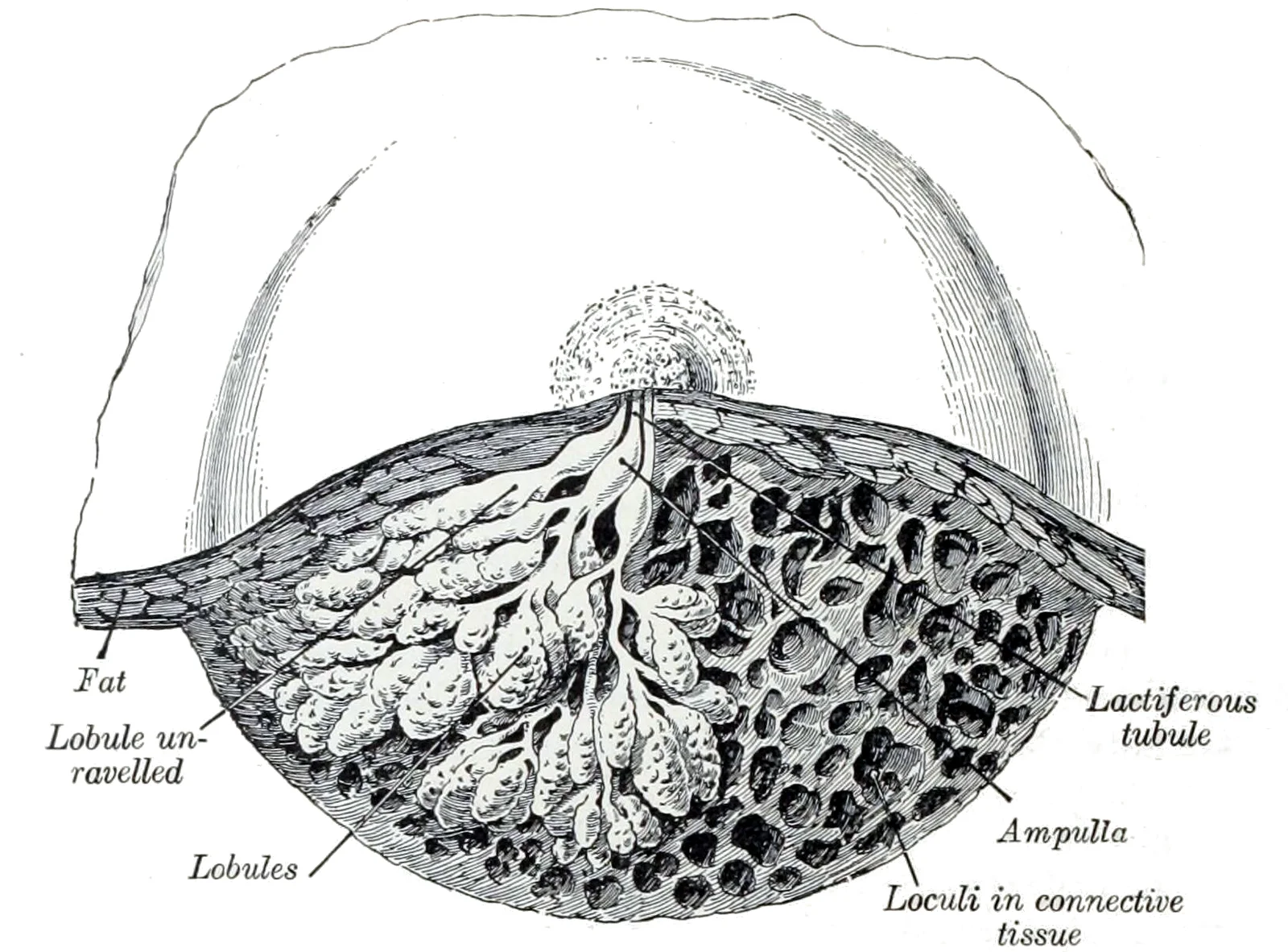
The pectoral region constitutes the anterior chest wall, bounded superiorly by the clavicle and inferiorly by the costal margin. It houses the breast, a modified sweat gland structure essential for lactation. The breast rests on the deep fascia covering the pectoralis major, serratus anterior, and external oblique muscles. Its boundaries extend from the second to the sixth rib laterally and the sternum medially. The breast tissue is supported by suspensory ligaments of Cooper, which are fibrous bands extending from the deep fascia to the skin. These ligaments maintain the structural integrity and position of the breast.
Development of the breast begins in puberty under hormonal influence. The glandular tissue proliferates, forming lobules and ducts. Variation in breast size and shape is common and depends on adipose tissue distribution. The axillary tail of Spence extends into the axilla, containing glandular tissue that can be clinically significant.
The shoulder joint, or glenohumeral joint, is a ball-and-socket articulation between the humeral head and the glenoid cavity of the scapula. This joint possesses the largest range of motion in the body. However, this mobility comes at the cost of stability due to limited structural support. The glenoid cavity is shallow, covering only about one third of the humeral head. Stability is enhanced by the glenoid labrum, a fibrocartilaginous lip deepening the socket.
DefinitionGlenohumeral Joint
A ball-and-socket joint formed by the humeral head and glenoid cavity, offering maximal mobility but reduced stability.
The articular capsule is thin and loose to permit extensive movement. Structural support is provided by intrinsic ligaments, including the coracohumeral ligament and three glenohumeral ligaments. These thickenings strengthen the superior and anterior capsule walls. The coracohumeral ligament runs from the coracoid process to the anterior humerus. The glenohumeral ligaments are located on the anterior aspect of the capsule.
| Ligament | Location | Function |
|---|---|---|
| Coracohumeral | Coracoid process to anterior humerus | Strengthens superior capsule |
| Glenohumeral | Anterior capsule | Strengthens anterior capsule |
The clavicle connects the sternum to the scapula, providing a rigid strut. The scapula provides attachment sites for numerous muscles. The boundaries are defined by the supraclavicular fossa above and the infraclavicular fossa below. The lateral border is the axilla. Understanding these boundaries is crucial for clinical examination and surgical approaches. The region contains major neurovascular structures, including the brachial plexus and axillary vessels. These structures pass through the region to supply the upper limb.
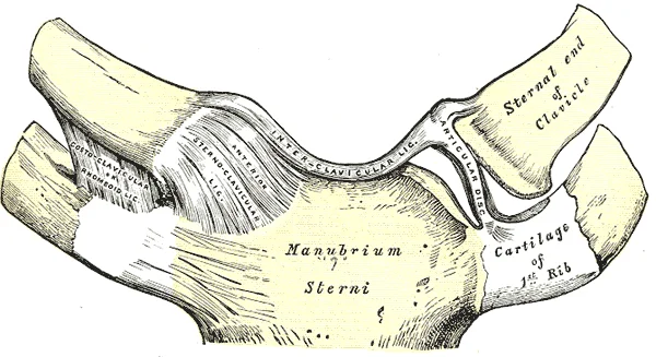
The sternum is centrally located, serving as the midline reference. The clavicles extend laterally from the sternum, forming the superior boundary of the thoracic inlet. The pectoralis major muscle is prominent, covering the upper anterior thorax. Its clavicular head originates from the medial half of the clavicle, while the sternocostal head arises from the sternum and upper costal cartilages. The deltoid muscle forms the lateral contour of the shoulder, overlapping the lateral aspect of the pectoralis major. The midclavicular line passes through the nipple, aiding in the localization of breast tissue. The axillary tail of Spence extends laterally into the axilla, a common site for breast tissue extension. The ribs are palpable beneath the skin, with the second rib being the most distinct landmark for counting intercostal spaces. The intercostal spaces are filled with intercostal muscles and neurovascular bundles. The superior vena cava drains most of the body superior to the diaphragm. The internal thoracic vein, also known as an internal mammary vein, drains the anterior surface of the chest wall and flows into the brachiocephalic vein. The cephalic vein begins in the antebrachium and drains blood from the superficial surface of the arm into the axillary vein. The remainder of the blood supply from the thorax drains into the azygos vein. Each intercostal vein drains muscles of the thoracic wall. These veins flow into the azygos vein, and with the smaller hemiazygos vein (hemi- = “half”) on the left of the vertebral column, drain blood from the thoracic region. The common iliac arteries provide blood to the pelvic region and ultimately to the lower limbs. They split into external and internal iliac arteries approximately at the level of the lumbar-sacral articulation. In females, they also provide blood to the uterus and vagina. The superior phrenic artery provides blood to the superior surface of the diaphragm. lists the arteries of the thoracic region. Many of the larger veins of the thoracic and abdominal region and upper limb are further represented in the flow chart in. summarizes the veins of the upper limbs. Bronchial veins carry approximately 13 percent of the blood that flows into the bronchial arteries; the remainder intermingles with the pulmonary circulation and returns to the heart via the pulmonary veins. [1] [3]
Figure 1Gray325
Long description
Sternoclavicular articulation. Anterior view.

Figure 1: Sternoclavicular articulation (anterior view).
Figure 2Gray1172
Long description
Dissection of the lower half of the mamma during the period of lactation. (Luschka.)

Figure 2: Dissection of the lower half of the mamma during the period of lactation (Luschka.).
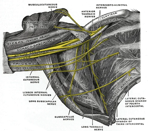
Figure 3Gray809
Long description
The right brachial plexus (infraclavicular portion) in the axillary fossa; viewed from below and in front. The Pectoralis major and minor muscles have been in large part removed; their attachments have been reflected. (Spalteholz.)

Figure 3: The right brachial plexus (infraclavicular portion) in the axillary fossa; viewed from below and in front.
The scapula lies posteriorly on the thoracic wall, spanning from the second to the seventh ribs. Its lateral corner features the glenoid fossa, a shallow cavity that articulates with the head of the humerus to form the glenohumeral joint. The acromion process extends anteriorly from the scapular spine to meet the clavicle. The coracoid process projects anteriorly and superiorly, serving as an attachment point for several muscles. These bony structures create the stable origin for the muscles that move the humerus[1]. This arrangement allows the head of the humerus to move in multiple directions within its ball-and-socket joint with the glenoid fossa of the scapula[1]. [2]
The muscles positioning the pectoral girdle are located on the thorax. The anterior thoracic muscles include the subclavius, pectoralis minor, and serratus anterior[1]. The posterior thoracic muscles are the trapezius, levator scapulae, rhomboid major, and rhomboid minor[1]. When the rhomboids contract, the scapula moves medially, pulling the shoulder and upper limb posteriorly[1]. The serratus anterior wraps around the lateral thorax, anchoring the scapula to the rib cage. These muscles ensure the stability required for upper limb function. [2]
The pectoral girdle provides a base for the arm's movement. The clavicle and scapula make up the pectoral girdle, which provides a stable origin for the muscles that move the humerus[1]. The muscles that position and stabilize the pectoral girdle are located on the thorax[1]. This anatomical arrangement supports the extensive range of motion available at the shoulder joint. [2]
| Group | Muscle | Action |
|---|---|---|
| Anterior | Subclavius | Stabilises clavicle |
| Anterior | Pectoralis minor | Depresses and protracts scapula |
| Anterior | Serratus anterior | Rotates scapula, anchors to ribs |
| Posterior | Trapezius | Elevates, retracts, rotates scapula |
| Posterior | Levator scapulae | Elevates scapula |
| Posterior | Rhomboid major | Retracts and rotates scapula |
| Posterior | Rhomboid minor | Retracts and rotates scapula |
The pectoral region is defined by the arrangement of muscles that cross the shoulder joint and the fascial sheets that separate the anterior thorax from the axilla. The two primary axial muscles involved in shoulder movement are the pectoralis major and the latissimus dorsi. These muscles work alongside scapular muscles to allow the arm to carry out different types of movements. [2]
The pectoralis major lies superficially, covering the superior anterior thorax, while the latissimus dorsi contributes to the posterior aspect of the axilla. This fascial layer is crucial for understanding the anatomical boundaries of the pectoral compartment, as it prevents the direct spread of infection or fluid between the axilla and the anterior chest wall.
The rotator cuff muscles, including the supraspinatus, infraspinatus, teres minor, and subscapularis, arise from the scapula and attach to the greater or lesser tubercles of the humerus. Their tendons encircle the head of the humerus and fuse with the articular capsule, forming the rotator cuff. This structure provides primary structural support for the glenohumeral joint, acting as dynamic ligaments to hold the humeral head in position. While the pectoral muscles are axial in origin, the scapular muscles provide the bulk of the shoulder’s mobility and stability. The deltoid, a thick muscle creating the rounded lines of the shoulder, is the major abductor of the arm. It also facilitates flexing, medial rotation, extension, and lateral rotation. The subscapularis originates on the anterior scapula and medially rotates the arm. The supraspinatus and infraspinatus, named for their locations superior and inferior to the spine of the scapula, abduct and laterally rotate the arm. The teres major, thick and flat, is inferior to the teres minor and extends the arm, assisting in adduction and medial rotation. [2] [3]
| Muscle | Origin | Insertion | Action | Nerve Supply |
|---|---|---|---|---|
| Pectoralis Major | Clavicle, sternum, ribs | Humerus | Adduction, flexion, medial rotation | Pectoral nerves |
| Latissimus Dorsi | Inferior thoracic vertebrae, iliac crest | Intertubercular groove | Extension, adduction, medial rotation | Thoracodorsal nerve |
| Deltoid | Clavicle, acromion, spine of scapula | Deltoid tuberosity of humerus | Abduction, flexion, extension, rotation | Axillary nerve |
| Subscapularis | Subscapular fossa | Lesser tubercle | Medial rotation | Subscapular nerves |
The clavipectoral fascia is a key landmark in the pectoral region, separating the pectoral muscles from the axillary vessels and nerves. This fascial layer creates a compartment that houses the cephalic vein and the thoracoacromial vessels. The pectoralis major, being the most superficial muscle of the anterior thorax, overlies the pectoralis minor and the serratus anterior. The latissimus dorsi, originating from the inferior back, crosses the posterior axillary fold to insert into the humerus. The relationship between these muscles and the fascia is critical for surgical approaches to the axilla, as the fascia must be incised to access the axillary contents. The rotator cuff muscles, by fusing with the joint capsule, provide dynamic stability that complements the static support of the ligaments. This integration of muscular and fascial structures ensures the shoulder’s wide range of motion while maintaining joint integrity.
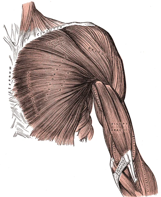
Figure 4Gray410
Long description
pectoralis major muscle. Labelled illustration from Wikimedia Commons, used under its open licence.

Figure 4: Pectoralis major muscle.
The pectoralis major is the most superficial muscle, originating from the clavicle, sternum, and costal cartilages, and inserting into the lateral lip of the intertubercular groove of the humerus. It is supplied by the medial and lateral pectoral nerves. Deep to it lies the pectoralis minor, originating from the third to fifth ribs and inserting into the coracoid process of the scapula, supplied by the medial pectoral nerve. The breast structure supply and drainage are visualised here; the breast receives arterial supply from perforating branches of the internal thoracic artery and lateral thoracic artery. Venous drainage follows the arterial supply into the axillary and internal thoracic veins. Lymphatic drainage primarily flows to the axillary lymph nodes, with some drainage to the parasternal nodes. The nerve supply to the skin of the anterior thorax is provided by intercostal nerves T2–T6. [3]
The muscles of the pectoral region are grouped by their anatomical compartment and function. The pectoralis major and minor, along with the subclavius, form the superficial and intermediate layers of the anterior chest wall. These muscles are crucial for upper limb movement and stability.
| Muscle | Origin | Insertion | Action | Nerve Supply |
|---|---|---|---|---|
| Pectoralis Major | Clavicle, sternum, costal cartilages | Lateral lip of intertubercular groove | Adducts and medially rotates humerus | Medial and lateral pectoral nerves |
| Pectoralis Minor | Ribs 3–5 | Coracoid process | Stabilises scapula, elevates ribs | Medial pectoral nerve |
| Subclavius | First rib | Inferior surface of clavicle | Depresses clavicle | Nerve to subclavius |
The arterial supply to the pectoral region is derived primarily from the subclavian artery, which itself arises from the aortic arch. The aortic arch gives rise to three major branches: the brachiocephalic artery, the left common carotid artery, and the left subclavian artery[1]. The left subclavian artery, named for its position under the clavicle, is classified as an elastic artery due to its proximity to the heart[1].
The thoracoacromial artery branches from the second part of the axillary artery, which is the continuation of the subclavian artery. This artery divides into pectoral, acromial, clavicular, and deltoid branches. The pectoral branch pierces the clavipectoral fascia to supply the pectoralis major and minor muscles. The internal thoracic artery also gives off anterior intercostal arteries, which anastomose with the posterior intercostal arteries arising from the aorta.
It provides branches to the anterior intercostal spaces. The thoracoacromial artery, arising from the axillary artery, has a pectoral branch that specifically targets the pectoral muscles. This branch enters the deep surface of the pectoralis major. The venous drainage generally parallels the arterial supply. Veins accompany the arteries, draining into the axillary vein and then the subclavian vein. The internal thoracic veins drain into the brachiocephalic veins. This parallel arrangement facilitates efficient blood flow and return.
The pectoralis major is supplied by the pectoral branch of the thoracoacromial artery and branches from the internal thoracic artery. The skin overlying the pectoral region is supplied by cutaneous branches from the intercostal nerves and vessels. The anastomoses between the internal thoracic and lateral thoracic arteries provide redundancy. This is clinically significant in cases of vascular compromise.
| Vessel | Origin | Course | Branches |
|---|---|---|---|
| Subclavian Artery | Aortic Arch | Passes under clavicle | Internal Thoracic, Thoracoacromial |
| Internal Thoracic | Subclavian | Descends lateral to sternum | Anterior Intercostals |
| Thoracoacromial | Axillary | Pierces clavipectoral fascia | Pectoral, Acromial, Clavicular, Deltoid |
The venous drainage mirrors this arterial pattern. The veins drain into the axillary vein, which becomes the subclavian vein. This ensures that deoxygenated blood from the upper limb and chest wall returns to the heart efficiently. The internal thoracic veins drain into the brachiocephalic veins. The lateral thoracic veins drain into the axillary vein. This system is crucial for maintaining hemodynamic stability in the upper thorax.
The venous system of the pectoral region operates on the principle of returning deoxygenated blood to the heart. Systemic veins return blood to the right atrium. Since the blood has already passed through the systemic capillaries, it will be relatively low in oxygen concentration. In many cases, there will be veins draining organs and regions of the body with the same name as the arteries that supplied these regions and the two often parallel one another. This is often described as a “complementary” pattern. However, there is a great deal more variability in the venous circulation than normally occurs in the arteries. [1]
DefinitionComplementary Venous Pattern
Veins often parallel the arteries that supplied the same region, though significant anatomical variation exists in the venous circulation.
The vein descends towards the diaphragm. It typically empties into the brachiocephalic vein. It drains the superficial tissues of the anterior chest and shoulder. The vein terminates by piercing the clavipectoral fascia to join the axillary vein. The axillary vein is the major collector of the upper limb and pectoral region. It becomes the subclavian vein at the lateral border of the first rib.
NoteClinical Variation
Keep this variation in mind when you move from the classroom to clinical practice.
Although the external and internal jugular veins are separate vessels, there are anastomoses between them close to the thoracic region. Blood from the external jugular vein empties into the subclavian vein. The subclavian vein receives blood from the upper limb and the pectoral region. It joins the internal jugular vein to form the brachiocephalic vein. The brachiocephalic veins unite to form the superior vena cava. The superior vena cava drains most of the veins superior to the diaphragm. It empties into the right atrium. The right atrium receives this deoxygenated blood. [1]
| Vein | Origin | Termination | Key Relations |
|---|---|---|---|
| Internal Thoracic | Superior epigastric | Brachiocephalic | Lateral to sternum |
| Cephalic | Dorsal venous network | Axillary | Deltopectoral groove |
| Basilic | Dorsal venous network | Axillary | Medial arm |
| External Jugular | Posterior auricular | Subclavian | Superficial neck |
As the basilic vein reaches the antecubital region, it gives off a branch called the median cubital vein that crosses at an angle to join the cephalic vein. The median cubital vein is the most common site for drawing venous blood in humans. This connection provides a direct superficial route for venous access. The basilic vein continues through the arm medially and superficially to the axillary vein. This pathway ensures efficient drainage from the upper limb into the central circulation. [1]
The pectoral region is innervated by a complex network of nerves that provide both motor control to the muscles of the chest wall and cutaneous sensation to the overlying skin. Understanding this supply is fundamental for clinical examinations and surgical procedures in the area.
Each structure lies in close relation to the major vessels of the axilla, particularly the axillary artery and its branches. The nerves are arranged in a predictable pattern relative to the pectoral muscles, allowing for systematic identification.
The motor supply to the pectoral muscles is derived primarily from the brachial plexus. The lateral pectoral nerve arises from the lateral cord and supplies the clavicular and sternocostal heads of the pectoralis major. These nerves ensure coordinated movement of the shoulder girdle.
Cutaneous sensation is provided by several nerves. The supraclavicular nerves, arising from the cervical plexus (C3-C4), supply the skin over the clavicle and upper pectoral region. The intercostobrachial nerve, a branch of the second intercostal nerve, supplies the skin of the axilla and the medial aspect of the arm. The anterior cutaneous branches of the intercostal nerves supply the skin over the lower pectoral region.
| Nerve | Origin | Muscles Supplied | Cutaneous Area |
|---|---|---|---|
| Lateral Pectoral | Lateral Cord of Brachial Plexus | Pectoralis Major | None |
| Medial Pectoral | Medial Cord of Brachial Plexus | Pectoralis Minor, Pectoralis Major | None |
| Supraclavicular | Cervical Plexus (C3-C4) | None | Skin over Clavicle and Upper Pectoral Region |
| Intercostobrachial | Second Intercostal Nerve | None | Skin of Axilla and Medial Arm |
| Anterior Cutaneous Branches | Intercostal Nerves | None | Skin over Lower Pectoral Region |
Lesions of these nerves can result in significant functional impairment. Damage to the lateral pectoral nerve may weaken shoulder adduction and medial rotation. Injury to the medial pectoral nerve can affect the stability of the shoulder joint. Sensory loss in the distribution of the intercostobrachial nerve is common after axillary lymph node dissection.
The integration of motor and sensory supply ensures the proper function of the pectoral region. The nerves work in concert to provide movement and sensation. Any disruption in this network can lead to clinical symptoms that require careful diagnosis. Understanding the anatomy from first principles allows for a deeper appreciation of the region's complexity.
The lymphatic drainage of the pectoral region is clinically significant due to its direct relationship with breast pathology and axillary surgery. Understanding the pathways is essential for staging malignancies and planning surgical interventions.
DefinitionAxillary Lymph Nodes
A group of lymph nodes located in the axilla that receive lymph from the upper limb, breast, and thoracic wall.
The breast lymphatics primarily drain into the axillary lymph nodes. These nodes are organised into five distinct groups based on their anatomical position relative to the pectoralis minor muscle. This organisation is critical for surgical clearance and radiotherapy planning. [1]
| Group | Anatomical Location |
|---|---|
| Pectoral (Anterior) | Along the lateral thoracic vessels, inferior to the pectoralis minor. |
| Subscapular (Posterior) | Along the subscapular vessels, posterior to the axillary vein. |
| Lateral | Along the lateral border of the axilla, accompanying the axillary vein. |
| Central | In the centre of the axillary fat, medial to the pectoralis minor. |
| Apical (Superior) | Above the upper border of the pectoralis minor, near the apex of the axilla. |
The subscapular nodes are located posteriorly, draining the back. The central nodes are situated in the axillary fat, acting as a convergence point. Finally, the apical nodes are found at the apex of the axilla, superior to the pectoralis minor, and drain into the subclavian trunk.
NoteClinical Relevance
Metastasis to the apical nodes often indicates advanced disease, as it suggests spread beyond the primary axillary basin.
Lymph from the pectoral nodes flows into the central nodes, and subsequently to the apical nodes. The apical nodes then drain into the subclavian lymphatic trunk. The right trunk drains into the right lymphatic duct, while the left trunk drains into the thoracic duct. This pathway explains why breast cancer can spread to the supraclavicular nodes.
Exam trapMedial Breast Drainage
The medial quadrants of the breast may drain to the parasternal nodes, not just the axillary nodes.
The parasternal nodes lie along the internal thoracic vessels. They receive lymph from the medial aspects of the breast and the anterior intercostal spaces. This pathway is particularly relevant for medial breast tumours.
| Feature | Description |
|---|---|
| Location | Along the internal thoracic artery and vein. |
| Drainage | Medial breast, anterior intercostal spaces, liver. |
| Termination | Bronchomediastinal trunk. |
Clinical pearlSurgical Landmark
The pectoralis minor muscle is the key landmark for dividing axillary nodes into levels I, II, and III.
This classification guides the extent of lymph node dissection.
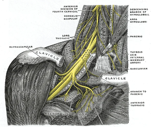
Figure 5Gray808
Long description
The right brachial plexus with its short branches, viewed from in front. The Sternomastoid and Trapezius muscles have been completely, the Omohyoid and Subclavius have been partially, removed; a piece has been sawed out of the clavicle; the Pectoralis muscles have been incised and reflected. (Spalte

Figure 5: The right brachial plexus with its short branches, viewed from in front.
We begin our exploration with the skin and superficial fascia, which contain the pectoral branches of the thoracoacromial artery and the lateral pectoral nerve. Deep to the fascia lies the pectoralis major muscle, a broad, thick muscle that forms the bulk of the anterior chest wall.
| Muscle | Origin | Insertion | Action | Nerve Supply |
|---|---|---|---|---|
| Pectoralis Major | Clavicular head: medial half of clavicle; Sternal head: sternum and upper costal cartilages; Abdominal head: aponeurosis of external oblique | Lateral lip of intertubercular groove of humerus | Adducts and medially rotates humerus; Protracts scapula | Lateral and medial pectoral nerves |
| Pectoralis Minor | Ribs 3–5 | Coracoid process of scapula | Depresses and protracts scapula; Stabilises scapula | Medial pectoral nerve |
The pectoralis minor lies deep to the pectoralis major. It originates from the third to fifth ribs and inserts onto the coracoid process of the scapula.
Deep to the pectoralis minor is the axillary space, which contains the axillary artery and vein, along with the three cords of the brachial plexus. The thoracoacromial artery has four branches: pectoral, acromial, clavicular and deltoid. The pectoral branch supplies the pectoralis major and minor muscles.
The lateral thoracic artery runs along the lateral border of the pectoralis minor and supplies the serratus anterior and the breast tissue. The thoracodorsal artery supplies the latissimus dorsi muscle.
This nerve is often at risk during axillary lymph node dissection.
This membrane helps to support the axillary vessels and nerves as they pass from the neck to the upper limb.
Understanding the pectoral region requires correlating surface landmarks with deep structures visible on imaging. The pectoralis major muscle forms the anterior boundary of the axilla and covers the upper ribs. On plain X-ray, the muscle mass appears as a soft tissue density overlying the upper thoracic cage. The clavicle is clearly visualised as a horizontal bone superior to the lung apex. The first rib is often obscured by the clavicle but can be traced laterally.
DefinitionPectoralis Major
A large, fan-shaped muscle forming the anterior wall of the axilla, originating from the clavicle, sternum, and cartilages of the first six ribs.
Computed Tomography (CT) provides cross-sectional detail essential for assessing trauma and pathology. In axial CT images, the pectoralis major lies anterior to the serratus anterior and the ribs. The pectoralis minor is situated deep to the major, attaching to the coracoid process. The axillary vessels are seen as circular structures within the axillary fat. The brachial plexus appears as a cluster of small round structures lateral to the axillary artery.
| Muscle | Origin | Insertion | Action | Nerve Supply |
|---|---|---|---|---|
| Pectoralis Major | Clavicle, sternum, ribs | Lateral lip of bicipital groove | Adducts and medially rotates humerus | Lateral and medial pectoral nerves |
| Pectoralis Minor | Ribs 3-5 | Coracoid process | Depresses shoulder | Medial pectoral nerve |
| Serratus Anterior | Ribs 1-8 | Medial border of scapula | Protracts scapula | Long thoracic nerve |
Magnetic Resonance Imaging (MRI) offers superior soft tissue contrast. It clearly delineates the muscle bellies and tendons. MRI is particularly useful for identifying muscle tears or tendon ruptures. The neurovascular bundle is well visualised, allowing assessment of nerve compression or vascular anomalies.
Ultrasound is dynamic and excellent for guiding interventions. It can visualise the pectoral muscles in real-time. The needle path for nerve blocks can be monitored. The axillary artery and vein are compressible and pulsatile. Lymph nodes in the axilla can be assessed for size and morphology.
The axillary artery and vein are located lateral to the chest wall. The ribs and intercostal muscles form the posterior boundary.
The pectoral region is defined by the pectoralis major and minor muscles, which form the anterior chest wall. It inserts into the lateral lip of the bicipital groove of the humerus. Its actions include flexion, adduction, and medial rotation of the arm. It is supplied by the medial and lateral pectoral nerves. The pectoralis minor originates from the third to fifth ribs. It inserts into the coracoid process of the scapula. Its action is to depress and protract the scapula. It is supplied by the medial pectoral nerve. [3]
| Muscle | Origin | Insertion | Action | Nerve Supply |
|---|---|---|---|---|
| Pectoralis Major | Clavicle, sternum, costal cartilages | Lateral lip of bicipital groove | Flexion, adduction, medial rotation | Medial and lateral pectoral nerves |
| Pectoralis Minor | Ribs 3-5 | Coracoid process | Depress and protract scapula | Medial pectoral nerve |
The pectoralis major is reflected laterally to reveal the underlying pectoralis minor. The axillary tail of Spence extends laterally into the axilla. This anatomical extension is clinically significant because breast tissue within this tail can be involved in malignancy. Understanding this spread is critical for staging breast cancer.
Injuries to the pectoral region often involve the pectoral nerves. A fracture of the surgical neck of the humerus may damage the axillary nerve, but the pectoral nerves are more vulnerable during axillary lymph node dissection. Nerve palsies result in weakness of adduction and medial rotation.
Applied anatomy of mastectomy involves the removal of breast tissue and axillary lymph nodes. The breast lies superficial to the pectoralis major fascia. Cancer spreads via lymphatics to the axillary nodes. The internal mammary nodes are also a site of spread. Clinicians must identify the intercostobrachial nerve during surgery to prevent sensory loss in the medial arm. This nerve arises from the second intercostal nerve. Preservation of this nerve improves postoperative quality of life.
The pectoral region is defined by the pectoralis major and minor muscles, which form the anterior wall of the axilla. Its fibres converge to insert on the lateral lip of the intertubercular groove of the humerus. This muscle acts to adduct and medially rotate the arm. It is innervated by the medial and lateral pectoral nerves. A common trap is confusing its action with that of the latissimus dorsi, which also medially rotates but extends the arm.
Its primary action is to depress and protract the scapula. It is innervated by the medial pectoral nerve. Injury to this muscle can alter scapular positioning, affecting shoulder mechanics.
The apex of the axilla is the cervico-axillary canal, which transmits neurovascular structures to the upper limb.
The brachial plexus emerges from the neck and passes through the axilla. It is formed by the ventral rami of C5 to T1. The cords of the brachial plexus are named relative to the axillary artery. The lateral cord gives rise to the musculocutaneous nerve and contributes to the median nerve. The medial cord gives rise to the ulnar nerve and contributes to the median nerve. The posterior cord gives rise to the radial and axillary nerves.
The axillary artery is the direct continuation of the subclavian artery. It begins at the lateral border of the first rib. It ends at the inferior border of the teres major muscle, where it becomes the brachial artery. The artery is divided into three parts by the pectoralis minor muscle. The first part gives off the superior thoracic artery. The second part gives off the thoracoacromial artery and the lateral thoracic artery. The third part gives off the subscapular artery, the anterior circumflex humeral artery, and the posterior circumflex humeral artery.
The subscapular artery is the largest branch of the axillary artery. It divides into the circumflex scapular artery and the thoracodorsal artery. The thoracodorsal artery supplies the latissimus dorsi muscle. This vessel is crucial for flap surgery in breast reconstruction.
It accompanies the axillary artery. It becomes the subclavian vein at the lateral border of the first rib. Thrombosis of this vein can cause upper limb swelling.
It is often damaged during axillary lymph node dissection, leading to numbness.
Understanding their course is vital for surgical approaches to the axilla.
Students frequently confuse the functional roles of ligaments within the pectoral and upper limb regions, particularly regarding stability versus movement. A primary error is assuming that all intracapsular ligaments provide significant mechanical support to the joint. For instance, the ligament of the head of the femur is often misinterpreted as a stabiliser. In reality, this intracapsular ligament is normally slack and does not provide any significant joint support, but it does provide a pathway for an important artery that supplies the head of the femur. This distinction is critical when assessing avascular necrosis risks, as the vascular supply is more vulnerable than the ligamentous integrity. [3]
Another frequent mistake involves the classification of joint types and their associated motions. Students often assume that the hip joint allows for a wide range of motion similar to the shoulder. However, the hip joint is a multiaxial ball-and-socket joint between the head of the femur and the acetabulum of the hip bone. The hip carries the weight of the body and thus requires strength and stability during standing and walking. For these reasons, its range of motion is more limited than at the shoulder joint. Confusing these limits can lead to errors in understanding gait mechanics and potential impingement syndromes. [3]
Misidentifying the specific ligaments supporting the lateral and medial aspects of joints is also common. At the elbow, the lateral side is supported by the radial collateral ligament. This arises from the lateral epicondyle of the humerus and then blends into the lateral side of the annular ligament. The annular ligament encircles the head of the radius. This ligament supports the head of the radius as it articulates with the radial notch of the ulna at the proximal radioulnar joint. Failing to distinguish the radial collateral ligament from the ulnar collateral ligament, which supports the medial elbow, leads to incorrect localisation of injury sites. [3]
Students also struggle with the complex movements of the temporomandibular joint (TMJ). It is a mistake to view TMJ movement as purely rotational. Movement at the TMJ during opening and closing of the mouth involves both gliding and hinge motions of the mandible. With the mouth closed, the mandibular condyle and articular disc are located within the mandibular fossa of the temporal bone. During opening of the mouth, the mandible hinges downward and at the same time is pulled anteriorly, causing both the condyle and the articular disc to glide forward from the mandibular fossa onto the downward projecting articular tubercle. Ignoring the gliding component results in an incomplete understanding of jaw pathology. [3]
Finally, there is confusion regarding the vascular and thermoregulatory roles of superficial versus deep veins. Students often overlook that superficial veins do not normally have direct arterial counterparts, but in addition to returning blood, they also make contributions to the maintenance of body temperature. When the ambient temperature is warm, more blood is diverted to the superficial veins where heat can be more easily dissipated to the environment. In colder weather, there is more constriction of the superficial veins and blood is diverted deeper where the body can retain more of the heat. Treating superficial veins solely as return pathways ignores their physiological importance in temperature regulation. [1]
Exam trapLigament Function
Do not assume all ligaments stabilise joints mechanically; some, like the ligament of the head of the femur, are slack and primarily vascular conduits.
Knowledge of the pectoral and associated regional anatomy is fundamental at the bedside for physical examination and clinical diagnosis. When assessing the shoulder or hip, understanding the limited range of motion of the hip joint is essential. The hip joint is a ball-and-socket joint whose motions are more restricted than at the shoulder to provide greater stability during weight bearing. This stability is provided by the iliofemoral, pubofemoral, and ischiofemoral ligaments which strongly support the hip joint in the upright, standing position. Clinicians use this knowledge to differentiate between ligamentous laxity and muscular weakness during gait analysis. [3]
In the operating theatre, precise identification of ligamentous structures is vital for surgical interventions involving the elbow and knee. The elbow is a uniaxial hinge joint that allows for flexion/extension of the forearm. It includes the humeroulnar joint and the humeroradial joint. The medial elbow is supported by the ulnar collateral ligament and the radial collateral ligament supports the lateral side. These ligaments prevent side-to-side movements and resist hyperextension of the elbow. Surgeons must preserve the annular ligament, which surrounds the head of the radius to hold it in place at the proximal radioulnar joint, to maintain rotational stability. [3]
On imaging, recognising the complex articulations of the knee is crucial for interpreting MRI and CT scans. The knee joint is the largest joint of the body. It actually consists of three articulations. The femoropatellar joint is found between the patella and the distal femur. The medial tibiofemoral joint and lateral tibiofemoral joint are located between the medial and lateral condyles of the femur and the medial and lateral condyles of the tibia. Radiologists look for disruptions in these specific articulations to diagnose fractures or meniscal tears. The knee functions primarily as a hinge joint, allowing flexion and extension of the leg. This action is generated by both rolling and gliding motions of the femur on the tibia. [3]
In examinations, students must accurately describe the ankle joint's structure and function. The ankle is formed by the talocrural joint. It consists of the articulations between the talus bone of the foot and the distal ends of the tibia and fibula of the leg. The superior aspect of the talus bone is square-shaped and has three areas of articulation. The top of the talus articulates with the inferior tibia. This is the portion of the ankle joint that carries the body weight between the leg and foot. The sides of the talus are firmly held in position by the articulations with the medial malleolus of the tibia and the lateral malleolus of the fibula, which prevent any side-to-side motion of the talus. [3]
Understanding the TMJ is also tested in oral and maxillofacial contexts. The temporomandibular joint is supported by an extrinsic ligament that anchors the mandible to the skull. This ligament spans the distance between the base of the skull and the lingula on the medial side of the mandibular ramus. Clinicians assess this ligament when evaluating temporomandibular disorders. The net result of TMJ movement is a forward and downward motion of the condyle and mandibular depression. [3]
Ankle Stability Assessment
- Assess Ankle InjuryPatient presents with pain
- Question: Check Medial Stability
Yes
- Test Deltoid LigamentResist eversion
No
- Check Lateral StabilityTest anterior talofibular ligament
The ligament of the head of the femur is slack and provides a vascular pathway, not mechanical support. The hip joint has a more limited range of motion than the shoulder to ensure stability during weight bearing. The elbow is a uniaxial hinge joint supported by ulnar and radial collateral ligaments. The knee consists of three articulations within a single capsule and requires a "screw home" mechanism for full extension. The ankle is a uniaxial hinge joint allowing only dorsiflexion and plantar flexion. Superficial veins contribute to body temperature regulation by diverting blood flow based on ambient temperature. The TMJ involves both hinge and gliding motions of the mandible. The annular ligament holds the head of the radius in place at the proximal radioulnar joint.
Answer each one in your head first, then open the answer.
What is the primary function of the ligament of the head of the femur?
Show answerHide answer
It provides a pathway for an important artery that supplies the head of the femur.Why is the hip joint's range of motion more limited than the shoulder?
Show answerHide answer
It carries the weight of the body and requires strength and stability during standing and walking.Which ligament supports the lateral side of the elbow?
Show answerHide answer
The radial collateral ligament.What motion does the talocrural joint allow?
Show answerHide answer
Dorsiflexion and plantar flexion of the foot.How do superficial veins help maintain body temperature?
Show answerHide answer
When warm, more blood is diverted to them for heat dissipation; when cold, they constrict to retain heat.
- Betts JG, Young KA, Wise JA, et al. Circulatory Pathways. OpenStax, Anatomy and Physiology 2e (CC BY-NC-SA 4.0). Accessed .
- Betts JG, Young KA, Wise JA, et al. Muscles of the Pectoral Girdle and Upper Limbs. OpenStax, Anatomy and Physiology 2e (CC BY-NC-SA 4.0). Accessed .
- Betts JG, Young KA, Wise JA, et al. Anatomy of Selected Synovial Joints. OpenStax, Anatomy and Physiology 2e (CC BY-NC-SA 4.0). Accessed .
Figure credits
- Figure 1: Henry Vandyke Carter, via Wikimedia Commons, Public domain, Public domain.
- Figure 2: Henry Vandyke Carter, via Wikimedia Commons, Public domain, Public domain.
- Figure 3: Henry Vandyke Carter, via Wikimedia Commons, Public domain, Public domain.
- Figure 4: Henry Vandyke Carter, via Wikimedia Commons, Public domain, Public domain.
- Figure 5: Henry Vandyke Carter, via Wikimedia Commons, Public domain, Public domain.
Educational content for medical students and doctors. It is not a substitute for clinical judgement or local guidelines.