Osteology of the upper limb
The upper limb skeleton comprises thirty bones across arm forearm and hand regions
The upper limb is divided into three regions: the arm between shoulder and elbow joints, the forearm between elbow and wrist joints, and the hand distal to the wrist[1]. Each upper limb contains a total of 30 bones[1]. The arm region contains the single humerus bone[1]. The forearm contains two bones: the ulna medially and the radius on the lateral (thumb) side[1]. The hand base contains eight carpal bones, the palm has five metacarpal bones, and the fingers and thumb contain 14 phalanx bones[1].
| Region | Bones | Count |
|---|---|---|
| Arm | Humerus | 1 |
| Forearm | Ulna, radius | 2 |
| Hand (carpals) | Scaphoid, lunate, triquetrum, pisiform, trapezium, trapezoid, capitate, hamate | 8 |
| The upper limb skeleton comprises the clavicle, scapula, humerus, radius, ulna, eight carpal bones, five metacarpals, and fourteen phalanges. Figure 2 shows the bones and bony landmarks of the upper limb in anterior view. The clavicle lies horizontally across the superior thorax, its medial end articulating with the manubrium at the sternoclavicular joint and its lateral end with the acromion at the acromioclavicular joint. The scapula rests on the posterior thoracic wall; its spine, acromion, a [1] | ||
| Hand (phalanges) | Proximal, middle, distal phalanges (thumb has two) | 14 |
| Total per limb | 30 |
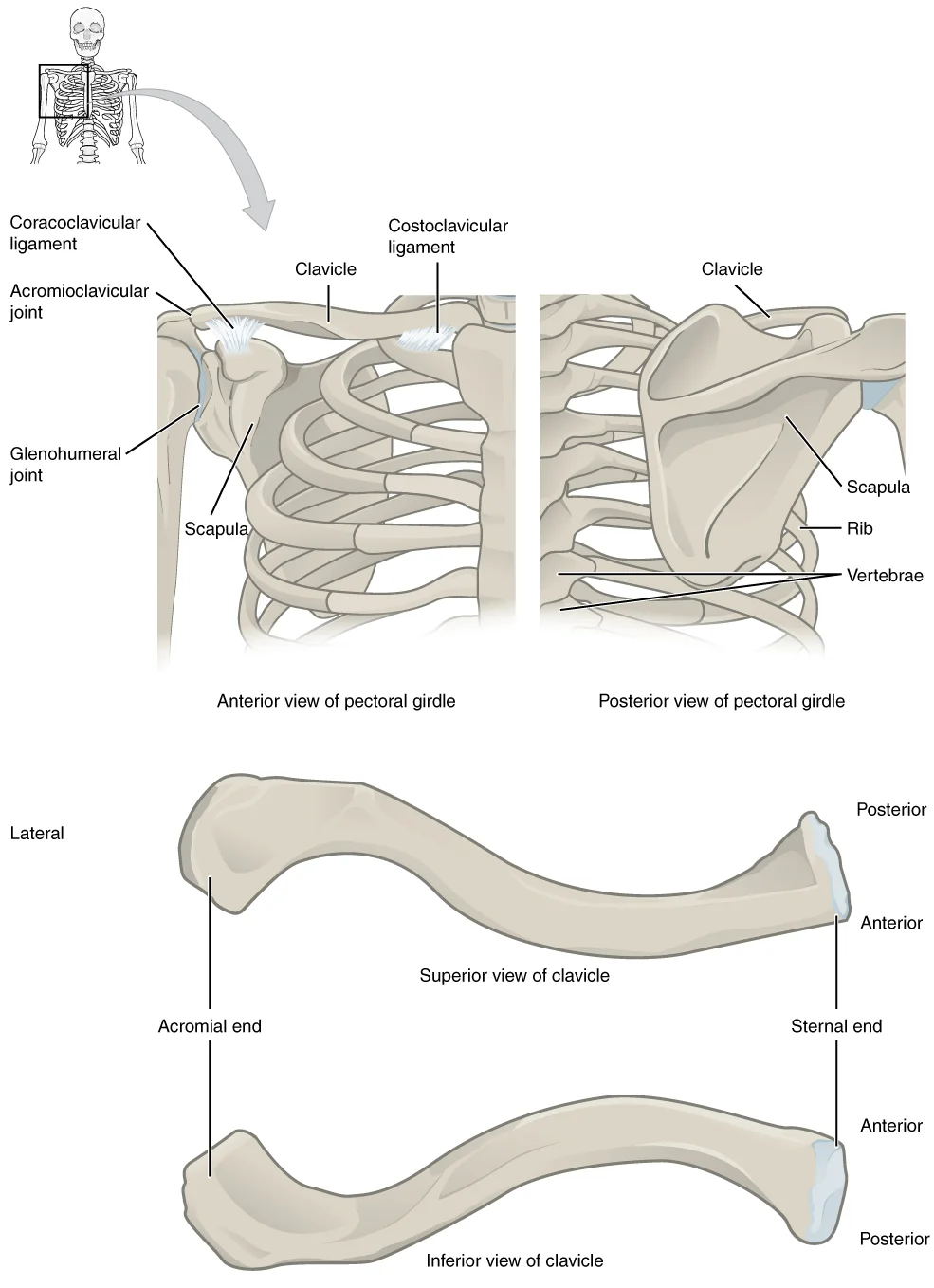
Figure 1802 Pectoral Girdle
Long description
The pectoral girdle consists of the clavicle and the scapula, which serve to attach the upper limb to the sternum of the axial skeleton.

Figure 1: The pectoral girdle consists of the clavicle and the scapula, which serve to attach the upper limb to the sternum of the axial skeleton.
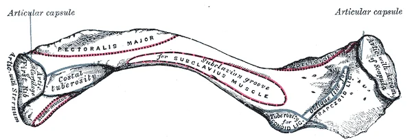
Figure 2Gray201
Long description
Left clavicle. Inferior surface. Labelled illustration from Wikimedia Commons, used under its open licence.

Figure 2: Left clavicle (inferior surface).
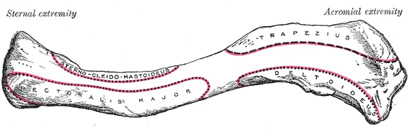
Figure 3Gray200
Long description
Left clavicle. Superior surface. Labelled illustration from Wikimedia Commons, used under its open licence.

Figure 3: Left clavicle (superior surface).
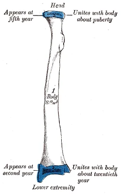
Figure 4Gray217
Long description
Plan of ossification of the radius. From three centers.

Figure 4: Plan of ossification of the radius (from three centers).
Figure 5Gray215
Long description
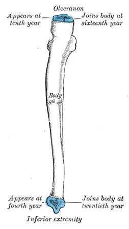
Plan of ossification of the ulna. From three centers.

Figure 5: Plan of ossification of the ulna (from three centers).
Figure 6Gray214
Long description
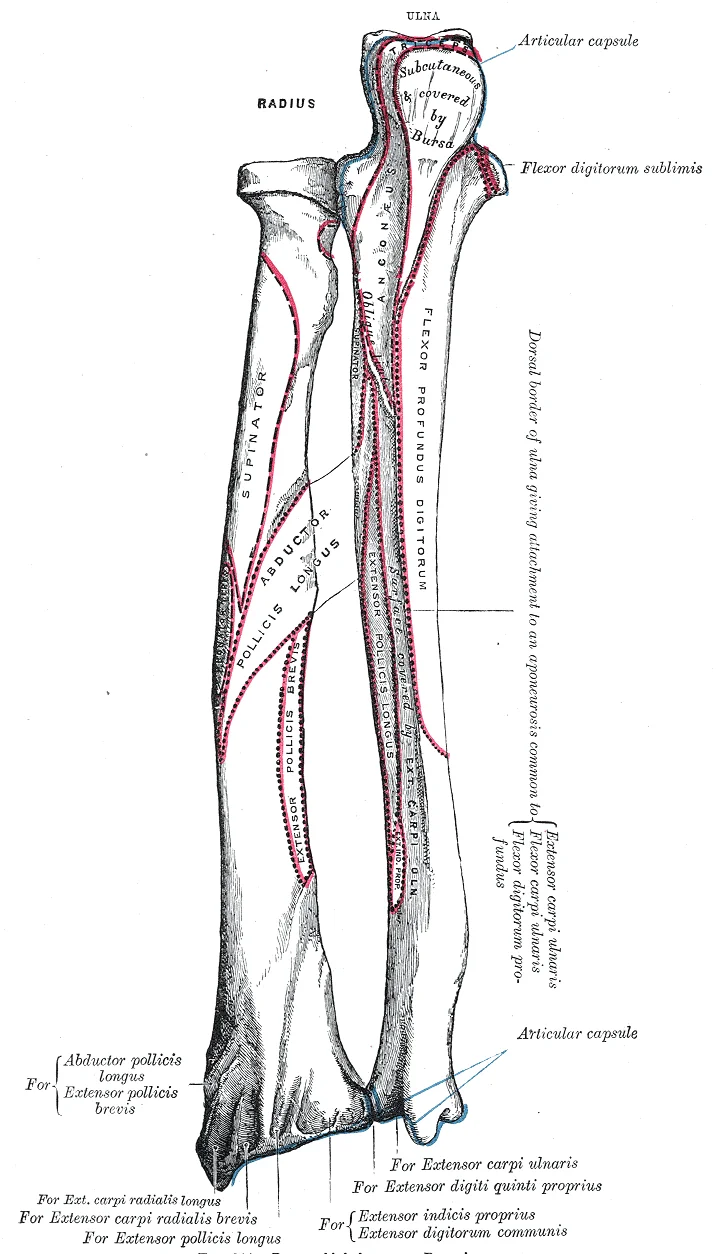
Bones of left forearm. Posterior aspect.

Figure 6: Bones of left forearm (posterior aspect).
Figure 7Gray213
Long description
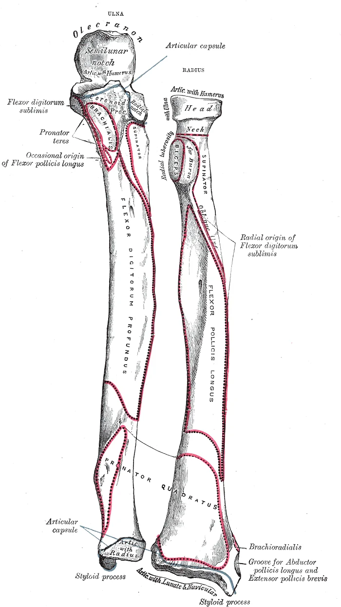
Bones of left forearm. Anterior aspect. Labelled illustration from Wikimedia Commons, used under its open licence.

Figure 7: Bones of left forearm (anterior aspect).
Figure 8Gray212
Long description
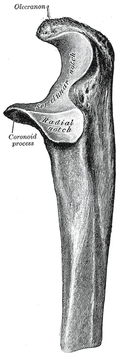
Upper extremity of left ulna. Lateral aspect.

Figure 8: Upper extremity of left ulna (lateral aspect).
Figure 9Gray210
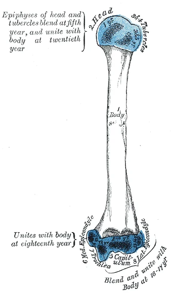
Long description
Plan of ossification of the humerus. Labelled illustration from Wikimedia Commons, used under its open licence.

Figure 9: Plan of ossification of the humerus.
Figure 10Gray209
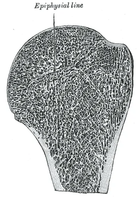
Long description
Longitudinal section of head of left humerus.

Figure 10: Longitudinal section of head of left humerus.
Figure 11Gray208
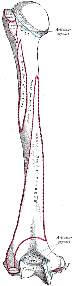
Long description
Left humerus. Posterior view. Labelled illustration from Wikimedia Commons, used under its open licence.

Figure 11: Left humerus (posterior view).
Figure 12Gray206
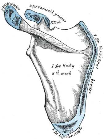
Long description
Plan of ossification of the scapula. From seven centers.

Figure 12: Plan of ossification of the scapula (from seven centers).
Figure 13Gray203
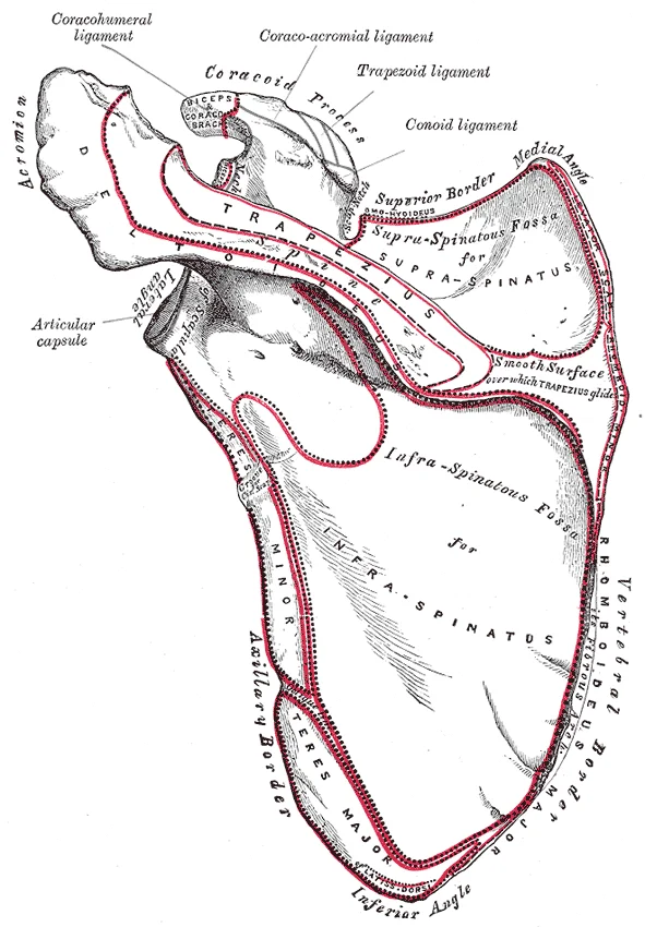
Long description
Left scapula. Dorsal surface. Labelled illustration from Wikimedia Commons, used under its open licence.

Figure 13: Left scapula (dorsal surface).
Figure 14Gray202
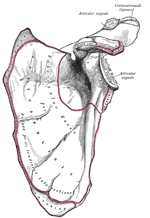
Long description
Left scapula. Costal surface. Labelled illustration from Wikimedia Commons, used under its open licence.

Figure 14: Left scapula (costal surface).
The forearm skeleton consists of the ulna and radius. The elbow joint involves the humeral trochlea articulating with the ulnar trochlear notch, and the capitulum with the radial head[1]. The proximal radioulnar joint is between the radial head and ulnar radial notch[1]. The proximal ulna features the olecranon process posteriorly, and the coronoid process with ulnar tuberosity anteriorly[1]. The proximal radius has a neck below the head and the radial tuberosity distally[1].
The scapula anchors the upper limb to the trunk as part of the pectoral girdle. It lies on the posterior shoulder, surrounded by muscles on both anterior (deep) and posterior (superficial) surfaces, and does not articulate with the thoracic ribs. This arrangement creates the scapulothoracic interface where the scapula glides over the thoracic wall. Scapular movements include elevation (upward movement) and depression (downward movement), protraction and retraction, and upward and downward rotation du [2][4]
Figure 15Gray204
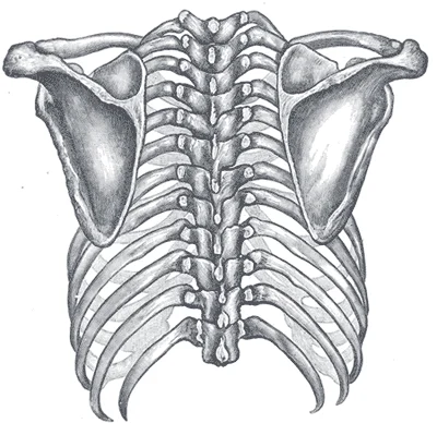
Long description
Posterior view of the thorax and shoulder girdle. (Morris.)

Figure 15: Posterior view of the thorax and shoulder girdle (Morris.).
DefinitionSurface anatomy
Surface anatomy identifies the bony landmarks that can be felt through the skin, providing a map for clinical examination and procedures.
The clavicle is the only long bone that lies in a horizontal position in the body. Its entire length is subcutaneous and easily palpated. The medial (sternal) end articulates with the manubrium at the sternoclavicular joint, the only bony articulation between the pectoral girdle and the axial skeleton. The lateral (acromial) end meets the acromion of the scapula at the acromioclavicular joint. The costoclavicular ligament anchors the sternal end to the first rib, and the coracoclavicular ligament links the clavicle to the coracoid process. [2]
The scapula lies on the posterior thoracic wall. Its spine runs transversely and ends laterally as the acromion, forming the bony tip of the shoulder. The coracoid process projects anteriorly from the superior border. The suprascapular notch sits lateral to the midpoint of the superior border. The superior angle lies between the medial and superior borders; the inferior angle, the most inferior portion of the scapula, sits between the medial and lateral borders. Superior rotation of the scapula [1][2]
The humerus consists of an upper extremity, a cylindrical and prismatic body, and a lower extremity. The upper extremity features a nearly hemispherical head directed upward, medialward, and backward to articulate with the glenoid cavity of the scapula, set off by the anatomical neck. Below the head lie the greater and lesser tubercles, separated by the bicipital groove. The surgical neck lies immediately distal to the tubercles and represents a frequent site of fracture. The body of the bone presents three borders—anterior, medial, and lateral—and three surfaces. The anterior border originates from the front of the greater tubercle and descends to the coronoid fossa, while the anterior border of the deltoid tuberosity receives the insertion of the deltoid muscle mid-shaft. Surface landmarks allow clinical identification: the greater tubercle forms the lateral prominence of the shoulder, the deltoid tuberosity is palpable along the mid-shaft, and the medial and lateral epicondyles are readily identified at the elbow joint. [1]
The radius and ulna each have an interosseous border for the interosseous membrane. The distal radius bears a styloid process laterally and an ulnar notch medially that articulates with the ulnar head to form the distal radioulnar joint. The distal radioulnar joint is found between the head of the ulna and the ulnar notch of the radius. The distal end of the radius articulates with the proximal carpal bones, but the ulna does not. [1]
The carpal bones form the wrist; the metacarpals constitute the palm; the phalanges make up the digits. Their dorsal surfaces are readily palpable.
Palpation sequence
- Start at sternal notchMove laterally along clavicle to acromioclavicular joint
- Follow acromion posteriorlyTrace scapular spine to medial border
- Palpate humeral headFeel greater tubercle under deltoid
- Identify epicondylesMedial and lateral at elbow
- Trace radius and ulnaStyloid processes at wrist
- Outline carpals, metacarpals, phalangesDorsal aspect of hand
The upper limb skeleton comprises the clavicle, scapula, humerus, radius, ulna, eight carpal bones, five metacarpals, and fourteen phalanges. The clavicle lies horizontally across the superior thorax, its medial end articulating with the manubrium at the sternoclavicular joint and its lateral end with the acromion at the acromioclavicular joint. The scapula rests on the posterior thoracic wall; its spine, acromion, a
DefinitionRadiocarpal joint
Formed by the distal radius, the scaphoid, lunate, and triquetrum; the ulna does not articulate directly with carpal bones. [1]
DefinitionMidcarpal joint
Articulation between the proximal and distal carpal rows; together with the radiocarpal joint, permits all wrist movements. [1]
| Row | Lateral to medial | Key feature |
|---|---|---|
| Proximal | Scaphoid, Lunate, Triquetrum, Pisiform | Pisiform articulates with anterior triquetrum [1] |
| The upper limb skeleton comprises the clavicle, scapula, humerus, radius, ulna, eight carpal bones, five metacarpals, and fourteen phalanges. Figure 2 shows the bones and bony landmarks of the upper limb in anterior view. The clavicle lies horizontally across the superior thorax, its medial end articulating with the manubrium at the sternoclavicular joint and its lateral end with the acromion at the acromioclavicular joint. The scapula rests on the posterior thoracic wall; its spine, acromion, a |
Clinical pearlCarpal tunnel boundaries
Floor and walls: carpal bones; roof: flexor retinaculum attached to trapezium, scaphoid, hamate, and pisiform. [1]
Wrist joint complex
- Distal radiusArticulates with scaphoid and lunate
- TriquetrumArticulates with fibrocartilaginous pad (triangular fibrocartilage complex)
- Question: Proximal row unity?
Yes
- Scaphoid, lunate, triquetrum move as unit via interosseous ligaments
No
- Pisiform excluded from radiocarpal joint
- Midcarpal jointProximal row articulates with distal row
- Distal rowArticulates with metacarpals 1–5 at carpometacarpal joints
The sternoclavicular joint is the only bony attachment of the pectoral girdle to the axial skeleton. It is a single, highly mobile synovial joint that anchors the medial end of the clavicle to the manubrium sterni and the first costal cartilage. This arrangement allows the entire pectoral girdle to operate independently on each side, providing the extensive mobility that underlies shoulder and upper limb movement. [2]
The acromioclavicular (AC) joint is a plane synovial joint between the acromial end of the clavicle and the acromion of the scapula. It transmits forces from the upper limb to the clavicle. The AC ligaments are relatively weak; a fall onto the elbow or outstretched hand can stretch or tear them, producing a moderate joint injury. The primary support comes from the coracoclavicular ligament, a very strong band that anchors the coracoid process to the inferior surface of the acromial end of the cl [2]
DefinitionCoracoclavicular ligament
A strong connective tissue band running from the coracoid process of the scapula to the inferior surface of the lateral clavicle; it is the main stabiliser of the acromioclavicular joint.
Figure 16Gray326
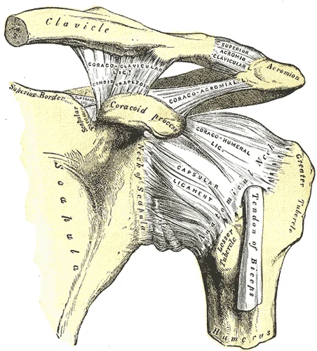
Long description
The left shoulder and acromioclavicular joints, and the proper ligaments of the scapula.

Figure 16: The left shoulder and acromioclavicular joints, and the proper ligaments of the scapula.
The shoulder joint is a multiaxial ball‑and‑socket synovial joint formed by the head of the humerus and the glenoid cavity of the scapula. It has little ligamentous support, which gives it the greatest range of motion of all synovial joints. Permitted movements include flexion‑extension, abduction‑adduction, circumduction, and medial (internal) and lateral (external) rotation. Rotation occurs as the humerus turns around its long axis: medial rotation brings the anterior surface of the arm toward the midline, while lateral rotation moves it away. [3] [4]
| Movement | Description |
|---|---|
| Flexion / Extension | Forward and backward movement in the sagittal plane |
| Abduction / Adduction | Movement away from and toward the midline in the coronal plane |
| Circumduction | Sequential combination of flexion, adduction, extension, abduction |
| Medial (internal) rotation | Anterior surface of humerus rotates toward midline |
| Lateral (external) rotation | Anterior surface of humerus rotates away from midline |
Repetitive overhead use can cause inflammation of bursae or tendons, glenoid labrum tears, or rotator cuff degeneration. Chronic inflammation may lead to adhesive capsulitis (“frozen shoulder”) due to scar tissue formation between the capsule and surrounding structures. [3]
Mechanism of inferior shoulder dislocation
- Upper limb fully abductedHumeral head levered against inferior capsule
- Question: Force applied to humerus?
Yes
- Inferior dislocation of humeral head
No
- Joint remains reduced
The joints of the upper limb are specialised for large ranges of motion, giving the limb great mobility for actions such as throwing or typing. Synovial joints share common features but each is structured for specific movements; the structural classification determines the movements allowed. Ball‑and‑socket joints provide the greatest range, while plane joints (e.g., acromioclavicular) allow only limited gliding motions that summate across adjacent joints. [3]
The upper limb skeleton comprises the clavicle, scapula, humerus, radius, ulna, eight carpal bones, five metacarpals, and fourteen phalanges. The clavicle lies horizontally across the superior thorax, its medial end articulating with the manubrium at the sternoclavicular joint and its lateral end with the acromion at the acromioclavicular joint. The scapula rests on the posterior thoracic wall; its spine, acromion, a
DefinitionFascial compartment
A fascial compartment is a closed space bounded by deep fascia and intermuscular septa that contains muscles sharing a common nerve supply and action.
| Compartment | Bony relations | Key contents | Nerve supply |
|---|---|---|---|
| Anterior arm | Humerus, medial intermuscular septum | Biceps brachii, brachialis, coracobrachialis | Musculocutaneous nerve |
| Posterior arm | Humerus, lateral intermuscular septum | Triceps brachii | Radial nerve |
| Anterior forearm | Radius, ulna, interosseous membrane | Flexor-pronator group | Median and ulnar nerves |
| Posterior forearm | Radius, ulna, interosseous membrane | Extensor-supinator group | Radial nerve (posterior interosseous) |
The shoulder joint capsule is reinforced by the coracohumeral ligament running from the coracoid process to the anterior humerus and by three glenohumeral ligaments on the anterior capsule. The glenoid cavity is deepened by the glenoid labrum, a fibrocartilaginous rim that increases the contact area for the humeral head, which is about one third larger than the cavity. The rotator cuff muscles (supraspinatus, infraspinatus, teres minor, subscapularis) blend with the capsule and act as dynamic ligaments, holding the humeral head in the glenoid cavity during movement. Movements of the forearm are described from the anatomical position: upright, upper limbs at sides, palms forward. In supination the radius and ulna are parallel; in pronation they cross forming an X-shape. Scapular upward rotation elevates the glenoid cavity during abduction, while downward rotation accompanies adduction. [3] [4]
Layers of fascia from superficial to deep
- SkinSuperficial fascia (subcutaneous tissue)
- Superficial fasciaContains fat, cutaneous vessels, nerves
- Deep fasciaBrachial fascia, antebrachial fascia, retinaculae
- Intermuscular septaMedial and lateral septa attach to humerus
- CompartmentsAnterior and posterior arm; anterior and posterior forearm
The rotator cuff muscles encircle the humeral head, while forearm muscles arise from the medial and lateral epicondyles. [1]
DefinitionRotator cuff
The rotator cuff is formed by the fused tendons of supraspinatus, infraspinatus, teres minor, and subscapularis, which stabilize the glenohumeral joint[1].
| Compartment | Muscle | Origin | Insertion | Action | Nerve Supply |
|---|---|---|---|---|---|
| Shoulder (rotator cuff) | Supraspinatus | Supraspinous fossa | Greater tubercle of humerus | Abduction (initiation) | Suprascapular nerve |
| Shoulder (rotator cuff) | Infraspinatus | Infraspinous fossa | Greater tubercle of humerus | Lateral rotation | Suprascapular nerve |
| Shoulder (rotator cuff) | Teres minor | Lateral border of scapula | Greater tubercle of humerus | Lateral rotation | Axillary nerve |
| Shoulder (rotator cuff) | Subscapularis | Subscapular fossa | Lesser tubercle of humerus | Medial rotation | Upper and lower subscapular nerves |
| Arm (anterior) | Biceps brachii | Supraglenoid tubercle (long head), coracoid process (short head) | Radial tuberosity, bicipital aponeurosis | Flex elbow, supinate forearm | Musculocutaneous nerve |
| Arm (anterior) | Brachialis | Anterior humerus | Ulnar tuberosity | Flex elbow | Musculocutaneous nerve |
| Arm (posterior) | Triceps brachii | Infraglenoid tubercle (long head), posterior humerus (lateral/medial heads) | Olecranon of ulna | Extend elbow | Radial nerve |
| Forearm (anterior) | Flexor carpi radialis | Medial epicondyle of humerus | Base of 2nd metacarpal | Flex and abduct wrist | Median nerve |
| Forearm (posterior) | Extensor carpi radialis longus | Lateral supracondylar ridge | Base of 2nd metacarpal | Extend and abduct wrist | Radial nerve |
| Hand (thenar) | Opponens pollicis | Flexor retinaculum, trapezium | 1st metacarpal | Oppose thumb | Median nerve |
Muscle compartments of the upper limb
- Shoulder girdleRotator cuff muscles from scapula to humerus
- Question: Arm compartment?
Anterior
- Biceps, brachialis, coracobrachialis – flex elbow, supinate
Posterior
- Triceps – extend elbow
- ForearmAnterior flexors from medial epicondyle; posterior extensors from lateral epicondyle
- Hand intrinsicsThenar, hypothenar, interossei, lumbricals
The deltoid tuberosity on the lateral humerus marks the deltoid insertion. The medial epicondyle gives rise to the powerful anterior forearm flexors, while the lateral epicondyle gives rise to the weaker posterior forearm muscles. The supraglenoid and infraglenoid tubercles provide attachments for muscles of the arm[1]. The scapular fossae provide large surface areas for the attachment of muscles that cross the shoulder joint[1]. [2]
| Digit | Number of phalanges | Phalanges |
|---|---|---|
| 1 (thumb) | 2 | Proximal, distal |
| 2–5 (index to little) | 3 each | Proximal, middle, distal |
| Ligament | Origin | Insertion | Function |
|---|---|---|---|
| Ulnar collateral ligament (anterior portion strongest) | Medial epicondyle of humerus | Medial side of proximal ulna | Resists hyperextension and valgus stress |
The ulnar artery, the larger terminal branch of the brachial artery, begins just distal to the elbow crease and courses obliquely toward the ulnar side of the forearm, reaching it midway between elbow and wrist. It then descends along the ulnar border, crosses the flexor retinaculum lateral to the pisiform bone, and divides into the superficial and deep palmar arches. In the proximal forearm the artery lies deep, covered by pronator teres, flexor carpi radialis, palmaris longus, and flexor digitorum superficialis, resting on brachialis and flexor digitorum profundus; the ulnar nerve runs medial to it. Its branches include the anterior and posterior ulnar recurrent arteries anastomosing around the medial epicondyle, the common interosseous artery dividing into anterior and posterior interosseous vessels, muscular branches, palmar and dorsal carpal branches, a deep palmar branch completing the deep palmar arch, and the superficial palmar arch typically completed by a branch from the radial artery. Anatomical variation in origin occurs in roughly one in thirteen individuals, with high origins often placing the artery superficial to the flexor musculature.
The venous drainage of the upper limb comprises superficial and deep systems that accompany the arterial supply. The superficial system lies in the subcutaneous fascia and includes the cephalic and basilic veins. The deep system consists of paired venae comitantes that run alongside the brachial, ulnar, and radial arteries. Both systems ultimately drain into the axillary vein, which becomes the subclavian vein at the lateral border of the first rib.
DefinitionSuperficial venous pattern
The cephalic vein ascends on the lateral aspect of the forearm and arm, pierces the clavipectoral fascia, and joins the axillary vein. The basilic vein runs medially, pierces the deep fascia at mid-arm, and unites with the venae comitantes of the brachial artery to form the axillary vein.
Clinical pearlCubital fossa connections
The median cubital vein links the cephalic and basilic veins in the cubital fossa, providing a common site for venepuncture. It crosses superficial to the bicipital aponeurosis and lies anterior to the brachial artery and median nerve.
| Vein | Course | Termination |
|---|---|---|
| Cephalic | Lateral forearm → lateral arm → deltopectoral groove | Axillary vein |
| Basilic | Medial forearm → medial arm → pierces deep fascia mid-arm | Brachial venae comitantes → axillary vein |
| Median cubital | Oblique across cubital fossa roof | Cephalic to basilic |
| Brachial venae comitantes | Paired, accompany brachial artery | Unite with basilic → axillary vein |
| Radial & ulnar venae comitantes | Paired, accompany respective arteries | Brachial venae comitantes |
Venous drainage pathway
- Capillaries of hand and forearmDrain into dorsal venous network and palmar arches
- Superficial systemCephalic (lateral) and basilic (medial) veins ascend in subcutaneous tissue
- Deep systemVenae comitantes accompany radial, ulnar, and brachial arteries
- ConfluenceBasilic joins brachial venae comitantes → axillary vein; cephalic drains directly into axillary vein
- Central drainageAxillary vein → subclavian vein → brachiocephalic vein → superior vena cava
The basilic vein courses medially, piercing the brachial fascia at the midpoint of the arm to join the paired brachial venae comitantes. The median cubital vein bridges the two superficial veins across the roof of the cubital fossa, superficial to the bicipital aponeurosis. Deep to the muscles, the radial and ulnar venae comitantes accompany their respective arteries and converge into the brachial venae comitantes. The axillary vein lies medial to the axillary artery, becoming the subclavian vein at the lateral border of the first rib. [1]
The upper limb is innervated by the brachial plexus, a complex network of nerves derived from the ventral rami of spinal nerves C5 to T1. This plexus organizes into roots, trunks, divisions, cords, and terminal branches. Understanding the motor and cutaneous supply is critical for localizing lesions. The major terminal branches include the musculocutaneous, axillary, radial, median, and ulnar nerves. Each nerve has a specific trajectory and territory.
The median nerve runs anteriorly, passing between the two heads of the pronator teres in the forearm. The ulnar nerve travels medially, passing posterior to the medial epicondyle of the humerus. This bony prominence is a common site for nerve compression. A mid-shaft humeral fracture places the radial nerve at risk due to its intimate contact with the radial groove. A medial epicondyle fracture may injure the ulnar nerve. A surgical neck fracture can damage the axillary nerve. [1]
The musculocutaneous nerve arises from the lateral cord. It pierces the coracobrachialis muscle and descends between the biceps brachii and brachialis. It provides motor supply to these anterior compartment muscles. It terminates as the lateral antebrachial cutaneous nerve, supplying skin on the lateral forearm.
The axillary nerve winds around the surgical neck of the humerus. It supplies the deltoid and teres minor muscles. It gives off the superior lateral cutaneous nerve of the arm, which innervates the skin over the lower deltoid.
The radial nerve is the largest branch of the brachial plexus. It supplies the triceps brachii, anconeus, brachioradialis, and extensor muscles of the forearm. Cutaneously, it supplies the posterior arm, posterior forearm, and the dorsal aspect of the lateral hand.
The median nerve passes through the cubital fossa and enters the forearm. It supplies most flexors in the anterior forearm. In the hand, it innervates the lateral two lumbricals, flexor pollicis brevis, and opponens pollicis. It provides cutaneous sensation to the palmar aspect of the lateral three and a half digits.
The ulnar nerve descends medially. It supplies the flexor carpi ulnaris and the medial half of the flexor digitorum profundus. In the hand, it innervates the medial two lumbricals, all interossei, adductor pollicis, and hypothenar muscles. It supplies the palmar and dorsal aspects of the medial one and a half digits.
| Nerve | Motor Supply | Cutaneous Supply |
|---|---|---|
| Musculocutaneous | Biceps brachii, Brachialis, Coracobrachialis | Lateral forearm |
| Axillary | Deltoid, Teres minor | Lower deltoid region |
| Radial | Triceps, Brachioradialis, Extensors | Posterior arm/forearm, Dorsal lateral hand |
| Median | Flexors (except FCU/medial FDP), Thenar | Palmar lateral 3.5 digits |
| Ulnar | FCU, Medial FDP, Intrinsic hand muscles | Palmar/Dorsal medial 1.5 digits |
Lesions present with specific deficits. Radial nerve injury causes wrist drop. Ulnar nerve injury leads to claw hand. Median nerve injury results in ape hand deformity. These patterns help clinicians localize the lesion anatomically.
The lymphatic drainage of the upper limb follows a predictable pattern, moving from superficial tissues to deep structures before entering the axillary nodes. This pathway is critical for understanding the spread of infection and malignancy in the region.
The superficial lymphatics accompany the superficial veins. They drain the skin and subcutaneous tissues of the hand, forearm, and arm. They pass medial to the biceps brachii muscle. A smaller number of vessels follow the cephalic vein. These drain into the deltopectoral nodes. These nodes lie between the deltoid and pectoralis major muscles. They are part of the infraclavicular group of axillary nodes.
DefinitionAxillary Nodes
The axillary lymph nodes are divided into five groups: pectoral, subscapular, lateral, central, and apical. They receive lymph from the upper limb, breast, and superficial chest wall.
Deep lymphatics accompany the deep arteries. They drain the muscles, joints, and bones of the upper limb. These vessels generally follow the brachial artery. They also follow the radial and ulnar arteries in the forearm. Some deep vessels from the elbow region drain into the supratrochlear nodes. These nodes lie above the medial epicondyle of the humerus. They receive lymph from the medial side of the forearm and hand.
The vessels terminate in the axillary lymph nodes.
Disruption of lymphatic drainage can lead to lymphoedema. This condition causes swelling of the limb. It is a common complication after axillary lymph node dissection. This surgery is often performed for breast cancer.
| Group | Location | Drainage Area |
|---|---|---|
| Lateral | Along basilic vein | Upper limb |
| Pectoral | Anterior axillary wall | Anterior chest wall, breast |
| Subscapular | Posterior axillary wall | Posterior chest wall, back |
| Central | Axillary fat | All previous groups |
| Apical | Apex of axilla | All previous groups, upper breast |
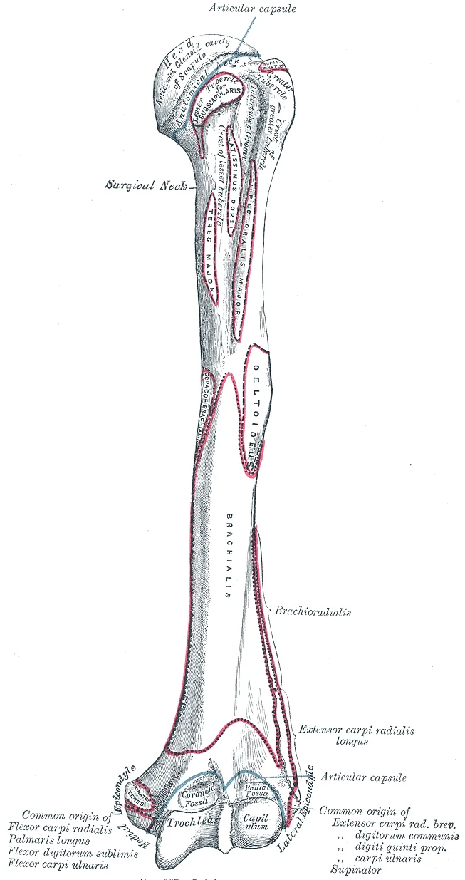
The distal humerus forms the proximal component of the elbow joint and therefore lies directly adjacent to the forearm bones. Two articulation areas on its distal end receive the ulna and radius respectively — the medial trochlea and the lateral capitulum — and together they create the hinge‑type elbow joint. [1]
The trochlea is a spindle‑shaped (pulley‑like) surface that articulates with the trochlear notch of the ulna. Immediately lateral to it, the capitulum is a small, knob‑like prominence that contacts the head of the radius. When the elbow is fully flexed, two shallow depressions sit just above these bony surfaces: the coronoid fossa superior to the trochlea receives the coronoid process of the ulna, and the radial fossa superior to the capitulum receives the radial head. These fossae prevent bony impingement during deep flexion. [1]
Posterior to the distal humerus lies the olecranon fossa, a deep depression that accommodates the olecranon process of the ulna when the elbow is extended. The brachial artery and median nerve pass anterior to the distal humerus in the cubital fossa, a triangular space bounded laterally by the brachioradialis, medially by the pronator teres, and superiorly by an imaginary line joining the epicondyles. [1]
Figure 17Gray207
Long description
Left humerus. Anterior view. Labelled illustration from Wikimedia Commons, used under its open licence.

Figure 17: Left humerus (anterior view).
Labeled structures include the trochlea (medial articulation), capitulum (lateral articulation), coronoid fossa, radial fossa, olecranon fossa, medial and lateral supracondylar ridges, and the cubital fossa with its neurovascular contents. The caption reads: “Anterior‑lateral view of the distal humerus showing articular surfaces, fossae and adjacent muscular and neurovascular landmarks.” [1]
DefinitionTrochlea
A spindle‑shaped medial articulation on the distal humerus that receives the ulna’s trochlear notch.
DefinitionCapitulum
A small lateral knob on the distal humerus that articulates with the radius head.
| Fossa | Position relative to articular surface | Receives |
|---|---|---|
| Coronoid fossa | Superior to trochlea | Coronoid process of ulna |
| Radial fossa | Superior to capitulum | Head of radius |
| Olecranon fossa | Posterior | Olecranon process of ulna |
These spatial relationships are essential for understanding elbow mechanics, recognising fracture patterns, and avoiding iatrogenic injury during procedures such as humeral shaft nailing or elbow arthroplasty.
Understanding the osteology of the upper limb requires correlating surface anatomy with internal structural integrity. Radiological imaging serves as the primary tool for this correlation, allowing clinicians to visualise bone density, alignment, and soft tissue relationships without invasive procedures. The choice of modality depends on the clinical question, whether assessing acute trauma, chronic degenerative change, or soft tissue pathology.
Plain words first, then the technical term. The humerus is the long bone of the upper arm. Its proximal end features the head, neck, and tubercles. The head articulates with the glenoid. The greater tubercle serves as the insertion for the supraspinatus, infraspinatus, and teres minor muscles. The lesser tubercle is the insertion for the subscapularis. The surgical neck is a common site for fracture, particularly in elderly patients with osteoporosis. [1]
The scapula is a flat, triangular bone. It has three borders and three angles. The glenoid cavity articulates with the humeral head. The acromion process extends laterally to form the acromioclavicular joint. [1]
The clavicle is the only bony connection between the upper limb and the axial skeleton. It is S-shaped, with a medial sternal end and a lateral acromial end. [1]
DefinitionKey Landmark
The surgical neck of the humerus is the region just distal to the greater and lesser tubercles, a frequent site of fracture. [1]
| Structure | Relation | Clinical Significance |
|---|---|---|
| Greater Tubercle | Lateral aspect of proximal humerus | Insertion for rotator cuff muscles |
| Lesser Tubercle | Anterior aspect of proximal humerus | Insertion for subscapularis |
| Surgical Neck | Distal to tubercles | Common fracture site |
| Acromion | Superior to glenoid | Forms roof of subacromial space |
| Coracoid Process | Anterior and inferior to glenoid | Attachment for coracobrachialis |
X-ray is the first-line investigation for suspected fractures. It provides excellent detail of cortical bone and joint alignment. CT scans offer superior resolution for complex intra-articular fractures. MRI is reserved for evaluating soft tissue structures. Ultrasound is useful for dynamic assessment of tendon integrity. [1]
NoteImaging Choice
Use X-ray for initial trauma assessment, CT for complex fractures, and MRI for soft tissue pathology.
The radius is positioned on the lateral side, corresponding to the thumb, while the ulna is medial. The head of the radius is a disc-shaped structure at the proximal end. Its small depression articulates with the capitulum of the humerus at the elbow joint. The smooth outer margin of the head articulates with the radial notch of the ulna at the proximal radioulnar joint. The neck of the radius is the narrowed region immediately below the expanded head. Inferior to this point on the medial side is the radial tuberosity, an oval-shaped bony protuberance serving as a muscle attachment point. The shaft of the radius is slightly curved and has a small ridge along its medial side. Understanding these landmarks is crucial for identifying fracture sites and assessing joint stability. [1] [3]
Injuries to the upper limb often involve these bony landmarks. The elbow joint is vulnerable to dislocation and fracture. The proximal radioulnar joint allows for rotation of the forearm. The radial tuberosity is the insertion site for the biceps brachii tendon. Damage here can lead to weakness in supination and flexion. Nerve palsies may result from compression or trauma near these structures. Injury to this nerve can cause wrist drop. The ulnar nerve passes behind the medial epicondyle of the humerus. Compression here leads to claw hand deformity.
Surface landmarks are essential for clinical procedures. The radial pulse is palpable at the wrist, lateral to the flexor carpi radialis tendon. This landmark is used for assessing circulation. The ulnar styloid process is prominent on the medial side of the wrist. It serves as a reference for reducing fractures. The olecranon of the ulna is palpable at the elbow. It forms the point of the elbow and is used to assess alignment.
Procedures such as intravenous cannulation often utilise these landmarks. The cephalic vein runs along the lateral aspect of the forearm, near the radius. The basilic vein runs along the medial aspect, near the ulna. Knowledge of the underlying bone structure helps avoid injury during deep injections or surgical approaches. Fractures of the radius and ulna are common, particularly in falls on an outstretched hand. Reduction and fixation require precise anatomical knowledge to restore function. [1]
The upper limb osteology and kinematics are defined by specific joint types and ligamentous constraints that dictate movement patterns. Understanding these principles is critical for clinical assessment.
Circumduction is a complex movement where one end of a body region remains stationary while the other describes a circle. This motion involves the sequential combination of flexion, abduction, extension, and adduction. It is found at biaxial condyloid and saddle joints, and at multiaxial ball-and-socket joints. Rotation occurs within the vertebral column, at a pivot joint, or at a ball-and-socket joint. At a pivot joint, one bone rotates in relation to another bone. This is a uniaxial joint, and thus rotation is the only motion allowed at a pivot joint. [4]
Clinical pearlPivot Joint Function
Rotation is the sole motion at pivot joints, such as the proximal radioulnar joint.
The proximal radioulnar joint is a pivot joint formed by the head of the radius and its articulation with the ulna. This joint allows for the radius to rotate along its length during pronation and supination movements of the forearm. Supination and pronation are the movements of the forearm that go between these two positions. Pronation is the motion that moves the forearm from the supinated (anatomical) position to the pronated (palm backward) position. This motion is produced by rotation of the radius at the proximal radioulnar joint, accompanied by movement of the radius at the distal radioulnar joint. [4] [3]
Exam trapPronation Mechanics
Pronation involves the radius crossing over the ulna, creating an X-shape, not just palm rotation.
This crossing over brings the radius and ulna into an X-shape position. Supination is the opposite motion, in which rotation of the radius returns the bones to their parallel positions and moves the palm to the anterior facing (supinated) position. [4]
| Structure | Type | Primary Function |
|---|---|---|
| Tibial collateral ligament | Extrinsic | Resists hyperextension or rotation of the extended knee joint |
| Fibular collateral ligament | Extrinsic | Resists hyperextension or rotation of the extended knee joint |
| Anterior cruciate ligament | Intracapsular | Resists hyperextension of the knee |
| Posterior cruciate ligament | Intracapsular | Prevents anterior sliding of the femur |
DefinitionCircumduction
Circumduction is the movement of a body region in a circular manner. [4]
NoteRotational Summation
Rotation of the neck or body is the twisting movement produced by the summation of the small rotational movements available between adjacent vertebrae.
Students frequently confuse the functional classifications of the limb joints, particularly regarding the shoulder and hip. A primary error is assuming that the shoulder’s extensive mobility implies stability. The glenohumeral joint is a ball-and-socket joint formed by the articulation between the head of the humerus and the glenoid cavity of the scapula. This joint has the largest range of motion of any joint in the body. However, this freedom of movement is due to the lack of structural support and thus the enhanced mobility is offset by a loss of stability. In contrast, the hip is a multiaxial ball-and-socket joint between the head of the femur and the acetabulum of the hip bone. The hip carries the weight of the body and thus requires strength and stability during standing and walking. For these reasons, its range of motion is more limited than at the shoulder joint. [3]
Another frequent mistake involves the mechanics of the knee. Students often overlook the "screw-home" mechanism required for full extension. The knee functions primarily as a hinge joint, allowing flexion and extension of the leg. This action is generated by both rolling and gliding motions of the femur on the tibia. Because of the shape of the medial femoral condyle, the knee must “screw home” to fully extend, and the tibia externally rotates to achieve proper alignment. Furthermore, some rotation of the leg is available when the knee is flexed, but not when extended. [3] [4]
Confusion also arises regarding the ankle’s ligamentous support and motion. The ankle is formed by the talocrural joint. It consists of the articulations between the talus bone of the foot and the distal ends of the tibia and fibula of the leg. The superior aspect of the talus bone is square-shaped and has three areas of articulation. The top of the talus articulates with the inferior tibia. This is the portion of the ankle joint that carries the body weight between the leg and foot. The sides of the talus are firmly held in position by the articulations with the medial malleolus of the tibia and the lateral malleolus of the fibula, which prevent any side-to-side motion of the talus. The ankle is thus a uniaxial hinge joint that allows only for dorsiflexion and plantar flexion of the foot. [3] [4]
Finally, students may misinterpret the role of the ligament of the head of the femur. Inside of the articular capsule, the ligament of the head of the femur (ligamentum teres) spans between the acetabulum and femoral head. This intracapsular ligament is normally slack and does not provide any significant joint support, but it does provide a pathway for an important artery that supplies the head of the femur. [3]
This anatomical knowledge is critical at the bedside for assessing joint integrity and function. When examining the temporomandibular joint, one must understand that movement at the TMJ during opening and closing of the mouth involves both gliding and hinge motions of the mandible. With the mouth closed, the mandibular condyle and articular disc are located within the mandibular fossa of the temporal bone. During opening of the mouth, the mandible hinges downward and at the same time is pulled anteriorly, causing both the condyle and the articular disc to glide forward from the mandibular fossa onto the downward projecting articular tubercle. The net result is a forward and downward motion of the condyle and mandibular depression. [3]
In the operating theatre, understanding ligamentous support guides surgical repair. The lateral side of the elbow is supported by the radial collateral ligament. This arises from the lateral epicondyle of the humerus and then blends into the lateral side of the annular ligament. The annular ligament encircles the head of the radius. This ligament supports the head of the radius as it articulates with the radial notch of the ulna at the proximal radioulnar joint. This is a pivot joint that allows for rotation of the radius during supination and pronation of the forearm. [3]
On imaging, recognising joint types aids in interpreting pathology. The elbow is a uniaxial hinge joint that allows for flexion/extension of the forearm. It includes the humeroulnar joint and the humeroradial joint. The medial elbow is supported by the ulnar collateral ligament and the radial collateral ligament supports the lateral side. These ligaments prevent side-to-side movements and resist hyperextension of the elbow. The proximal radioulnar joint is a pivot joint that allows for rotation of the radius during pronation/supination of the forearm. The annular ligament surrounds the head of the radius to hold it in place at this joint. [3]
In examinations, precise terminology is tested. Note that extension of the thigh beyond the anatomical (standing) position is greatly limited by the ligaments that support the hip joint. Knee flexion is the bending of the knee to bring the foot toward the posterior thigh, and extension is the straightening of the knee. Flexion and extension movements are seen at the hinge, condyloid, saddle, and ball-and-socket joints of the limbs. It helps to remember that supination is the motion you use when dorsiflexion and plantar flexion are the only movements available at the ankle joint. [4]
The glenohumeral joint has the largest range of motion but least stability. The hip joint is stable and weight-bearing with limited range of motion. The knee requires a "screw-home" mechanism for full extension. The ankle is a uniaxial hinge joint allowing only dorsiflexion and plantar flexion. The elbow comprises hinge and pivot components for flexion and rotation. The ligamentum teres supplies blood but provides minimal stability to the hip. Supination is functionally likened to scooping soup. The TMJ involves both hinge and gliding motions.
Answer each one in your head first, then open the answer.
What joint has the largest range of motion but lacks stability?
Show answerHide answer
Glenohumeral jointWhat motion is described as scooping up soup?
Show answerHide answer
SupinationWhat happens to the tibia during knee screw-home mechanism?
Show answerHide answer
It externally rotatesWhat are the only movements at the ankle joint?
Show answerHide answer
Dorsiflexion and plantar flexionWhat does the ligamentum teres primarily provide?
Show answerHide answer
A pathway for an artery
- Betts JG, Young KA, Wise JA, et al. Bones of the Upper Limb. OpenStax, Anatomy and Physiology 2e (CC BY-NC-SA 4.0). Accessed .
- Betts JG, Young KA, Wise JA, et al. The Pectoral Girdle. OpenStax, Anatomy and Physiology 2e (CC BY-NC-SA 4.0). Accessed .
- Betts JG, Young KA, Wise JA, et al. Anatomy of Selected Synovial Joints. OpenStax, Anatomy and Physiology 2e (CC BY-NC-SA 4.0). Accessed .
- Betts JG, Young KA, Wise JA, et al. Types of Body Movements. OpenStax, Anatomy and Physiology 2e (CC BY-NC-SA 4.0). Accessed .
Figure credits
- Figure 1: OpenStax College, via Wikimedia Commons, CC BY 3.0, CC BY 3.0.
- Figure 2: Henry Vandyke Carter, via Wikimedia Commons, Public domain, Public domain.
- Figure 3: Henry Vandyke Carter, via Wikimedia Commons, Public domain, Public domain.
- Figure 4: Henry Vandyke Carter, via Wikimedia Commons, Public domain, Public domain.
- Figure 5: Henry Vandyke Carter, via Wikimedia Commons, Public domain, Public domain.
- Figure 6: Henry Vandyke Carter, via Wikimedia Commons, Public domain, Public domain.
- Figure 7: Henry Vandyke Carter, via Wikimedia Commons, Public domain, Public domain.
- Figure 8: Henry Vandyke Carter, via Wikimedia Commons, Public domain, Public domain.
- Figure 9: Henry Vandyke Carter, via Wikimedia Commons, Public domain, Public domain.
- Figure 10: Henry Vandyke Carter, via Wikimedia Commons, Public domain, Public domain.
- Figure 11: Henry Vandyke Carter, via Wikimedia Commons, Public domain, Public domain.
- Figure 12: Henry Vandyke Carter, via Wikimedia Commons, Public domain, Public domain.
- Figure 13: Henry Vandyke Carter, via Wikimedia Commons, Public domain, Public domain.
- Figure 14: Henry Vandyke Carter, via Wikimedia Commons, Public domain, Public domain.
- Figure 15: Henry Vandyke Carter, via Wikimedia Commons, Public domain, Public domain.
- Figure 16: Henry Vandyke Carter, via Wikimedia Commons, Public domain, Public domain.
- Figure 17: Henry Vandyke Carter, via Wikimedia Commons, Public domain, Public domain.
Educational content for medical students and doctors. It is not a substitute for clinical judgement or local guidelines.