Applied abdominal anatomy
Anatomical study of the abdominal cavity, its boundaries, and regional divisions.
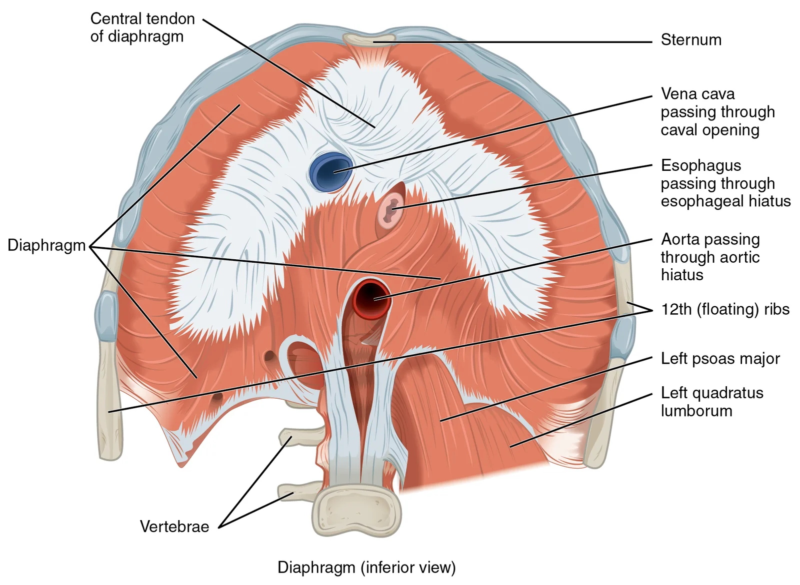
The abdominal region is the space between the thoracic cavity and the pelvic region. It is bounded superiorly by the diaphragm, a dome-shaped muscle that separates the thoracic and abdominal cavities[1]. The superior surface of the diaphragm is convex, forming the floor of the thorax, while the inferior surface is concave, creating the curved roof of the abdominal cavity[1].
Figure 11113 The Diaphragm
Long description
The diaphragm separates the thoracic and abdominal cavities.

Figure 1: The diaphragm separates the thoracic and abdominal cavities.
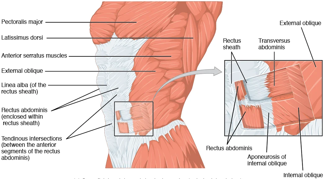
The anterolateral abdominal wall is formed by four pairs of muscles that meet at the anterior midline[1]. These are divided into four groups: the external obliques, the internal obliques, the transversus abdominis, and the rectus abdominis[1].
The posterior limit is defined by the vertebral column and the retroperitoneal space. The abdominal aorta, the largest artery in the body, descends through the diaphragm via the aortic hiatus. It remains to the left of the vertebral column, embedded in adipose tissue behind the peritoneal cavity. The aorta formally ends at approximately the level of the fourth lumbar vertebra (L4), where it bifurcates into the two common iliac arteries. [2] [1]
The inferior boundary is the pelvic inlet, leading toward the perineum. The perineum is a diamond-shaped space located inferior to the pelvic diaphragm (levator ani and coccygeus)[1]. It is bounded anteriorly by the pubic symphysis, posteriorly by the coccyx, and laterally by the ischial tuberosities[1].
| Muscle | Origin | Insertion | Action | Nerve Supply |
|---|---|---|---|---|
| External Oblique | Lower 8 ribs | Linea alba, iliac crest | Compress abdomen | Intercostal nerves |
| Internal Oblique | Thoracolumbar fascia, iliac crest | Linea alba, ribs | Compress abdomen | Intercostal nerves |
| Transversus Abdominis | Costal cartilages, thoracolumbar fascia | Linea alba, iliac crest | Compress abdomen | Intercostal nerves |
| Rectus Abdominis | Pubic symphysis, pubic crest | Xiphoid process, costal cartilages | Flex vertebral column | Intercostal nerves |
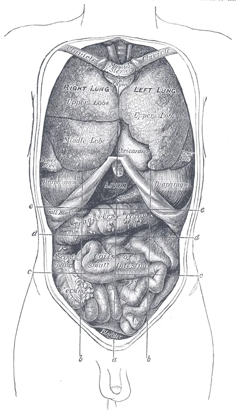
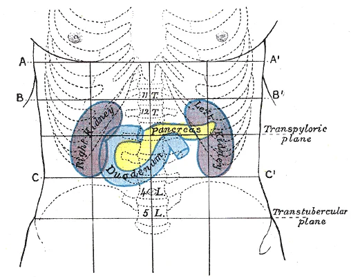
The abdomen is divided into regions using anatomical planes. A key landmark is the transpyloric plane. Organs within the cavity are positioned relative to the vertebrae; for example, the left kidney is located at about the T12 to L3 vertebrae. The right kidney is positioned slightly lower due to displacement by the liver. [3]
NoteRetroperitoneal Space
Certain structures, such as the kidneys and the abdominal aorta, are located behind the peritoneum, anchored to the posterior abdominal wall.
Figure 2Gray1034
Long description
Front view of the thoracic and abdominal viscera. a. Median plane. b b. Lateral planes. c c. Trans tubercular plane. d d. Subcostal plane. e e. Transpyloric plane.

Figure 2: Front view of the thoracic and abdominal viscera (a).
Palpable landmarks of the anterior abdominal wall begin with the xiphoid process at the inferior sternum, the costal margins sweeping down laterally, and the iliac crests forming the lower boundary. The pubic symphysis and pubic tubercles lie midline inferiorly, with the anterior superior iliac spines (ASIS) palpable at the lateral ends of the inguinal ligaments. The umbilicus sits at the L3–L4 level in most adults. The rectus abdominis muscles form the vertical "six-pack" ridges, and their lateral borders are the linea semilunaris; the tendinous intersections cross them transversely. The linea alba runs midline between the two rectus sheaths.
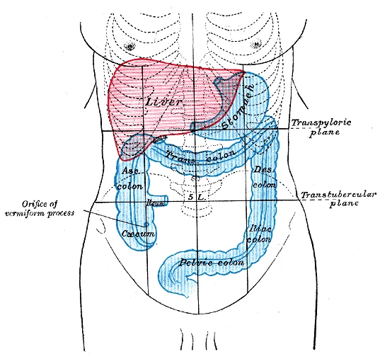
Surface markings of organs are drawn from these landmarks. The liver's upper border projects at the right 5th intercostal space in the midclavicular line, and its lower edge runs along the right costal margin to the epigastrium. The gallbladder fundus marks at the junction of the right costal margin and the lateral border of rectus abdominis (the 9th costal cartilage). The appendix projects from the caecum at McBurney's point, one-third of the way from the ASIS to the umbilicus.
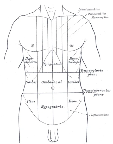
The abdomen is divided into four quadrants by the median plane and the transumbilical plane, and into nine regions by two vertical (midclavicular) and two horizontal (subcostal and transtubercular) planes: right and left hypochondrium, epigastrium, right and left lumbar, umbilical, right and left iliac fossa, and hypogastrium. These regions guide clinical localisation of pain and masses.
Figure 3Gray1225
Long description
Front of abdomen, showing surface markings for duodenum, pancreas, and kidneys. A A’. Plane through joint between body and xiphoid process of sternum. B B’. Plane midway between A A’ and transpyloric plane. C C’. Plane midway between transpyloric and transtubercular planes.

Figure 3: Front of abdomen, showing surface markings for duodenum, pancreas, and kidneys (a a’).
Surface markings guide imaging. Ultrasound uses the liver as an acoustic window for the gallbladder and right kidney; the spleen for the left kidney. On CT, the aorta and inferior vena cava are seen retroperitoneally, parallel to each other, with the IVC lying just beneath the parietal peritoneum[1]. Hernias emerge at weak points: inguinal (above the inguinal ligament, medial to the ASIS), femoral (below the ligament), and umbilical. Surgical incisions respect muscle fibre directions: [2]
The abdominal skeleton provides structural support and attachment points for muscles while protecting vital organs. The posterior abdominal wall is formed by the lumbar vertebrae, parts of the ilia of the hip bones, psoas major and iliacus muscles, and quadratus lumborum muscle. This bony framework plays a key role in stabilizing the rest of the body and maintaining posture. [1]
Figure 4Gray1221
Long description
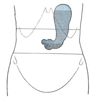
With the patient in the erect posture. Labelled illustration from Wikimedia Commons, used under its open licence.

Figure 4: With the patient in the erect posture.
The kidneys lie on either side of the spine in the retroperitoneal space between the parietal peritoneum and the posterior abdominal wall, well protected by muscle, fat, and ribs.
The lumbar vertebrae (L1-L5) form the central bony support of the posterior abdominal wall. The iliac crests of the hip bones form the lateral borders, with the anterior superior iliac spine being an important landmark for surgical incisions. The sacrum and coccyx complete the posterior bony framework.
The abdominal aorta terminates when it bifurcates into the two common iliac arteries at the level of the fourth lumbar vertebra. This bifurcation point is an important anatomical landmark that can be palpated clinically. The kidneys are positioned at approximately the T12 to L3 vertebral levels, with the right kidney typically positioned slightly lower than the left due to the liver's presence superiorly. [1]
Figure 5Gray1226
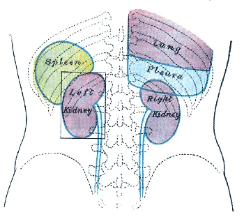
Long description
Back of lumbar region, showing surface markings for kidneys, ureters, and spleen. The lower portions of the lung and pleura are shown on the right side.

Figure 5: Back of lumbar region, showing surface markings for kidneys, ureters, and spleen.
Figure 6Gray1036
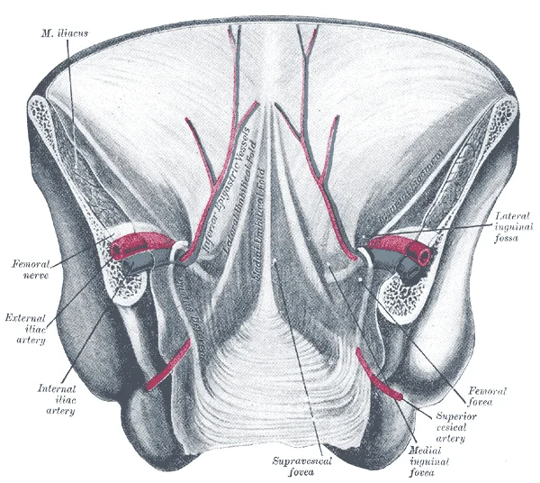
Long description
Posterior view of the anterior abdominal wall in its lower half. The peritoneum is in place, and the various cords are shining through. (After Joessel.)

Figure 6: Posterior view of the anterior abdominal wall in its lower half.
The abdominal wall is built from concentric layers that can be remembered from superficial to deep: skin, superficial fascia, deep fascia, muscle, transversalis fascia, extraperitoneal fat and parietal peritoneum. Each layer has a specific mechanical role and a predictable behaviour in disease, which is why surgeons and radiologists rely on fascial planes to interpret the spread of pus, blood, urine or tumour.
The lymphatic drainage of the abdominal wall is divided into superficial and deep components. This distinction is clinically significant for understanding the spread of skin cancers or infections. Deep lymphatics from the abdominal muscles and parietal peritoneum follow the arteries and drain into the para-aortic or iliac nodes. [1] [1]
Deep to the superficial fascia lies a thin deep fascia that is poorly developed over the abdomen, unlike the strong fascia lata of the thigh. The main investing layer is the aponeurosis of the external oblique, which forms the inguinal ligament at its lower free edge, running from the anterior superior iliac spine to the pubic tubercle. The internal oblique and transversus abdominis aponeuroses contribute to the rectus sheath, which encloses the rectus abdominis muscle. Above the arcuate line (m [2]
Figure 7Gray1227
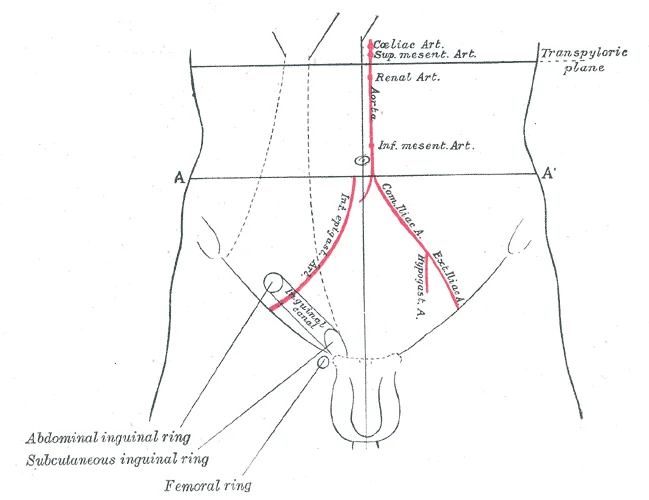
Long description
Front of abdomen, showing surface markings for arteries and inguinal canal.

Figure 7: Front of abdomen, showing surface markings for arteries and inguinal canal.
The arcuate line is marked, showing the transition in the rectus sheath.
Clinical pearlClinical pearl
Below the arcuate line, the posterior rectus sheath is absent, so a Spigelian hernia (through the linea semilunaris) or a suprapubic incisional hernia may present with only peritoneum and transversalis fascia as coverings.
| Muscle | Origin | Insertion | Action | Nerve supply |
|---|---|---|---|---|
| External oblique | Lower 8 ribs | Linea alba, iliac crest, inguinal ligament | Compresses abdomen, flexes trunk | Thoracoabdominal nerves (T7–T11), subcostal (T12) |
| Internal oblique | Thoracolumbar fascia, iliac crest, inguinal ligament | Lower 3 ribs, linea alba | Compresses abdomen, flexes trunk | Thoracoabdominal nerves (T7–T11), iliohypogastric (L1), ilioinguinal (L1) |
| Transversus abdominis | Lower 6 ribs, thoracolumbar fascia, iliac crest, inguinal ligament | Linea alba, pubic crest | Compresses abdomen | Thoracoabdominal nerves (T7–T11), iliohypogastric (L1), ilioinguinal (L1) |
| Rectus abdominis | Pubic symphysis, pubic crest | Xiphoid process, costal cartilages 5–7 | Flexes trunk, compresses abdomen | Thoracoabdominal nerves (T7–T11), subcostal (T12) |
Abdominal wall layers from superficial to deep
- Skin
- Superficial fascia (Camper + Scarpa)
- Deep fascia (thin)
- Muscle layer (external oblique, internal oblique, transversus abdominis)
- Transversalis fascia
- Extraperitoneal fat
- Parietal peritoneum
Figure 8Gray388
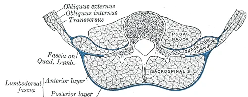
Long description
Diagram of a transverse section of the posterior abdominal wall, to show the disposition of the lumbodorsal fascia.

Figure 8: Diagram of a transverse section of the posterior abdominal wall, to show the disposition of the lumbodorsal fascia.
Figure 91112 Muscles of the Abdomen Anterolateral
Long description
Caption: (a) The anterior abdominal muscles include the medially located rectus abdominis, which is covered by a sheet of connective tissue called the rectus sheath. On the flanks of the body, medial to the rectus abdomi

Figure 9: Caption: (a) The anterior abdominal muscles include the medially located rectus abdominis, which is covered by a sheet of connective tissue called the rectus sheath.
The abdominal muscles can be grouped into anterior, lateral, and posterior compartments based on their location and function.
| Compartment | Muscle | Origin | Insertion | Action | Nerve Supply |
|---|---|---|---|---|---|
| Anterior | Rectus abdominis | Pubic crest and symphysis | Xiphoid process, costal cartilages 5-7 | Flexion of trunk, compression of abdomen | Anterior rami of T7-L1 |
| External oblique | Lower 8 ribs | Iliac crest, linea alba, pubic tubercle | Flexion and rotation of trunk, compression of abdomen | Anterior rami of T7-L1 | |
| Internal oblique | Thoracolumbar fascia, iliac crest, inguinal ligament | Costal cartilages 7-12, linea alba, pubic crest | Flexion and rotation of trunk, compression of abdomen | Anterior rami of T7-L1 | |
| Transversus abdominis | Lower 6 ribs, thoracolumbar fascia, iliac crest, inguinal ligament | Linea alba, pubic crest | Compression of abdomen, increases intra-abdominal pressure | Anterior rami of T7-L1 | |
| Lateral | Quadratus lumborum | Iliac crest, iliolumbar ligament | Transverse processes of L1-L4 | Lateral flexion of trunk, stabilizes pelvis | Anterior rami of T12-L4 |
| Posterior | Psoas major | Transverse processes of T12-L5 | Lesser trochanter of femur | Flexion of thigh at hip, lateral flexion of trunk | Anterior rami of L1-L3 |
| Iliacus | Iliac fossa | Lesser trochanter of femur | Flexion of thigh at hip | Femoral nerve (L2-L4) |
The rectus abdominis muscles are a pair of long, linear muscles that originate at the pubic crest and symphysis, extending the length of the trunk. These muscles are segmented by three transverse bands of collagen fibers called the tendinous intersections, creating the appearance of "six-pack abs" when hypertrophied. The linea alba is a white, fibrous band formed by the bilateral rectus sheaths that join at the anterior midline of the body, enclosing the rectus abdominis muscles. [1]
The diaphragm is a dome-shaped muscle that plays a crucial role in breathing. Its central tendon is fused to the inferior surface of the pericardial sac and the inferior surfaces of the pleural membranes. The skeletal muscle portions of the diaphragm insert into the central tendon while originating from the xiphoid process of the sternum anteriorly, the inferior six ribs and their cartilages laterally, and the lumbar vertebrae and 12th ribs posteriorly. [1]
When the diaphragm contracts and flattens, it increases the volume inside the pleural cavities, decreasing pressure and allowing air to flow into the lungs. The intercostal muscles span the spaces between the ribs and assist in breathing by changing the shape of the rib cage. [1]
The linea alba appears as a vertical midline structure where the rectus sheaths meet. The internal oblique muscles lie beneath the external obliques, with fibers running perpendicular to them. The transversus abdominis is the deepest of the lateral abdominal muscles, with fibers running horizontally.
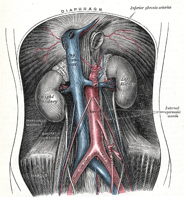
Figure 10Gray531
Long description
The abdominal aorta and its branches. Labelled illustration from Wikimedia Commons, used under its open licence.

Figure 10: The abdominal aorta and its branches.
The abdominal aorta is the direct continuation of the thoracic aorta, beginning at the aortic hiatus of the diaphragm at the level of the T12 vertebra and ending at the level of L4, where it bifurcates into the right and left common iliac arteries. It lies slightly to the left of the midline, anterior to the vertebral bodies, and is enveloped by the prevertebral plexus of nerves and lymphatics. As it descends, it gives off both parietal and visceral branches. [2]
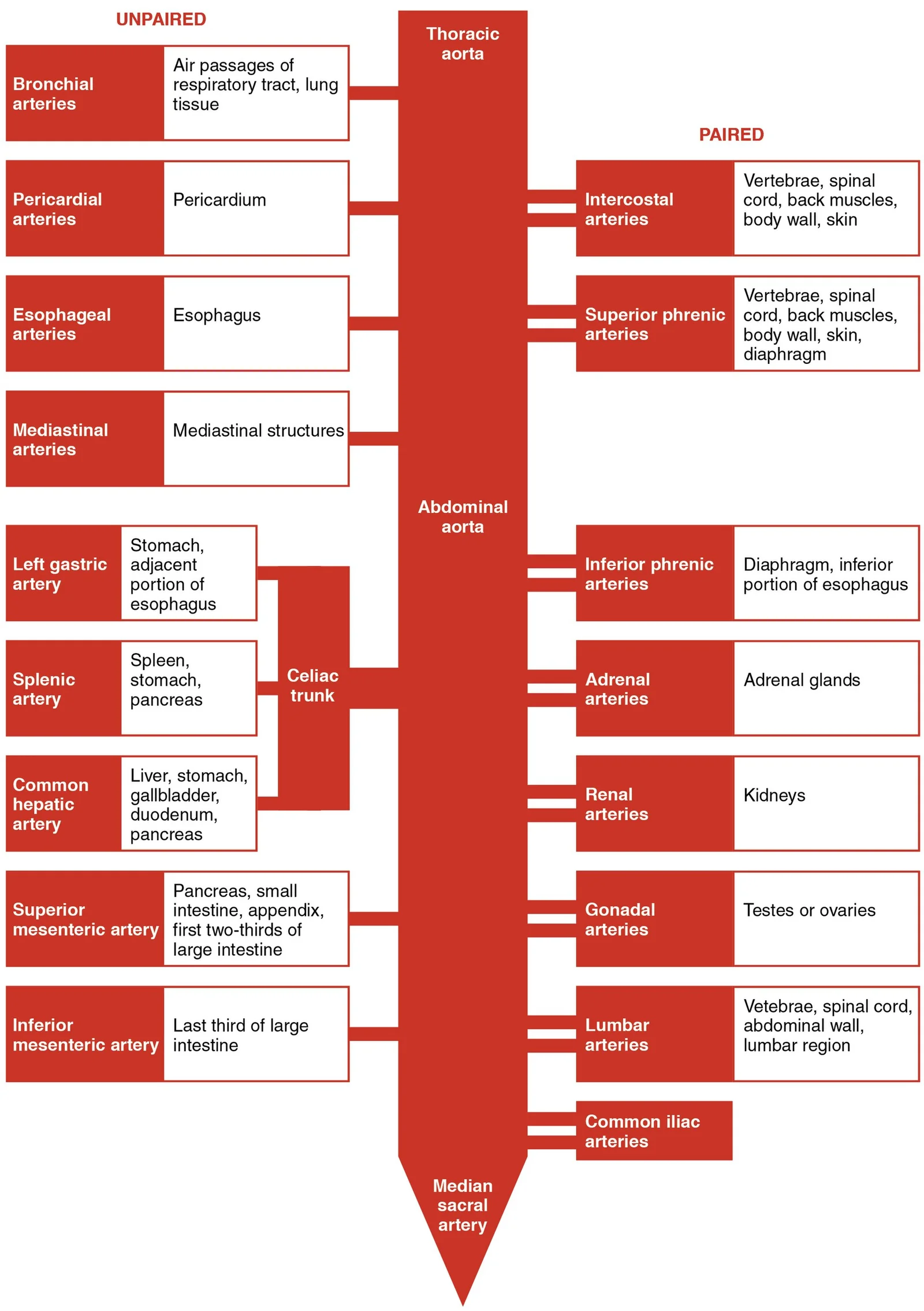
Figure 112125 Thoracic Abdominal Arteries Chart
Long description
The flow chart summarizes the distribution of the major branches of the aorta into the thoracic and abdominal regions.

Figure 11: The flow chart summarizes the distribution of the major branches of the aorta into the thoracic and abdominal regions.
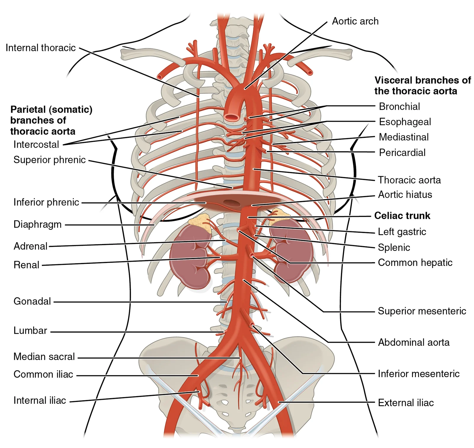
Figure 122124 Thoracic Abdominal Arteries
Long description
The thoracic aorta gives rise to the arteries of the visceral and parietal branches.

Figure 12: The thoracic aorta gives rise to the arteries of the visceral and parietal branches.
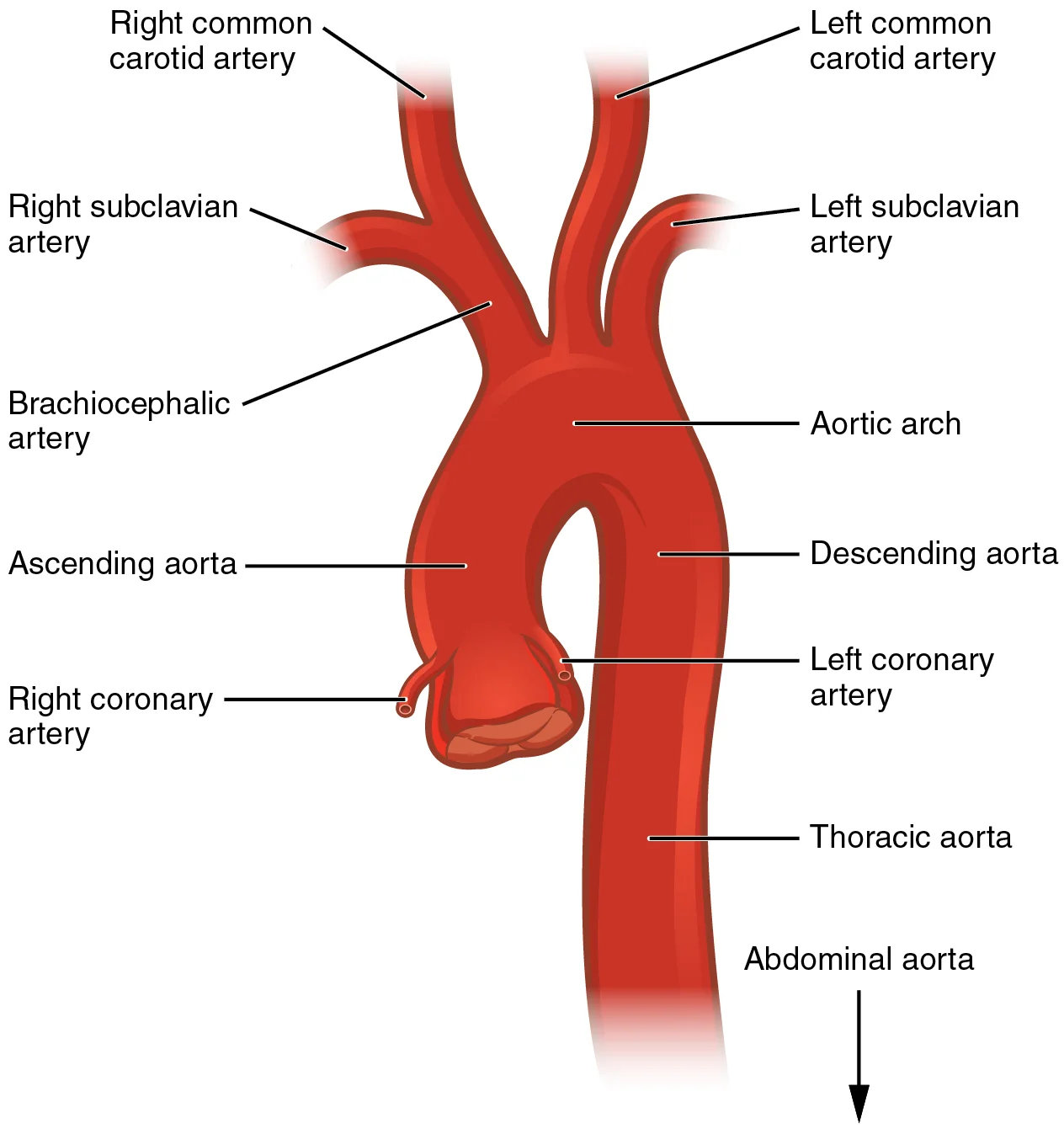
Figure 132121 Aorta
Long description
The aorta has distinct regions, including the ascending aorta, aortic arch, and the descending aorta, which includes the thoracic and abdominal regions.

Figure 13: The aorta has distinct regions, including the ascending aorta, aortic arch, and the descending aorta, which includes the thoracic and abdominal regions.
Figure 14Gray1224
Long description
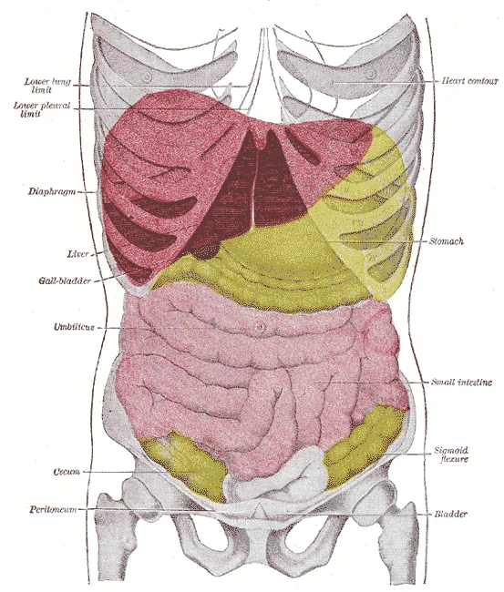
Topography of thoracic and abdominal viscera.

Figure 14: Topography of thoracic and abdominal viscera.
Figure 15Gray1121
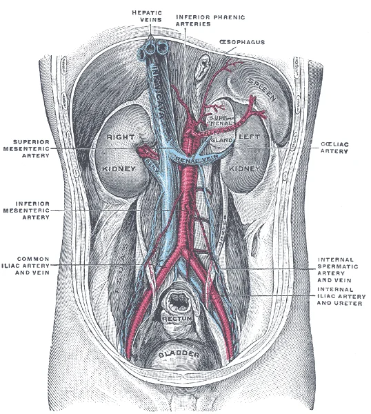
Long description
Posterior abdominal wall, after removal of the peritoneum, showing kidneys, suprarenal capsules, and great vessels. (Corning).

Figure 15: Posterior abdominal wall, after removal of the peritoneum, showing kidneys, suprarenal capsules, and great vessels (Corning).
The venous drainage of the abdomen is a low-pressure, high-volume system that returns blood from the gastrointestinal tract, spleen, pancreas, gallbladder and the paired retroperitoneal organs to the inferior vena cava (IVC). Unlike the arterial tree, which is largely symmetrical, the venous side shows a striking asymmetry: the left renal vein crosses the midline anterior to the aorta, and the gonadal veins drain asymmetrically (left into the left renal vein, right directly into the IVC). This a [1]
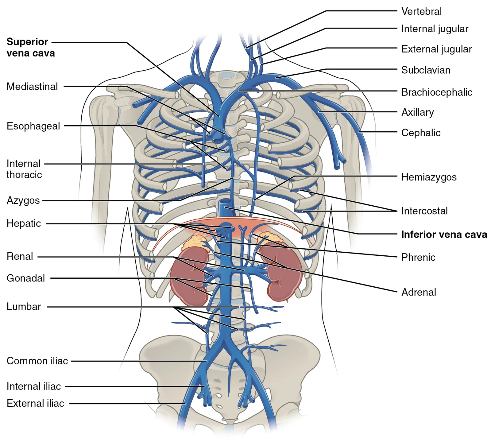
Figure 162132 Thoracic Abdominal Veins
Long description
Veins of the thoracic and abdominal regions drain blood from the area above the diaphragm, returning it to the right atrium via the superior vena cava.

Figure 16: Veins of the thoracic and abdominal regions drain blood from the area above the diaphragm, returning it to the right atrium via the superior vena cava.
The key functional principle is the hepatic portal system. Blood from the abdominal digestive tract, spleen and pancreas does not return directly to the IVC. Instead, it drains into the portal vein, which carries it to the liver for processing before the blood finally reaches the IVC via the hepatic veins. The portal vein is formed behind the neck of the pancreas by the union of the splenic vein (from the spleen and left side of the colon) and the superior mesenteric vein (from the small intestine and right colon). The inferior mesenteric vein (from the left colon and rectum) usually joins the splenic vein, though it may join the superior mesenteric vein or the junction itself.
The splenic vein runs horizontally along the posterior surface of the pancreas, receiving the short gastric veins and the left gastro-omental vein. The superior mesenteric vein lies to the right of the superior mesenteric artery, ascending anterior to the third part of the duodenum. The inferior mesenteric vein ascends on the left, behind the peritoneum, to join the splenic vein. The portal vein then ascends to the porta hepatis, where it divides into left and right branches. The right branch receives the cystic vein from the gallbladder.
The systemic venous drainage of the abdominal wall and retroperitoneum is simpler. The IVC is formed at the level of L5 by the union of the common iliac veins, ascends to the right of the aorta, and receives the renal veins, right gonadal vein, and hepatic veins before piercing the diaphragm at T8. The azygos and hemiazygos veins drain the posterior abdominal wall and ascend into the thorax. Clinically, portal-systemic anastomoses (e.g., at the oesophagus, rectum, and umbilicus) become dilated in portal hypertension, producing varices, haemorrhoids, and caput medusae respectively.
Figure 17Gray848
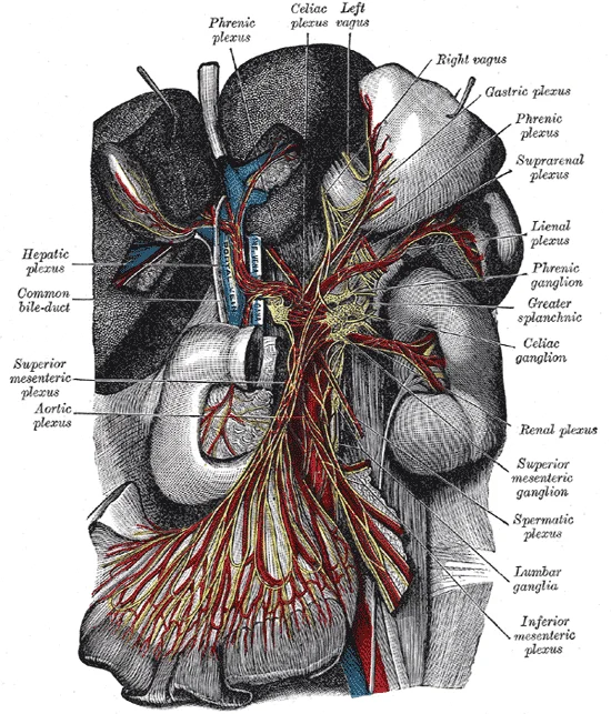
Long description
The celiac ganglia with the sympathetic plexuses of the abdominal viscera radiating from the ganglia. (Toldt.)

Figure 17: The celiac ganglia with the sympathetic plexuses of the abdominal viscera radiating from the ganglia (Toldt.).
Figure 18Gray838
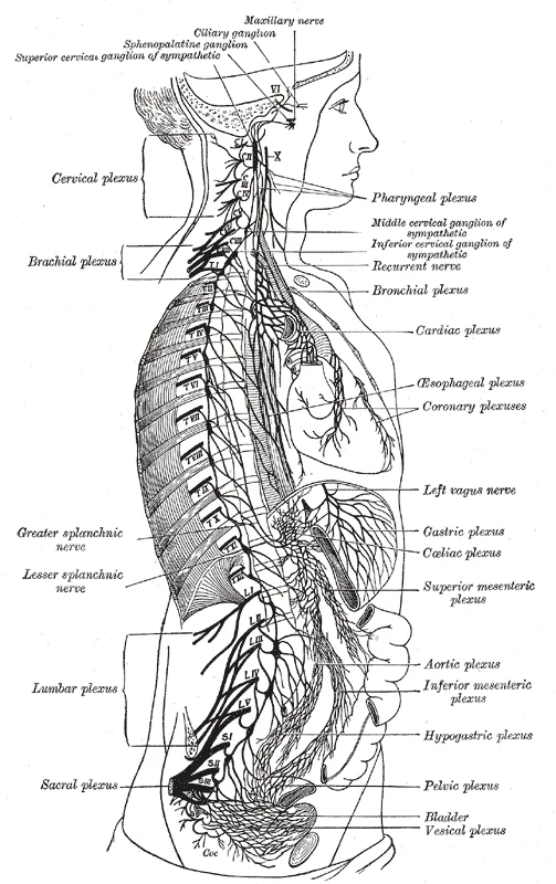
Long description
The right sympathetic chain and its connections with the thoracic, abdominal, and pelvic plexuses (After Schwalbe)

Figure 18: The right sympathetic chain and its connections with the thoracic, abdominal, and pelvic plexuses (After Schwalbe).
The anterior abdominal wall is supplied segmentally by the lower six thoracic nerves and the first lumbar nerve. These are the thoraco-abdominal nerves, the continuation of the T7–T11 intercostal nerves beyond the costal margin, and the subcostal nerve (T12). Each nerve runs in the neurovascular plane between the internal oblique and transversus abdominis muscles, giving off a lateral cutaneous branch that pierces the internal and external oblique muscles to supply the skin of the flank. The ant
The subcostal nerve (T12) lies below the twelfth rib.
The motor supply follows the same segmental pattern. The lower six thoracic nerves innervate the external oblique, internal oblique, transversus abdominis, and rectus abdominis. The pyramidalis muscle, when present, is supplied by T12. The cremaster muscle receives fibres from the genital branch of the genitofemoral nerve (L1–L2). The diaphragm, although a muscle of the abdominal wall, is supplied by the phrenic nerves (C3–C5), which descend through the thorax and reach the diaphragm from above; the phrenic nerve also carries sensory fibres from the central part of the diaphragm, while the peripheral diaphragm is supplied by the lower six intercostal nerves. The diaphragm has three openings for structures passing between thorax and abdomen: the inferior vena cava passes through the caval opening, the esophagus and attached nerves pass through the esophageal hiatus, and the aorta, thoracic duct, and azygous vein pass through the aortic hiatus of the posterior diaphragm. [1]
The autonomic supply to the abdominal viscera is separate. The vagus nerves (parasympathetic) travel with the esophagus through the esophageal hiatus and supply the foregut and midgut. The sympathetic supply arises from the T5–L2 spinal segments, relays in the coeliac, superior mesenteric, and inferior mesenteric ganglia, and accompanies the arteries. Pain from the abdominal viscera is referred to the dermatomes of the corresponding spinal segments: foregut pain to the epigastrium (T5–T9), midgut pain to the periumbilical region (T9–T11), and hindgut pain to the hypogastrium (T11–L1). Somatic pain from the parietal peritoneum is sharp and localised, carried by the same segmental nerves that supply the overlying muscles and skin.
| Muscle | Origin | Insertion | Action | Nerve supply |
|---|---|---|---|---|
| External oblique | Lower eight ribs | Linea alba, pubic tubercle, iliac crest | Compresses abdomen, flexes and rotates trunk | T7–T11, T12 |
| Internal oblique | Thoracolumbar fascia, iliac crest, inguinal ligament | Lower three ribs, linea alba | Compresses abdomen, flexes and rotates trunk | T7–T11, T12 |
| Transversus abdominis | Lower six ribs, thoracolumbar fascia, iliac crest, inguinal ligament | Linea alba, pubic crest | Compresses abdomen | T7–T11, T12 |
| Rectus abdominis | Pubic symphysis, pubic crest | Xiphoid process, costal cartilages 5–7 | Flexes trunk, compresses abdomen | T7–T12 |
| Pyramidalis | Pubic crest | Linea alba | Tenses linea alba | T12 |
A useful clinical rule is that the T10 dermatome is at the level of the umbilicus, T7 at the xiphisternum, and L1 at the inguinal ligament. In a patient with a spinal cord injury, the sensory level can be mapped by testing these dermatomes. In pregnancy, the enlarging uterus stretches the anterior abdominal wall, and the lateral cutaneous branches of T10–T12 may be compressed, causing pain over the flanks. In older adults, loss of muscle tone in the transversus abdominis and internal oblique contributes to abdominal wall laxity and an increased risk of inguinal hernia; the ilioinguinal nerve (L1) is at risk during hernia repair, and injury causes numbness over the base of the scrotum or labium majus.
The lymphatic system of the abdomen serves as a critical drainage network, removing interstitial fluid and transporting immune cells. Understanding this drainage is essential for tracing the spread of malignancies and infections. The drainage patterns are largely determined by embryological origin and anatomical position relative to the peritoneal cavity.
DefinitionCisterna Chyli
A dilated sac at the lower end of the thoracic duct, receiving lymph from the intestinal and lumbar trunks.
The lymphatic drainage of the abdominal wall is divided into superficial and deep components. This distinction is clinically significant for understanding the spread of skin cancers or infections. Deep lymphatics from the abdominal muscles and parietal peritoneum follow the arteries and drain into the para-aortic or iliac nodes. [1]
| Region | Primary Drainage Nodes | Clinical Relevance |
|---|---|---|
| Foregut | Coeliac nodes | Gastric and pancreatic cancers |
| Midgut | Superior mesenteric nodes | Appendicitis and small bowel tumours |
| Figure 8 (F-B1-2366-8) illustrates the lymphatic drainage of applied abdominal anatomy. This diagram presents an anterior view of the retroperitoneal and intraperitoneal structures. The orientation shows the liver superiorly, the stomach centrally, and the intestines inferiorly. Key labelled structures include the coeliac nodes, superior mesenteric nodes, inferior mesenteric nodes, and para-aortic nodes. The coeliac nodes lie anterior to the abdominal aorta, just below the diaphragm, receiving d | ||
| Gonads | Para-aortic nodes | Testicular and ovarian metastases |
| Structure | Origin | Course | Termination |
|---|---|---|---|
| Thoracic Duct | Cisterna Chyli | Pass through aortic hiatus | Left venous angle |
| Right Lymphatic Duct | Right jugular, subclavian, bronchomediastinal trunks | Drains right upper quadrant | Right venous angle |
The clinical importance of these pathways cannot be overstated. Surgeons must identify and ligate specific lymphatic channels during oncological resections to ensure complete removal of potentially metastatic tissue. For example, in a gastrectomy, the coeliac nodes are dissected to stage gastric cancer accurately. Similarly, in colorectal surgery, the mesenteric nodes are examined to determine the extent of disease spread. Knowledge of these anatomical relationships allows for precise surgical planning and improved patient outcomes.
Figure 19Gray1222
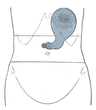
Long description
With the patient lying down. Labelled illustration from Wikimedia Commons, used under its open licence.

Figure 19: With the patient lying down.
Figure 20Gray1125
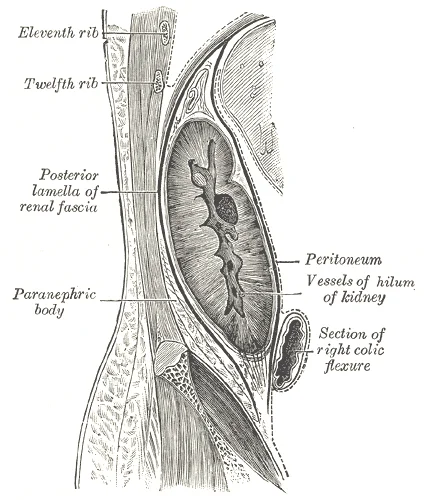
Long description
Sagittal section through posterior abdominal wall, showing the relations of the capsule of the kidney. (After Gerota).

Figure 20: Sagittal section through posterior abdominal wall, showing the relations of the capsule of the kidney (After gerota).
The abdomen is a cavity, not an empty space; it is packed with viscera, vessels, nerves and connective tissue, all held in specific relations. Understanding these relations is the key to clinical examination and surgical approach. The peritoneal cavity is a potential space within the abdomen, lined by peritoneum, and contains the abdominal organs. The retroperitoneal space lies behind the peritoneum and contains the kidneys, ureters, pancreas, duodenum (except its first part), and major vessels.
The abdominal aorta enters through the aortic hiatus at T12 and descends anterior to the vertebral bodies, slightly left of midline. It is related anteriorly to the pancreas, duodenum, and root of the mesentery; posteriorly to the lumbar vertebrae and anterior longitudinal ligament; and laterally to the sympathetic trunks and ureters. Its branches include the coeliac trunk (T12), superior mesenteric artery (L1), renal arteries (L1–L2), gonadal arteries (L2), inferior mesenteric artery (L3), and terminal bifurcation into common iliac arteries at L4. The inferior vena cava lies to its right, and
The portal vein forms behind the neck of the pancreas from the union of the superior mesenteric and splenic veins. It passes upward, posterior to the duodenum and bile duct, and enters the liver at the porta hepatis. It carries nutrient-rich blood from the gut to the liver. The hepatic artery and bile duct accompany it, forming the portal triad. The portal system is unique: a capillary bed (gut) drains into a vein that forms a second capillary bed (liver sinusoids).
The kidneys lie retroperitoneally, from T12 to L3, with the right kidney slightly lower due to the liver. Each kidney is related superiorly to the adrenal gland. The renal artery, a branch of the aorta, divides into segmental arteries, then interlobar arteries that pass through the renal columns to reach the cortex; these branch into arcuate arteries, cortical radiate arteries, and afferent arterioles. The renal vein lies anterior to the artery and drains into the inferior vena cava; the left re [3]
The inguinal canal is an oblique passage, about 4 cm long, between the deep ring (midway between anterior superior iliac spine and pubic tubercle) and the superficial ring (just above and lateral to the pubic tubercle). Its walls are: anterior—external oblique aponeurosis reinforced laterally by internal oblique; posterior—transversalis fascia reinforced medially by the conjoint tendon; roof—internal oblique and transversus abdominis; floor—inguinal ligament. In the male, the canal contains the [1]
On axial CT at the level of vertebra L1, viewed from below, the aorta appears as a round structure anterior to the vertebral body, with the inferior vena cava to its right. The right and left kidneys are positioned laterally, with the pancreas crossing anterior to the aorta, and the spleen located posterolaterally on the left side.
On sagittal ultrasound through the right upper quadrant, the liver lies superiorly, the right kidney posteriorly, and the gallbladder inferiorly. The diaphragm appears as a curved echogenic line above the liver. The portal vein can be seen branching within the liver, with the hepatic artery positioned anteriorly to it. The common bile duct appears as an anechoic structure anterior to the portal vein. In children, the liver is relatively larger and more echogenic compared to adults, while the gallbladder may appear smaller. During pregnancy, the gallbladder may appear dilated due to hormonal effects.
On coronal MRI at the level of the renal hila, both kidneys are visible with their pelves medially. The aorta and inferior vena cava lie centrally, with the psoas muscles flanking the vertebral column. The pancreas appears as a structure with intermediate signal intensity, crossing anterior to the aorta. The spleen demonstrates homogeneous high signal on T2-weighted images. In older adults, the renal cortex may show decreased signal intensity on T1-weighted images due to decreased fat content.
On CT, the aorta typically shows a calcified wall in older adults, while the inferior vena cava appears oval and collapses with Valsalva maneuver. The renal arteries arise at approximately L1-L2 level, with the right artery typically longer than the left due to the aorta's position. On ultrasound, the pancreas is echogenic relative to the liver, and the common bile duct is seen anterior to the portal vein, with a normal diameter of less than 6mm in adults. On MRI, the renal cortex is brighter than the medulla on T1-weighted images, while the perirenal fat shows high signal on T1 and low signal on fat-suppressed sequences. The portal vein appears as a dark structure on T1-weighted images and bright on T2-weighted images.
Figure 21Gray1220
Long description
Nine abdominal regions, as illustrated in the 1918 edition of Anatomy of the Human Body

Figure 21: Nine abdominal regions, as illustrated in the 1918 edition of Anatomy of the Human Body.
The abdominal wall is a layered, dynamic structure that protects the viscera and generates intra-abdominal pressure. From superficial to deep, the layers are skin, superficial fascia (Camper's and Scarpa's), deep fascia, muscle, transversalis fascia, extraperitoneal fat and peritoneum. The anterolateral muscles—external oblique, internal oblique, transversus abdominis and rectus abdominis—each have a distinct fibre direction, which is clinically relevant when assessing a cough impulse or a surgical incision.
Deep to the superficial fascia lies a thin deep fascia that is poorly developed over the abdomen, unlike the strong fascia lata of the thigh. The main investing layer is the aponeurosis of the external oblique, which forms the inguinal ligament at its lower free edge, running from the anterior superior iliac spine to the pubic tubercle. The internal oblique and transversus abdominis aponeuroses contribute to the rectus sheath, which encloses the rectus abdominis muscle. Above the arcuate line (m
The inguinal canal is an oblique passage, about 4 cm long, between the deep ring (midway between anterior superior iliac spine and pubic tubercle) and the superficial ring. Its walls are: anterior—external oblique aponeurosis reinforced laterally by internal oblique; posterior—transversalis fascia reinforced medially by the conjoint tendon; roof—internal oblique and transversus abdominis; floor—inguinal ligament. In the male, the canal contains the
The posterior abdominal wall is dominated by the psoas major, quadratus lumborum and iliacus. The psoas major arises from T12–L5 vertebral bodies and discs, passes under the inguinal ligament, and inserts into the lesser trochanter; it is the principal flexor of the hip and is overlapped by the ureter, which crosses its anterior surface at the pelvic brim. The quadratus lumborum runs from the iliac crest to the 12th rib and transverse processes of L1–L4; it fixes the 12th rib during inspiration [1]
The abdominal aorta enters at the aortic hiatus and bifurcates, just left of midline. Its main branches—coeliac trunk, superior mesenteric artery, renal arteries (right longer and passing behind the IVC), and inferior mesenteric artery—are all related to vertebral levels that can be palpated or imaged. The IVC forms to the right of the aorta, ascends, and pierces the diaphragm. The ureters cross the pelvic brim at the bifurcation of the common iliac arteries, a common site for stone impaction. [2] [1]
| Muscle | Origin | Insertion | Action | Nerve supply |
|---|---|---|---|---|
| External oblique | Lower 8 ribs | Iliac crest, linea alba, inguinal ligament | Flexes/rotates trunk, compresses abdomen | T7–T12, iliohypogastric, ilioinguinal |
| Internal oblique | Thoracolumbar fascia, iliac crest, inguinal ligament | Lower 3 ribs, linea alba, pubic crest | Flexes/rotates trunk, compresses abdomen | T7–T12, iliohypogastric, ilioinguinal |
| Transversus abdominis | Lower 6 ribs, thoracolumbar fascia, iliac crest, inguinal ligament | Linea alba, pubic crest | Compresses abdomen | T7–T12, iliohypogastric, ilioinguinal |
| Rectus abdominis | Pubic crest, symphysis | Xiphoid, 5th–7th costal cartilages | Flexes trunk, compresses abdomen | T7–T12 |
The rectus abdominis lies centrally within its sheath; the external oblique is the most superficial lateral muscle, its fibres running downwards and forwards; the internal oblique lies beneath it with fibres running upwards and forwards; the transversus abdominis is deepest, with horizontal fibres.
The aorta is central, the IVC to its right. The ureters cross the psoas and then the pelvic brim, anterior to the common iliac vessels.
These are the facts that exam questions return to when they test the arterial anatomy of the abdomen, pelvis and the wider systemic circulation.
DefinitionTrunk
A trunk is an occasional, very large artery that gives rise to several smaller arteries; the celiac trunk is the classic example, giving rise to the left gastric, common hepatic and splenic arteries. [2]
Clinical pearlCeliac branches
Learn the celiac trunk as three branches — left gastric, common hepatic and splenic — because questions often ask which named vessel is not a branch of it.
Clinical pearlGonadal arteries
Each gonadal artery supplies a gonad and is called an ovarian artery in the female or a testicular (internal spermatic) artery in the male; both arise inferior to the renal arteries and are generally retroperitoneal. [2]
Exam trapOvarian versus testicular
The ovarian artery is considerably shorter than the testicular artery, which travels outside the body cavity to the testes and forms one component of the spermatic cord. [2]
Clinical pearlUterine and vaginal supply
The uterine arteries and the vaginal arteries, which distribute blood to the vagina, are branches of the internal iliac artery — the central arterial supply of the female pelvis. [2]
| Artery | Origin | Branches or territory |
|---|---|---|
| Celiac trunk | A very large artery, or trunk, giving rise to several smaller arteries | Left gastric, common hepatic and splenic arteries |
| Inferior mesenteric artery | Approximately 5 cm superior to the common iliac arteries | Distal segment of the large intestine, including the rectum |
| Gonadal artery | Inferior to the renal arteries; generally retroperitoneal | Ovary, uterine tube and uterus, or testis |
Students often draw absorbed nutrients passing from the gut capillaries straight into the inferior vena cava. That is wrong: portal systems begin and end in capillaries. The initial capillaries from the stomach, small intestine, large intestine and spleen lead to the hepatic portal vein, which ends in specialised capillaries within the liver, the hepatic sinusoids. [2]
DefinitionPortal system
A portal system is a circulation that begins and ends in capillaries; the hepatic portal vein carries blood from the gut and spleen to the hepatic sinusoids of the liver.
Drawing both gonadal veins into the inferior vena cava is a classic error. The right gonadal vein empties directly into the inferior vena cava, while the left gonadal vein empties into the left renal vein. [2]
Labelling every nephron juxtamedullary is a mistake. Some nephrons have a short loop of Henle that does not dip beyond the cortex; these are the cortical nephrons. About 15 per cent of nephrons have long loops of Henle that extend deep into the medulla and are called juxtamedullary nephrons. [3]
Equating the cortex with the renal corpuscles alone is another slip. All of the renal corpuscles, as well as both the proximal convoluted tubules (PCTs) and the distal convoluted tubules, are found in the cortex.
| Nephron | Loop of Henle | Key fact |
|---|---|---|
| Cortical | Short; does not dip beyond the cortex | Corpuscles and convoluted tubules lie in the cortex |
| Juxtamedullary | Long; extends deep into the medulla | About 15 per cent of nephrons |
Reversing the outflow pathway is easy under pressure. Filtrate is gathered by collecting ducts that drain into the minor calyces, which merge to form major calyces; it then proceeds to the renal pelvis and finally the ureters. [3]
Assuming that every abdomen follows the diagram is unsafe, because individual variations in circulation patterns are not uncommon. [2]
Treating the pelvic floor as one muscle loses marks. The pelvic diaphragm spans anteriorly to posteriorly from the pubis to the coccyx, comprising the levator ani and the ischiococcygeus, and its openings include the anal canal, the urethra and, in females, the vagina. [1]
The portal route explains why the liver receives gut-derived blood before the rest of the body: capillaries of the stomach, small intestine, large intestine and spleen drain through the hepatic portal vein into the hepatic sinusoids. In liver disease this routing underlies the abnormal handling of absorbed substances. In a man with a scrotal swelling, the asymmetric drainage matters, because the left gonadal vein enters the left renal vein while the right enters the inferior vena cava directly. Women with urinary or faecal incontinence are examined with the pelvic diaphragm and its openings in mind. [2]
Figure 22Gray1223
Long description
Front of abdomen, showing surface markings for liver, stomach, and great intestine.

Figure 22: Front of abdomen, showing surface markings for liver, stomach, and great intestine.
Kidney and ureteric surgery follows the collecting system in order: collecting ducts open into minor calyces, which merge into major calyces, then the renal pelvis and the ureter. Pelvic floor repair restores the sheet formed by the levator ani and ischiococcygeus while protecting the anal canal, the urethra and, in women, the vagina. Operations near the porta hepatis protect the hepatic portal vein, through which all gut and splenic blood passes before the liver.
In a dissected kidney the cortex appears lighter in colour than the rest of the organ, which helps orientation. Contrast studies display the collecting system from calyces to ureter, and cross-sectional imaging traces the hepatic portal vein to its gut tributaries. Each gonadal vein can be followed to its own termination. [3]
Spot tests repeatedly probe the same anchors: where the left gonadal vein ends, how many nephrons are juxtamedullary, which muscles form the pelvic diaphragm, the order of the collecting system, and the capillary-to-capillary definition of a portal system. Candidates who can redraw the table and work through the flowchart below should manage most items.
| Vessel | Begins at | Ends at |
|---|---|---|
| Right gonadal vein | Testis or ovary | Inferior vena cava |
| Left gonadal vein | Testis or ovary | Left renal vein |
| Hepatic portal vein | Stomach, small intestine, large intestine, spleen | Hepatic sinusoids |
Sorting a nephron
- StartFind the loop of Henle of the nephron in the question.
- Question: Does the loop extend deep into the medulla?
Yes
- Juxtamedullary nephronLong loop; about 15 per cent of nephrons.
No
- Cortical nephronShort loop that does not dip beyond the cortex.
- Absorbed nutrients and certain wastes reach the liver first through the hepatic portal vein, not the systemic veins directly.
- The right gonadal vein opens directly into the inferior vena cava, whereas the left gonadal vein opens into the left renal vein.
- Cortical nephrons have short loops of Henle that remain in the cortex; juxtamedullary nephrons, about 15 per cent of nephrons, send long loops deep into the medulla.
- All renal corpuscles and both convoluted tubules lie in the cortex, which appears lighter than the rest of the kidney. [3]
- Filtrate passes from collecting ducts to minor calyces, major calyces, renal pelvis and ureters.
- The pelvic diaphragm spans from pubis to coccyx and comprises the levator ani and ischiococcygeus, transmitting the anal canal, the urethra and, in females, the vagina. [1]
- Circulation patterns show individual variation, so textbook diagrams are guides rather than guarantees.
Answer each one in your head first, then open the answer.
Which vein receives blood from the stomach, small intestine, large intestine and spleen?
Show answerHide answer
The hepatic portal vein, which ends in specialised capillaries within the liver, the hepatic sinusoids.Where does the left gonadal vein terminate?
Show answerHide answer
In the left renal vein; the right gonadal vein empties directly into the inferior vena cava.What proportion of nephrons are juxtamedullary?
Show answerHide answer
About 15 per cent; these nephrons have long loops of Henle that extend deep into the medulla.Which muscles form the pelvic diaphragm?
Show answerHide answer
The levator ani and the ischiococcygeus; the sheet spans from the pubis to the coccyx.Give the order of the collecting system from collecting duct to ureter.
Show answerHide answer
Collecting ducts drain into minor calyces, which merge to form major calyces; the filtrate then proceeds to the renal pelvis and finally the ureters.
- Betts JG, Young KA, Wise JA, et al. Axial Muscles of the Abdominal Wall, and Thorax. OpenStax, Anatomy and Physiology 2e (CC BY-NC-SA 4.0). Accessed .
- Betts JG, Young KA, Wise JA, et al. Circulatory Pathways. OpenStax, Anatomy and Physiology 2e (CC BY-NC-SA 4.0). Accessed .
- Betts JG, Young KA, Wise JA, et al. Gross Anatomy of the Kidney. OpenStax, Anatomy and Physiology 2e (CC BY-NC-SA 4.0). Accessed .
Figure credits
- Figure 1: OpenStax, via Wikimedia Commons, CC BY 4.0, CC BY 4.0. resized.
- Figure 2: Henry Vandyke Carter, via Wikimedia Commons, Public domain, Public domain.
- Figure 3: Henry Vandyke Carter, via Wikimedia Commons, Public domain, Public domain.
- Figure 4: Henry Vandyke Carter, via Wikimedia Commons, Public domain, Public domain.
- Figure 5: Henry Vandyke Carter, via Wikimedia Commons, Public domain, Public domain.
- Figure 6: Henry Vandyke Carter, via Wikimedia Commons, Public domain, Public domain.
- Figure 7: Henry Vandyke Carter, via Wikimedia Commons, Public domain, Public domain.
- Figure 8: Henry Vandyke Carter, via Wikimedia Commons, Public domain, Public domain.
- Figure 9: OpenStax College, via Wikimedia Commons, CC BY 3.0, CC BY 3.0.
- Figure 10: Henry Vandyke Carter, via Wikimedia Commons, Public domain, Public domain.
- Figure 11: OpenStax College, via Wikimedia Commons, CC BY 3.0, CC BY 3.0. resized.
- Figure 12: OpenStax College, via Wikimedia Commons, CC BY 3.0, CC BY 3.0. resized.
- Figure 13: OpenStax College, via Wikimedia Commons, CC BY 3.0, CC BY 3.0.
- Figure 14: Henry Vandyke Carter, via Wikimedia Commons, Public domain, Public domain.
- Figure 15: Henry Vandyke Carter, via Wikimedia Commons, Public domain, Public domain.
- Figure 16: OpenStax College, via Wikimedia Commons, CC BY 3.0, CC BY 3.0. resized.
- Figure 17: Henry Vandyke Carter, via Wikimedia Commons, Public domain, Public domain.
- Figure 18: Henry Vandyke Carter, via Wikimedia Commons, Public domain, Public domain.
- Figure 19: Henry Vandyke Carter, via Wikimedia Commons, Public domain, Public domain.
- Figure 20: Henry Vandyke Carter, via Wikimedia Commons, Public domain, Public domain.
- Figure 21: Henry Vandyke Carter, via Wikimedia Commons, Public domain, Public domain.
- Figure 22: Henry Vandyke Carter, via Wikimedia Commons, Public domain, Public domain.
Educational content for medical students and doctors. It is not a substitute for clinical judgement or local guidelines.