Bony pelvis
The bony pelvis forms a ring connecting the axial skeleton to the lower limbs
The pelvis is a basin-shaped ring of bones that connects the axial skeleton to the lower limbs. It consists of two hip bones, the sacrum and the coccyx. Each hip bone forms from the fusion of the ilium, ischium and pubis. The right and left hip bones meet anteriorly at the pubic symphysis and articulate posteriorly with the sacrum at the sacroiliac joints. [1]
Figure 1Gray320
Long description
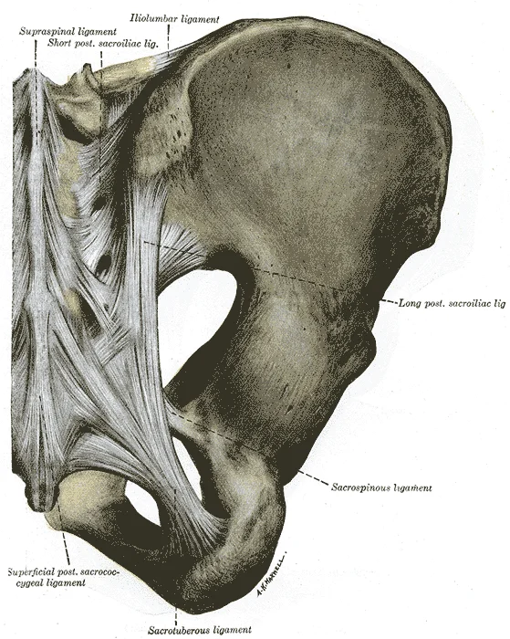
Articulatios of pelvis. Posterior view. (Quain.)

Figure 1: Articulatios of pelvis (posterior view).
Figure 2Gray319
Long description
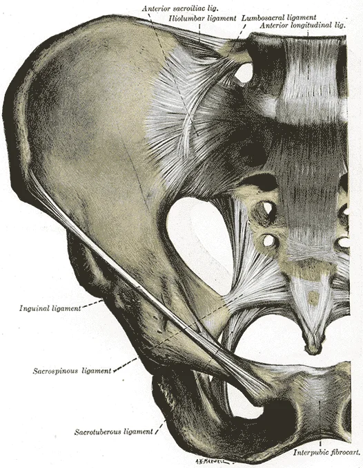
Articulations of pelvis. Anterior view. (Quain.)

Figure 2: Articulations of pelvis (anterior view).
The space enclosed by the bony pelvis divides into two regions. The broad superior region is the greater pelvis (false pelvis), bounded laterally by the fan-like upper part of the hip bone. It contains portions of the small and large intestines. The narrow inferior region is the lesser pelvis (true pelvis), which contains the bladder and other pelvic organs. These regions are separated by the pelvic brim, also called the pelvic inlet. [1]
Figure 3Gray239
Long description
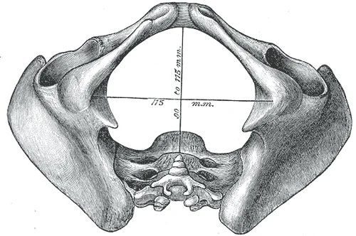
Diameters of inferior aperture of lesser pelvis (female).

Figure 3: Diameters of inferior aperture of lesser pelvis (female).
Figure 4Gray238
Long description
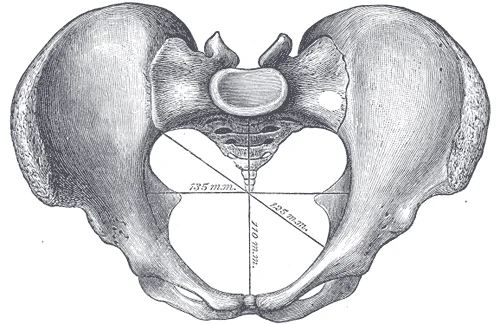
Diameters of superior aperture of lesser pelvis (female).

Figure 4: Diameters of superior aperture of lesser pelvis (female).
The pelvic inlet is defined by a line running from the upper margin of the pubic symphysis anteriorly, along the pectineal line of the pubis and the arcuate line of the ilium, to the sacral promontory posteriorly. Because the pelvis tilts anteriorly, the lesser pelvis is oriented from anterosuperior (inlet) to posteroinferior (outlet). [1]
The pubic symphysis is a secondary cartilaginous joint uniting the two pubic bodies, with a fibrocartilaginous disc between them. The pubic arch is formed by the pubic symphysis, the pubic bodies and the inferior pubic rami. The inferior pubic ramus joins the ischial ramus to form the ischiopubic ramus, and the medial convergence of the two ischiopubic rami creates the subpubic angle. [1]
The female greater sciatic notch is broader, the ischial tuberosities are farther apart, and the pelvic outlet is larger. The female sacrum is wider, shorter and less curved, with a less prominent promontory, giving the pelvic inlet a more oval shape. These differences allow the hip bone to be the most accurate bone for sex determination. [1]
DefinitionPelvic inlet and outlet
The pelvic inlet (brim) separates the greater from the lesser pelvis; the pelvic outlet is the inferior opening of the lesser pelvis. [1]
| Feature | Male | Female |
|---|---|---|
| Subpubic angle | Less than 70 degrees | Greater than 80 degrees |
| Greater sciatic notch | Narrower, deeper | Broader, shallower |
| Ischial tuberosities | Closer together | Farther apart |
| Sacrum | Longer, narrower, more curved | Wider, shorter, less curved |
| Pelvic inlet shape | Heart-shaped | Oval or rounded |
| Lesser pelvis | Narrow, deep, tapering | Wider, shallower |
Pelvic cavity divisions
- Bony pelvisTwo hip bones, sacrum, coccyx
- Question: Pelvic brim (inlet)?
Superior
- Greater pelvis (false pelvis)Contains intestines
Inferior
- Lesser pelvis (true pelvis)Contains bladder, reproductive organs
- Question: Pelvic outlet?
Anterior
- Inferior pubic symphysis
Posterior
- Ischiopubic ramus, ischial tuberosity, sacrotuberous ligament, coccyx tip
The bony pelvis is the entire structure formed by the two hip bones, the sacrum, and the coccyx attached inferiorly to the sacrum[1]. Each hip bone is a single bone in the adult, but it develops from three separate bones that fuse together during the late teenage years: the ilium, ischium, and pubis[1]. These three regions remain distinct in the adult hip bone and serve as the primary landmarks for surface anatomy[1].
DefinitionBony pelvis
The bony pelvis consists of two hip bones, the sacrum, and the coccyx, forming a weight‑bearing ring that transfers load from the vertebral column to the lower limbs.
The ilium forms the large, fan‑like superior portion of the hip bone[1]. Its superior margin is the iliac crest, which can be palpated along the waistline[1]. The anterior end of the iliac crest is the anterior superior iliac spine (ASIS), an important bony landmark felt at the anterolateral hip[1]. Inferior to the ASIS lies the anterior inferior iliac spine (AIIS)[1]. Posteriorly, the iliac crest curves downward to end as the posterior superior iliac spine (PSIS), often visible as a dimple on the lower back[1]. Below the PSIS is the posterior inferior iliac spine (PIIS)[1]. The auricular surface of the ilium articulates with the sacrum to form the sacroiliac joint[1]. The medial surface of the upper ilium forms the iliac fossa, bounded inferiorly by the arcuate line[1]. The posterior margin of the ilium presents the greater sciatic notch[1].
Figure 5Gray236
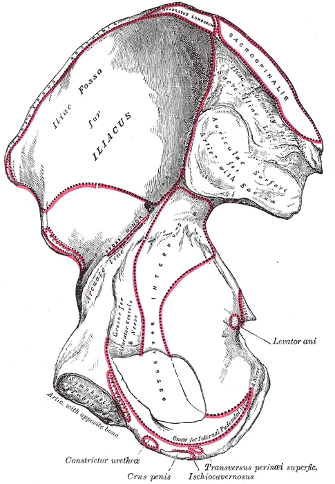
Long description
Right hip bone. Internal surface. Labelled illustration from Wikimedia Commons, used under its open licence.

Figure 5: Right hip bone (internal surface).
The ischium forms the posterolateral portion of the hip bone[1]. Its most prominent feature is the ischial tuberosity, a large roughened area that bears body weight when sitting and can be felt by wiggling the pelvis on a chair[1]. Projecting superiorly and anteriorly from the tuberosity is the ischial ramus[1]. Above the tuberosity, the posterior margin of the ischium forms the lesser sciatic notch, separated from the greater sciatic notch by the ischial spine[1].
The pubis forms the anterior portion of the hip bone. The pubic tubercles are palpable on either side of the pubic symphysis[1]. In the anatomical position, the anterior superior iliac spines and the pubic tubercles lie in the same vertical plane[1].
Figure 6Gray241
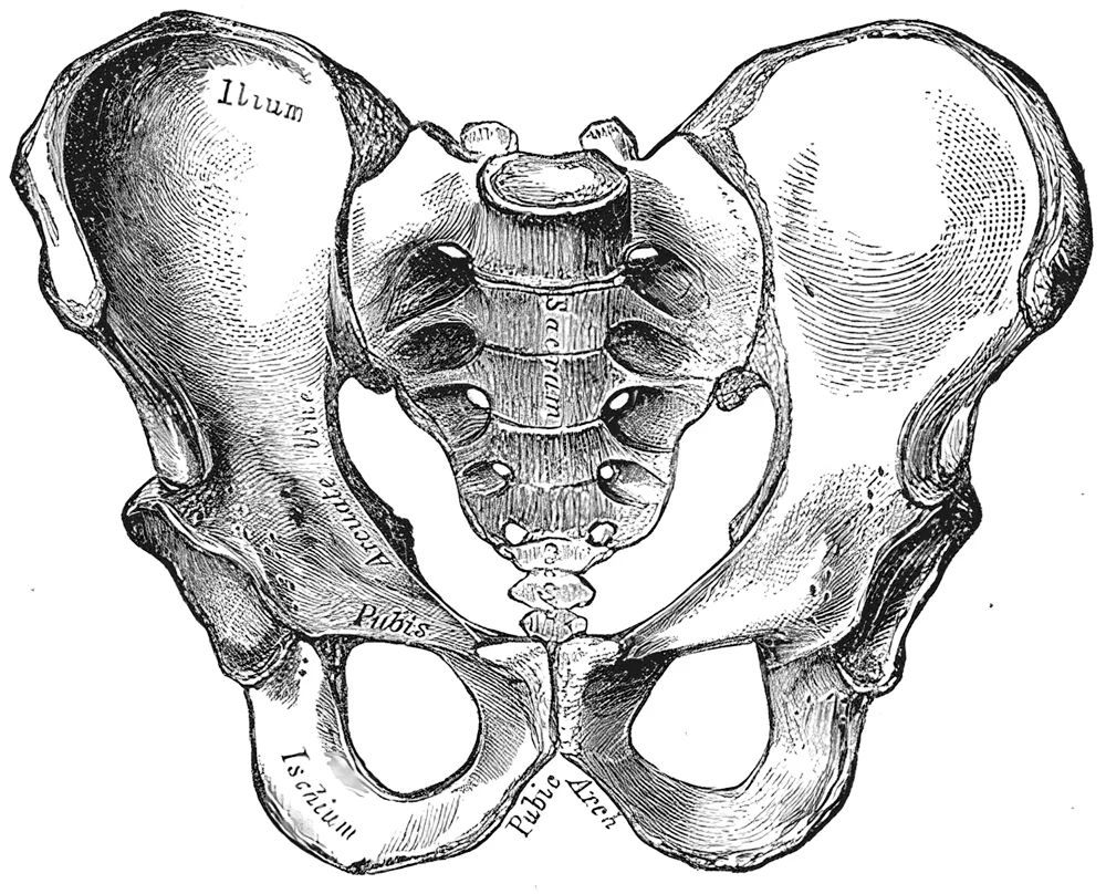
Long description
Male pelvis. Labelled illustration from Wikimedia Commons, used under its open licence.

Figure 6: Male pelvis.
The sacrum articulates with the ilium at the auricular surfaces to form the sacroiliac joints[1]. The coccyx is attached inferiorly to the sacrum[1]. The anterior (internal) surface of the sacrum faces forward and downward when standing[1].
| Landmark | Location | Clinical note |
|---|---|---|
| Iliac crest | Superior margin of ilium, waistline | Site for bone marrow biopsy |
| Anterior superior iliac spine (ASIS) | Anterior end of iliac crest | Reference for inguinal ligament, femoral artery |
| Anterior inferior iliac spine (AIIS) | Inferior to ASIS | Origin of rectus femoris |
| Posterior superior iliac spine (PSIS) | Posterior end of iliac crest | Dimple of Venus; sacroiliac joint level |
| Posterior inferior iliac spine (PIIS) | Inferior to PSIS | Ligament attachment |
| Ischial tuberosity | Inferior ischium | Weight‑bearing when sitting; hamstring origin |
| Ischial spine | Between greater and lesser sciatic notches | Landmark for pudendal nerve block |
| Pubic tubercle | Lateral to pubic symphysis | Inguinal ligament attachment |
| Sacral promontory | Anterior margin of sacral base | Pelvic inlet measurement |
The iliac crests arch across the top, with the ASIS at each anterolateral corner. The AIIS sits just below each ASIS. The pubic tubercles flank the pubic symphysis in the midline. The coccyx extends inferiorly from the sacrum.
Identifying pelvic landmarks by palpation
- StartPlace hands on waist to feel iliac crests
- Question: Move anteriorly along crest?
Yes
- Feel ASIS at anterior end
- Press inferior to ASIS for AIIS
No
- Move posteriorly along crest to PSIS dimples
- Press inferior to PSIS for PIIS
- Sit and wiggle to feel ischial tuberosities
- Palpate pubic tubercles lateral to symphysis
Figure 7Gray242
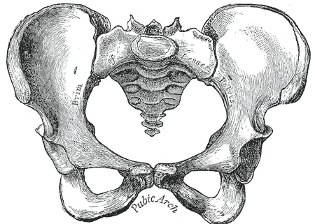
Long description
Female pelvis. Labelled illustration from Wikimedia Commons, used under its open licence.

Figure 7: Female pelvis.
The bony pelvis consists of four bones: the two hip bones, the sacrum, and the coccyx. Together they form a sturdy basin-like ring that transmits body weight from the vertebral column to the lower limbs.
The hip bone is formed by the fusion of three distinct regions: the superior ilium, the posteroinferior ischium, and the anterior pubis. These three areas converge centrally to form a deep, cup-shaped cavity called the acetabulum on the lateral side, which articulates with the femoral head to form the hip joint. Superiorly, the ilium forms a fan-like region that creates the largest part of the hip bone. The ischium supports the body weight when sitting. Anteriorly, the pubis curves medially to join the opposite pubis at a specialized midline joint termed the pubic symphysis. Between the ischium and pubis lies the obturator foramen, a large anteroinferior opening largely filled by connective tissue that serves for muscle attachments on both its internal and external surfaces. [1]
Figure 8Gray237
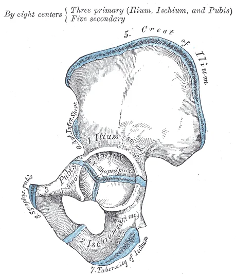
Long description
Plan of ossification of the hip bone. The three primary centers unite through a Y-shaped piece about puberty. Epiphyses appear about puberty, and unite about twenty-fifth year.

Figure 8: Plan of ossification of the hip bone.
Figure 9Gray235
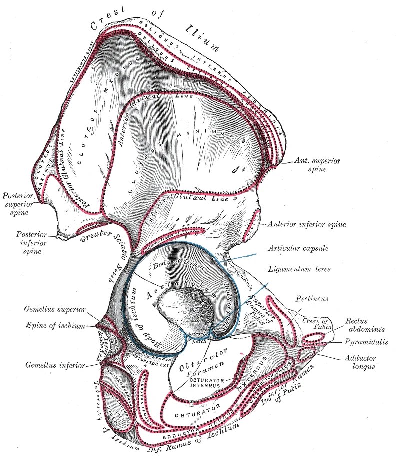
Long description
Right hip bone. External surface. Labelled illustration from Wikimedia Commons, used under its open licence.

Figure 9: Right hip bone (external surface).
Figure 10Gray321
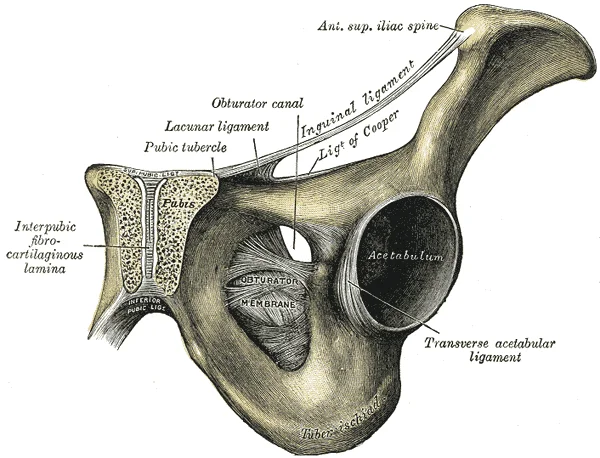
Long description
Symphysis pubis exposed by a coronal section.

Figure 10: Symphysis pubis exposed by a coronal section.
The sacrum lies centrally at the back of the pelvis, wedged between the two hip bones. It is firmly united to the ilium at the largely immobile sacroiliac joint. This joint is supported by strong ligaments, specifically the anterior sacroiliac ligament on the anterior side and the posterior sacroiliac ligament on the posterior side. Additional stability is provided by two major ligaments spanning the sacrum and the hip bone: the sacrospinous ligament, which runs from the sacrum to the ischial spine, and the sacrotuberous ligament, which runs from the sacrum to the ischial tuberosity. These ligaments help to support and immobilize the sacrum as it carries the weight of the body. Furthermore, the sacrospinous and sacrotuberous ligaments contribute to the formation of the greater and lesser sciatic foramina on the posterolateral sides of the pelvis, through which key structures exit for the lower limb. The coccyx forms the small, terminal tailbone segment situated at the very base of the sacrum. [1]
Figure 11Gray240
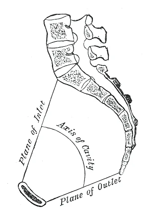
Long description
Median sagittal section of pelvis. Labelled illustration from Wikimedia Commons, used under its open licence.

Figure 11: Median sagittal section of pelvis.
Figure 12Gray324
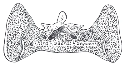
Long description
Coronal section of posterior sacral segment.

Figure 12: Coronal section of posterior sacral segment.
Figure 13Gray323
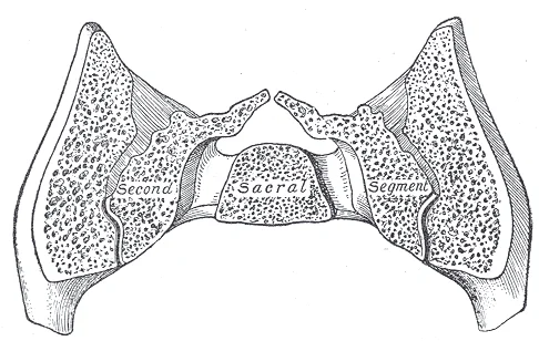
Long description
Coronal section of middle sacra segment.

Figure 13: Coronal section of middle sacra segment.
Figure 14Gray322
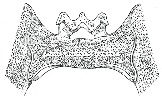
Long description
Coronal section of anterior sacral segment.

Figure 14: Coronal section of anterior sacral segment.
DefinitionPelvic Inlet and Outlet
The pelvic inlet defines the upper aperture of the lesser pelvis, whereas the pelvic outlet forms the lower bony circumference bounded by the pubic arch, ischial tuberosities, and coccyx.
Obstetric measurements evaluate the dimensions of the birth canal, differentiating between the male and female pelvis. The female pelvis is typically wider, shallower, and adapted for childbearing, featuring a rounded pelvic inlet and a larger subpubic angle, whereas the male pelvis is narrower, deeper, and more funnel-shaped with prominent bony landmarks.
The joints of the bony pelvis include the sacroiliac joints, the pubic symphysis, and the lumbosacral and sacrococcygeal joints. The sacroiliac joint is a strong, weight-bearing diarthrodial joint formed between the auricular surfaces of the sacrum and the ilium. It permits minimal gliding and rotation movements. The pubic symphysis is a secondary cartilaginous joint (symphysis) between the two pubic bones, united by an interpubic fibrocartilaginous disc that allows negligible movement.
The ligaments supporting the pelvic joints are exceptionally strong. The sacroiliac joints are stabilised by anterior sacroiliac, posterior sacroiliac, and interosseous sacroiliac ligaments. Additional stability is provided by the sacrotuberous ligament, which runs from the ischial tuberosity to the sacrum and coccyx, and the sacrospinous ligament, running from the ischial spine to the lateral margin of the sacrum and coccyx.
Superiorly, the iliolumbar ligament extends from the transverse process of the fifth lumbar vertebra to the iliac crest. Inferiorly and posteriorly, the broad sacrotuberous ligament lies superficial to the smaller, triangular sacrospinous ligament as both span the greater and lesser sciatic notches. The pubic symphysis is bridged anteriorly by the superior pubic ligament and the arcuate pubic ligament inferiorly. [1]
Significant pelvic differences exist between males and females. The male pelvis is generally narrow, heavy, and deep, with a heart-shaped pelvic inlet, a narrow subpubic angle of less than 70 degrees, and a prominent sacral promontory. Conversely, the female pelvis is wide, shallow, and light, adapted for childbirth, featuring an oval or round pelvic inlet, a wide subpubic angle of 90 to 100 degrees, and a less pronounced sacral promontory. [1]
Obstetric pelvic measurements assess the capacity of the birth canal. The anteroposterior diameter of the pelvic inlet, known as the conjugate vera, measures at least 11 centimetres from the upper margin of the pubic symphysis to the sacral promontory. The transverse diameter of the pelvic inlet is the widest distance between the linea terminalis, measuring approximately 13 centimetres. The interspinous diameter, measured between the ischial spines at the pelvic outlet, is the narrowest transverse dimension of the pelvis at 10 centimetres.
Clinical pearlObstetric measurements
The diagonal conjugate is measured clinically during a vaginal examination by reaching the sacral promontory with the tip of the middle finger and noting the point on the hand or glove touching the inferior margin of the pubic symphysis.
The bony pelvis is enveloped by a complex arrangement of fascial layers that define distinct anatomical compartments. These layers are critical for understanding the spread of infection, fluid, and neoplastic disease within the pelvic region. The internal surface of the pelvic bones is lined by the parietal pelvic fascia, which is continuous with the transversalis fascia superiorly and the endopelvic fascia inferiorly. This fascial lining provides a smooth surface for the pelvic viscera and serves as an attachment point for various ligaments and muscles.
Radiotherapy can lead to direct bone necrosis; usually this is incomplete and a reactive inflammatory reaction ensues. [2]
The pelvic compartments are divided into the true pelvis and the false pelvis. The true pelvis is bounded by the pelvic bones and contains the pelvic viscera. The false pelvis is bounded by the iliac wings and contains the abdominal viscera. The fascia plays a crucial role in maintaining the integrity of these compartments. It prevents the spread of infection from one compartment to another. However, in cases of severe trauma or infection, the fascia can be breached, leading to widespread pelvic sepsis.
Clinical pearlClinical Relevance
The parietal pelvic fascia is a key landmark in pelvic surgery, providing a plane for dissection and helping to identify neurovascular structures.
This line serves as an attachment for the obturator membrane. The fascia also provides attachment for the levator ani muscle, which forms the floor of the pelvis. The levator ani is innervated by the nerve to the levator ani, a branch of the sacral plexus.
| Structure | Location | Relation |
|---|---|---|
| Parietal Pelvic Fascia | Lines internal pelvic surface | Continuous with transversalis fascia |
| Obturator Fascia | Covers obturator internus | Forms lateral pelvic wall |
| Endopelvic Fascia | Surrounds pelvic viscera | Contains neurovascular bundles |
| Levator Ani Fascia | Covers levator ani | Forms pelvic floor |
DefinitionEndopelvic Fascia
A layer of connective tissue that surrounds the pelvic viscera and contains neurovascular structures.
These structures are crucial for the innervation and blood supply of the perineum.
Exam trapCommon Error
Confusing the parietal pelvic fascia with the endopelvic fascia can lead to errors in surgical dissection and understanding of pelvic anatomy.
The fascia of the bony pelvis is a complex and dynamic structure that plays a vital role in maintaining the integrity of the pelvic compartment. It provides support to the pelvic viscera, contains neurovascular structures, and serves as a plane for surgical dissection. Understanding the anatomy of the pelvic fascia is essential for medical students and clinicians alike.
The muscles of the bony pelvis are organised into distinct compartments, each with specific origins, insertions, actions, and nerve supplies. Understanding these groupings is essential for clinical anatomy and surgical approaches.
The gluteal muscles are located on the lateral aspect of the pelvis. They are primarily responsible for hip movement and stability.
| Muscle | Origin | Insertion | Action | Nerve Supply |
|---|---|---|---|---|
| Gluteus maximus | Ilium, sacrum, coccyx | Iliotibial tract, gluteal tuberosity | Extends and laterally rotates hip | Inferior gluteal nerve |
| Gluteus medius | Outer surface of ilium | Greater trochanter | Abducts and medially rotates hip | Superior gluteal nerve |
| Gluteus minimus | Outer surface of ilium | Greater trochanter | Abducts and medially rotates hip | Superior gluteal nerve |
The gluteus maximus is the most superficial layer. It covers the deeper gluteus medius and minimus. The gluteus medius lies lateral to the ilium. The gluteus minimus is deep to the medius. These muscles attach to the greater trochanter of the femur. Their action stabilises the pelvis during walking.
The iliopsoas is the primary flexor of the hip joint. It consists of two muscles that merge before insertion.
| Muscle | Origin | Insertion | Action | Nerve Supply |
|---|---|---|---|---|
| Iliacus | Iliac fossa | Lesser trochanter | Flexes hip | Femoral nerve |
| Psoas major | Transverse processes of lumbar vertebrae | Lesser trochanter | Flexes hip | Ventral rami of L1-L3 |
The iliacus originates from the iliac fossa. The psoas major originates from the lumbar vertebrae. Both muscles converge to insert on the lesser trochanter. This common insertion allows for powerful hip flexion. The femoral nerve supplies the iliacus. The ventral rami of L1-L3 supply the psoas major.
These muscles are part of the deep gluteal group. They act as lateral rotators of the hip.
| Muscle | Origin | Insertion | Action | Nerve Supply |
|---|---|---|---|---|
| Obturator internus | Inner surface of obturator membrane | Greater trochanter | Laterally rotates hip | Nerve to obturator internus |
| Piriformis | Anterior surface of sacrum | Greater trochanter | Laterally rotates hip | Nerve to piriformis |
The obturator internus originates from the obturator membrane. The piriformis originates from the sacrum. Both insert on the greater trochanter. They stabilise the hip joint during movement. The nerve to the obturator internus supplies the first muscle. The nerve to the piriformis supplies the second.
The obturator internus is deeper still. These muscles are crucial for lateral rotation. Their nerve supplies are distinct. This anatomy is vital for surgical safety.
The main arterial supply to the bony pelvis is delivered by the internal iliac artery, which arises from the common iliac artery at the level of the lumbosacral disc and bifurcates near the pelvic brim into anterior and posterior divisions.
DefinitionInternal iliac artery
The primary vessel supplying the pelvic walls, viscera, perineum, and gluteal region.
The posterior division of the internal iliac artery runs posteriorly against the pelvic wall and gives off three main parietal branches: the iliolumbar artery, lateral sacral arteries, and superior gluteal artery. The anterior division continues inferiorly and gives off multiple visceral and parietal branches, including the obturator artery, internal pudendal artery, and inferior gluteal artery.
| Division | Branch | Main Distribution |
|---|---|---|
| Posterior | Superior gluteal | Gluteal muscles and hip |
| Posterior | Iliolumbar | Iliacus and psoas major |
| Anterior | Obturator | Medial compartment of thigh |
| Anterior | Internal pudendal | Perineum and external genitalia |
The common iliac artery descends from the abdomen before splitting into the external iliac and internal iliac arteries. The internal iliac artery lies deep within the pelvic cavity, closely related to the ureter anteriorly and the internal iliac vein posteriorly. Tracing the posterior division leads to the superior gluteal artery passing out of the greater sciatic foramen above the piriformis muscle.
The venous drainage of the bony pelvis is a complex network that mirrors the arterial supply, ultimately converging into the internal iliac veins. This system is critical for understanding pelvic pathology, as infections or malignancies can spread via these valveless channels. The primary vessels involved are the internal iliac veins, which receive blood from the pelvic viscera and the pelvic wall.
The internal iliac veins are formed by the union of tributaries that drain the pelvic organs and walls. They run alongside the internal iliac arteries and terminate by joining the external iliac veins to form the common iliac veins. The right and left common iliac veins then unite to form the inferior vena cava. This drainage pathway ensures that deoxygenated blood from the pelvis returns to the heart efficiently. [1]
The internal iliac veins receive several key tributaries, which can be categorized into parietal and visceral groups. The parietal veins drain the pelvic wall and lower limbs, while the visceral veins drain the pelvic organs. [1]
| Structure | Origin | Course | Termination |
|---|---|---|---|
| Inferior Gluteal Vein | Gluteal region | Runs with inferior gluteal artery | Internal iliac vein |
| Obturator Vein | Obturator canal | Runs with obturator artery | Internal iliac vein |
| Internal Pudendal Vein | Perineum | Runs with internal pudendal artery | Internal iliac vein |
| Middle Rectal Vein | Rectum | Runs with middle rectal artery | Internal iliac vein |
| Vesical Veins | Bladder | Form vesical plexus | Internal iliac vein |
| Uterine Veins | Uterus | Form uterine plexus | Internal iliac vein |
| Vaginal Veins | Vagina | Form vaginal plexus | Internal iliac vein |
The pelvic region contains several important venous plexuses, which are interconnected networks of veins. These plexuses allow for collateral circulation and provide alternative pathways for blood flow if primary vessels are obstructed.
The prostatic venous plexus in males and the uterine venous plexus in females are particularly significant. These plexuses communicate with the vertebral venous plexus, which extends from the skull to the pelvis. This connection is clinically relevant because it provides a route for the spread of metastatic cancer cells from pelvic organs to the vertebral column and brain, bypassing the pulmonary circulation. [1]
Understanding this anatomy is essential for diagnosing and treating pelvic venous disorders.
The bony pelvis is innervated by a complex network of nerves derived from the lumbosacral plexus. These nerves provide both motor supply to the muscles of the pelvic floor and lower limb, and cutaneous sensation to the perineum and medial thigh. Understanding this supply is critical for interpreting neurological deficits in pelvic trauma or surgery. [2]
The sciatic nerve is the largest branch, exiting the pelvis through the greater sciatic foramen below the piriformis muscle. The superior gluteal nerve exits above the piriformis.
The motor and cutaneous supply can be summarised in the table below.
| Nerve | Motor Supply | Cutaneous Supply |
|---|---|---|
| Sciatic | Hamstrings, muscles of leg and foot | Posterior thigh, leg, and foot |
| Pudendal | Perineal muscles, external sphincters | Perineum, external genitalia |
| Superior Gluteal | Gluteus medius, minimus, tensor fasciae latae | Lateral thigh (via lateral cutaneous nerve) |
| Inferior Gluteal | Gluteus maximus | None specific |
| Nerve | Origin | Course | Branches | Termination |
|---|---|---|---|---|
| Sciatic | L4-S3 | Exits greater sciatic foramen | Tibial, Common fibular | Below knee |
| Pudendal | S2-S4 | Loops ischial spine | Inferior rectal, Perineal, Dorsal penile/clitoral | Perineum |
Lesions of these nerves result in specific clinical signs. A lesion of the sciatic nerve causes weakness in knee flexion and foot movement. A lesion of the pudendal nerve leads to perineal sensory loss and sphincter dysfunction. The superior gluteal nerve lesion results in a Trendelenburg gait due to gluteus medius weakness.
DefinitionSciatic Nerve
The largest nerve in the body, arising from the sacral plexus, supplying the posterior thigh and all muscles below the knee.
Exam trapPudendal Nerve
Often injured in childbirth; remember it loops around the ischial spine, not the ischial tuberosity.
The bony pelvis contains a complex network of lymph nodes and channels that drain the pelvic viscera, pelvic walls, and lower limbs. Understanding this drainage pathway is essential for staging pelvic malignancies and predicting their patterns of metastasis.
Lymph from the pelvic structures ascends primarily through three major chains of lymph nodes that cluster around the principal branches of the internal iliac artery. The external iliac lymph nodes lie along the external iliac vessels, receiving lymph from the lower limb, lower abdominal wall, and parts of the pelvic viscera. The internal iliac lymph nodes surround the internal iliac vessels and drain the pelvic organs, perineum, and gluteal region. The sacral lymph nodes sit on the anterior surface of the sacrum, draining the posterior pelvic wall, rectum, and prostate or cervix.
Efferent vessels from these three primary groups converge upon the common iliac lymph nodes, which are arranged around the common iliac artery from the bifurcation of the aorta to the pelvic brim. From the common iliac chain, lymph ascends to the lumbar aortic lymph nodes situated along the abdominal aorta and inferior vena cava.
DefinitionSentinel Node Concept
The first lymph node in the lymphatic basin that receives drainage from a primary tumour, dictating the initial site of potential metastasis.
| Node Group | Anatomical Relations | Main Drainage Areas |
|---|---|---|
| External iliac | Along external iliac vessels | Lower limb, lower abdominal wall |
| Internal iliac | Around internal iliac artery | Pelvic viscera, perineum |
| Sacral | Anterior surface of sacrum | Posterior pelvis, rectum |
| Common iliac | Along common iliac artery | Primary pelvic nodes |
Pelvic Lymphatic Flow
- Pelvic Viscera and Lower LimbInitial lymph collection
- Question: Which pathway?
Viscera
- Internal Iliac NodesAlong internal iliac vessels
Limb/Wall
- External Iliac NodesAlong external iliac vessels
- Common Iliac NodesConvergence at pelvic brim
- Lumbar Aortic NodesAscent to abdomen
The bony pelvis forms a robust ring that supports the axial skeleton and protects the pelvic viscera. Its structural integrity relies on the precise articulation of the sacrum with the ilia at the sacroiliac joints, and the anterior union at the pubic symphysis. Understanding the spatial relationships within this ring is critical for interpreting trauma and surgical approaches.
The sacrospinous and sacrotuberous ligaments convert the sciatic notches into the greater and lesser sciatic foramina, which serve as passageways for neurovascular structures. [1]
The pelvic cavity is divided into the false pelvis (greater pelvis) and the true pelvis (lesser pelvis). The false pelvis is bounded by the iliac wings and contains loops of the small intestine. The true pelvis houses the bladder, rectum, and reproductive organs. The pelvic floor, formed by the levator ani and coccygeus muscles, supports these viscera. Its primary action is to support the pelvic viscera and maintain continence.
Injuries to the bony pelvis often involve complex fracture patterns. Complete fractures extend through both cortices and may be associated with a medial spike, incomplete fractures involve only the lateral cortex. Such fractures can disrupt the stability of the pelvic ring, leading to significant haemorrhage due to the rich vascular supply of the pelvic bones and surrounding soft tissues. The internal iliac artery and its branches are particularly vulnerable in posterior pelvic ring injuries. [2]
It is important to realise that what appears like increased fluid signal on MRI and is therefore colloquially called “bone marrow oedema” is in fact granulation tissue with fibrovascular ingrowth. Oedema-like bone marrow changes therefore represent a tissue reaction to a variety of stimuli; trabecular microfractures leading to bone repair is one of them. This distinction is vital for accurate diagnosis and management, as it reflects the body's healing response rather than simple fluid accumulation. [2]
Clinical pearlClinical Pearl
Always distinguish between true oedema and granulation tissue on MRI to avoid misinterpreting healing fractures as active pathology.
The neurovascular structures passing through the sciatic foramina are of paramount importance. The sciatic nerve, the largest nerve in the body, exits the pelvis through the greater sciatic foramen, inferior to the piriformis muscle. It provides motor innervation to the posterior thigh muscles and sensory innervation to the leg and foot. The pudendal nerve, responsible for perineal sensation and motor control of the external sphincters, also exits via the greater sciatic foramen and re-enters the pelvis through the lesser sciatic foramen.
| Muscle | Origin | Insertion | Action | Nerve Supply |
|---|---|---|---|---|
| Levator Ani | Pubis, Obturator Internus | Coccyx, Anococcygeal Raphe | Support viscera, maintain continence | Nerve to Levator Ani |
| Coccygeus | Sacrospinous Ligament | Coccyx, Sacrum | Support viscera | Nerve to Coccygeus |
| Piriformis | Sacrum | Greater Trochanter | Lateral rotation of hip | Nerve to Piriformis |
X-ray imaging of the bony pelvis provides a primary overview of skeletal integrity, showing cortical outlines, joint spaces, and trabecular patterns. Computed tomography, abbreviated as CT, offers superior bony detail compared to standard radiography. CT is more sensitive than radiography but remains less sensitive than magnetic resonance imaging, known as MRI, or bone scintigraphy.
Magnetic resonance imaging provides exceptional soft tissue contrast and detects early bone marrow edema and occult trabecular disruption. Ultrasound has specific applications in musculoskeletal assessment, though it is not routinely performed for primary pelvic stress assessment.
DefinitionImaging hierarchy
Computed tomography provides high spatial resolution for bone architecture, while magnetic resonance imaging excels at detecting early marrow pathology.
The anterior superior iliac spine lies at the superior aspect of the iliac wing, projecting lateral to the pelvic brim and serving as the attachment site for the inguinal ligament. The pubic symphysis lies in the midline inferiorly, joining the two pubic bones via a fibrocartilaginous disc. The ischial tuberosity lies posteroinferiorly, supporting body weight in the seated position and relating medially to the obturator foramen. The femoral head lies superolaterally, articulating within the acetabulum, which is formed by the fusion of the ilium, ischium, and pubis.
| Modality | Primary advantage | Relative sensitivity |
|---|---|---|
| Radiography | Cortical alignment and fractures | Lowest |
| Computed tomography | Fine bony architecture | Moderate |
| Magnetic resonance imaging | Marrow edema and soft tissue | High |
Pelvic Imaging Workflow
- Clinical suspicion of bony injuryAssess pain pattern and activity history.
- Question: Initial X-ray negative?
Yes
- Proceed to MRI or CT for occult injury
No
- Confirm fracture and manage
Clinical assessment of the pelvis relies on distinct surface landmarks. Clinicians palpate the anterior superior iliac spine, the pubic tubercle, and the ischial tuberosity to align pelvic radiographs and assess bony symmetry. The internal surface of the upper ilium features the shallow depression called the iliac fossa. The inferior margin of this space is formed by the arcuate line of the ilium, which is the ridge created by the pronounced change in curvature between the upper and lower portions of the ilium. Posteriorly, the large, inverted U-shaped indentation located on the margin of the lower ilium is called the greater sciatic notch. This notch transmits major neurovascular structures exiting the true pelvis into the gluteal region, making it a critical landmark during surgical exposures and in interpreting pelvic radiographs. [1]
Injuries to the pelvic ring commonly result from high-energy trauma such as motor vehicle collisions, whereas insufficiency fractures occur in osteoporotic bone under normal physiological loads. Colloquially, the term stress fracture is also used instead of fatigue fractures; however, stress fracture encompasses both fatigue and insufficiency fractures in clinical definitions. In the early phase of a stress fracture, a cortical radiolucency without a periosteal reaction or callus formation might In later stages cortical irregularity or disruption and later still callus formation may become visible [2]
Exam trapRadiographic limitations
Initial radiographs have a sensitivity of less than 50 percent for stress fracture, with some authors describing sensitivities as low as 10 percent, meaning normal films never exclude early injuries. [2]
In the operating theatre, precise anatomical knowledge is vital for surgical approaches. Recognising the shallow lesser sciatic notch and the greater sciatic notch allows surgeons to navigate around these structures safely. During hip replacement or pelvic fixation, understanding the biomechanics of the femoral shaft is crucial.
The iliac fossa is seen as the smooth concavity on the anteromedial aspect of the upper ilium, bounded inferiorly by the arcuate line of the ilium. The greater sciatic notch appears as the large, inverted U-shaped indentation on the posterior margin of the lower ilium, situated directly above the ischial spine.
Pelvic Stress Injury Evaluation
- Clinical presentation with pelvic or groin painAssess risk factors such as osteoporosis or repetitive loading.
- Question: Are initial radiographs positive?
Yes
- Confirm fracture and manage accordingly
No
- Recognize sensitivity is less than 50 percent
- Order advanced cross-sectional imaging like MRI to detect marrow oedema or cortical radiolucency
This in turn makes the bone vulnerable to stress fractures. [2]
The differentiation between a fatigue and an insufficiency fracture is not always easy, as both can occur at the same time in the same patient and fatigue as well as insufficiency can contribute to any fracture. In clinical practice, the use of the term “stress fracture” relieves the radiologist from committing himself to a more specific diagnosis of a fatigue or an insufficiency fracture. It is often not possible to differentiate between the two without further information. Therefore, a comprehensive history including activity levels and bone health status is required for accurate classification. [2]
Atypical stress fractures are not common. When evaluating femoral fractures, only 7–10 % of femoral fractures are located below the lesser trochanter; of these 75 % are due to major trauma reducing the incidence to 2-3 %. This low incidence highlights that subtrochanteric fractures are predominantly traumatic rather than stress-related in the general population. [2]
Longitudinal stress fractures of the patella are less common and are due to compression against the femoral condyles. [2]
Clinical pearlKey Distinction
Fatigue fractures result from abnormal stress on normal bone, whereas insufficiency fractures result from normal stress on abnormal bone.
Exam trapDiagnostic Caution
The differentiation between a fatigue and an insufficiency fracture is not always easy, as both can occur at the same time in the same patient and fatigue as well as insufficiency can contribute to any fracture.
DefinitionAtypical Fracture Prevalence
Atypical stress fractures are not common.
NoteFemoral Anatomy
Only 7–10 % of femoral fractures are located below the lesser trochanter.
DefinitionPatellar Fracture Mechanism
Transverse stress fractures are due to traction stress. [2]
Exam trapPatellar Risk
In particular transverse patella fractures have a high risk of becoming complete fractures. [2]
NoteLongitudinal Patellar Fracture
Longitudinal stress fractures of the patella are less common and are due to compression against the femoral condyles.
Clinical pearlSite Specificity
Patella and tibia plateau stress fractures are more commonly due to insufficiency fractures. [2]
DefinitionRadiological Terminology
The use of the term “stress fracture” relieves the radiologist from committing himself to a more specific diagnosis of a fatigue or an insufficiency fracture.
NoteDiagnostic Limitation
It is often not possible to differentiate between the two without further information.
Exam trapTrauma vs Stress
Of these 75 % are due to major trauma reducing the incidence to 2-3 %.
Clinical pearlDual Aetiology
The differentiation between a fatigue and an insufficiency fracture is not always easy, as both can occur at the same time in the same patient and fatigue as well as insufficiency can contribute to any fracture.
Students frequently confuse the anatomical boundaries of the hip bone with the functional units of the pelvis. A primary error is misidentifying the ischium as the primary weight-bearing structure during standing. The ischium is the posterolateral portion of the hip bone, and while it supports body weight when sitting via the expanded ischial tuberosity, it is not the main load-bearing element in upright posture. Another frequent mistake involves the relationship between the sciatic notches. Students often overlook the ischial spine, which separates the greater and lesser sciatic notches, leading to errors in identifying the path of the piriformis muscle or the sciatic nerve. [1]
In the context of stress fractures, a major pitfall is assuming that early imaging is definitive. Bone and periosteal reactions do not occur until several weeks into the disease process, which explains the delayed occurrence of radiographically visible changes in stress fractures. Consequently, relying solely on plain radiographs in the acute phase often leads to a false negative diagnosis. In relatively acute stress fractures, one may see a cortical lucency without a periosteal reaction or callus formation, which should not be dismissed as normal. [2]
A critical diagnostic trap is misclassifying atypical femoral fractures (AFF). These are not simple insufficiency fractures. The American Society for Bone and Mineral Research criteria require the fracture to be located along the femoral diaphysis from just distal to the lesser trochanter to just proximal to the supracondylar flare. Furthermore, at least four of five Major Features must be present for diagnosis. A common error is ignoring the origin of the fracture line. The fracture line origin The fracture line originates at the lateral cortex and is substantially transverse in its orientation [2]
The authors are aware of cases where atypical femoral fractures were misdiagnosed as “normal” insufficiency fractures leading to continued or even newly instigated bisphosphonate treatment and eventually to overt fracture. [2]
Changes in the collagen make-up affect its mechanical strength and elasticity. [2]
Knowledge of the bony pelvis is essential at the bedside for localising pain. When a patient presents with buttock pain that worsens with sitting, the clinician must consider the ischial tuberosity, which supports body weight when sitting. Understanding that the ischial ramus projects anteriorly and superiorly helps in palpating the ischial tuberosity and assessing for tenderness indicative of avulsion injuries or bursitis.
Nevertheless it is clear that the benefit of bisphosphonate use by far outweighs the risk of atypical femoral fractures [2]
Localized periosteal or endosteal thickening of the lateral cortex is present at the fracture site [2]
Students must know that once a patient has developed a stress fracture there is increased risk of further stress fractures in the same limb and the contralateral side. This is [2]
Diagnosis of Atypical Femoral Fracture
- Patient presents with thigh painMinimal or no trauma history
- The bony pelvis is a weight-bearing ring composed of two hip bones, the sacrum, and the coccyx, which connects the axial skeleton to the lower limbs.
- The pelvic cavity is divided into the greater pelvis, which contains portions of the intestines, and the lesser pelvis, which houses the bladder and other pelvic organs, separated by the pelvic brim.
- The female pelvis is wider and shallower than the male pelvis, featuring a subpubic angle greater than 80 degrees and an oval-shaped inlet to facilitate childbirth.
- The sacroiliac joint is a strong synovial joint between the ilium and sacrum, while the pubic symphysis is a secondary cartilaginous joint uniting the two pubic bodies.
- The internal iliac artery is the primary arterial supply to the pelvis, dividing into anterior and posterior divisions that provide parietal and visceral branches to pelvic structures.
- Venous drainage from the pelvis converges into the internal iliac veins, which contain valveless channels and plexuses that allow for collateral circulation and potential spread of metastasis.
- The sciatic nerve is the largest nerve in the body, exiting the pelvis through the greater sciatic foramen below the piriformis muscle to supply the posterior thigh and lower limb.
- The parietal pelvic fascia lines the internal surface of the pelvic bones and serves as a key surgical landmark for dissection and identifying neurovascular structures.
Answer each one in your head first, then open the answer.
What anatomical structure separates the greater pelvis from the lesser pelvis?
Show answerHide answer
The pelvic brim, also known as the pelvic inlet, separates the broad superior greater pelvis from the narrow inferior lesser pelvis.How does the shape of the pelvic inlet differ between male and female pelvises?
Show answerHide answer
The male pelvic inlet is heart-shaped, whereas the female pelvic inlet is oval or rounded to accommodate childbirth.Which specific ligaments contribute to the formation of the greater and lesser sciatic foramina?
Show answerHide answer
The sacrospinous and sacrotuberous ligaments contribute to the formation of the greater and lesser sciatic foramina on the posterolateral sides of the pelvis.What is the clinical significance of the communication between pelvic venous plexuses and the vertebral venous plexus?
Show answerHide answer
This communication provides a route for metastatic cancer cells from pelvic organs to spread to the vertebral column and brain, bypassing the pulmonary circulation.Why is the hip bone considered the most accurate bone for sex determination?
Show answerHide answer
The hip bone exhibits significant sexual dimorphism, with the female pelvis being wider and shallower and the male pelvis being narrower and deeper, allowing for reliable differentiation.
- Betts JG, Young KA, Wise JA, et al. The Pelvic Girdle and Pelvis. OpenStax, Anatomy and Physiology 2e (CC BY-NC-SA 4.0). Accessed .
- Tins BJ, Garton M, Cassar-Pullicino VN, et al. Stress fracture of the pelvis and lower limbs including atypical femoral fractures-a review. Insights into imaging 2015;6(1):97-110. doi:10.1007/s13244-014-0371-z. Accessed .
Figure credits
- Figure 1: Henry Vandyke Carter, via Wikimedia Commons, Public domain, Public domain.
- Figure 2: Henry Vandyke Carter, via Wikimedia Commons, Public domain, Public domain.
- Figure 3: Henry Vandyke Carter, via Wikimedia Commons, Public domain, Public domain.
- Figure 4: Henry Vandyke Carter, via Wikimedia Commons, Public domain, Public domain.
- Figure 5: Henry Vandyke Carter, via Wikimedia Commons, Public domain, Public domain.
- Figure 6: Henry Vandyke Carter, via Wikimedia Commons, Public domain, Public domain.
- Figure 7: Henry Vandyke Carter, via Wikimedia Commons, Public domain, Public domain.
- Figure 8: Henry Vandyke Carter, via Wikimedia Commons, Public domain, Public domain.
- Figure 9: Henry Vandyke Carter, via Wikimedia Commons, Public domain, Public domain.
- Figure 10: Henry Vandyke Carter, via Wikimedia Commons, Public domain, Public domain.
- Figure 11: Henry Vandyke Carter, via Wikimedia Commons, Public domain, Public domain.
- Figure 12: Henry Vandyke Carter, via Wikimedia Commons, Public domain, Public domain.
- Figure 13: Henry Vandyke Carter, via Wikimedia Commons, Public domain, Public domain.
- Figure 14: Henry Vandyke Carter, via Wikimedia Commons, Public domain, Public domain.
Educational content for medical students and doctors. It is not a substitute for clinical judgement or local guidelines.