Pelvic walls and floor
Anatomical boundaries and muscular architecture of the pelvic walls and floor.
The pelvic region serves as the structural foundation for the lower limbs, connecting the axial skeleton to the appendicular system. Unlike the pectoral girdle, which allows significant movement, the pelvic girdle is designed primarily for stability and support. This stability arises from its robust connection to the sacrum at the base of the axial skeleton. The pelvic girdle attaches the legs to the axial skeleton, with the hip joint marking the convergence of the pelvic girdle and the leg. [1]
The bony pelvis is divided into two distinct regions by the pelvic brim, also known as the pelvic inlet. The broad, superior region, defined laterally by the large, fan-like portion of the upper hip bone, is called the greater pelvis. The inferior region is the lesser pelvis, or true pelvis, which is narrow and rounded, containing the bladder and other pelvic organs. The pelvic brim is defined anteriorly by the upper margin of the pubic symphysis and posteriorly by the sacral promontory, al [3]
The pelvic walls are formed by specific muscles that line the inner surface of the bony pelvis. The obturator internus and piriformis muscles are key components of the lateral pelvic walls. These muscles, along with the levator ani and coccygeus, contribute to the structural integrity of the pelvic region. The pelvic floor is a muscular sheet that defines the inferior portion of the pelvic cavity. It comprises the pelvic diaphragm, which spans from the pubis anteriorly to the coccyx posteriorly. [2]
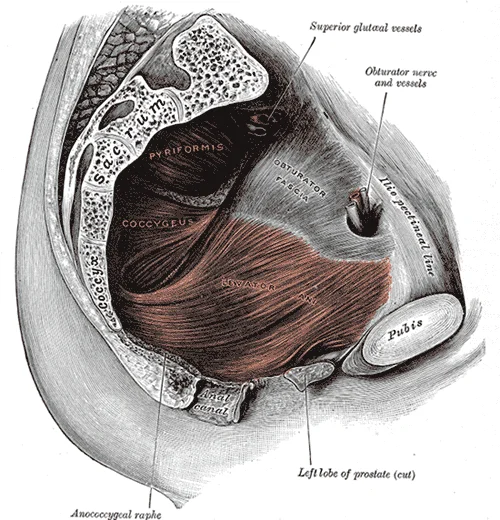
Figure 1Gray404
Long description
Left Levator ani from within. Labelled illustration from Wikimedia Commons, used under its open licence.

Figure 1: Left Levator ani from within.
DefinitionPelvic Diaphragm
The pelvic diaphragm spans anteriorly to posteriorly from the pubis to the coccyx and comprises the levator ani and the ischiococcygeus. [2]
This resistance aids defecation by applying pressure to the colon and assists childbirth by supporting the uterus. It is divided into the urogenital triangle anteriorly an [2]
| Structure | Primary Function |
|---|---|
| Obturator Internus | Lateral rotation of the femur |
| Piriformis | Lateral rotation of the femur |
| Levator Ani | Supports pelvic viscera |
| Ischiococcygeus | Pulls coccyx anteriorly |
The bony pelvis is divided into two distinct regions by the pelvic brim, also known as the pelvic inlet. The superior region is the greater pelvis, or false pelvis, which is broad and occupied by portions of the small and large intestines. The inferior region is the lesser pelvis, or true pelvis, which is narrow and rounded, containing the bladder and other pelvic organs. The pelvic brim is defined by a line formed by the upper margin of the pubic symphysis anteriorly, and the pectineal line of the pubis, the arcuate line of the ilium, and the sacral promontory [3]
The figure labels the iliac crest running between the ASIS and PSIS, the iliac fossa medial to the crest, the arcuate line crossing the ilium, the sacrum and coccyx posteriorly, the pubic symphysis and pubic tubercles anteriorly, the ischial tuberosities inferolaterally, and the greater sciatic notch on the posterior ilium. The inguinal ligament stretches from the ASIS to the pubic tubercle, marking the femoral triangle’s superior border. The sacroiliac joints lie deep to the PSIS, palpable as dimples over the posterior superior iliac spines. [2][3]
| Landmark | Location | Palpable? | Passage |
|---|---|---|---|
| Anterior superior iliac spine (ASIS) | Anterior end of iliac crest | Yes | P54 |
| Posterior superior iliac spine (PSIS) | Posterior end of iliac crest | Yes (dimples) | P54 |
| Pubic tubercle | Lateral end of pubic crest | Yes | P45 |
| Ischial tuberosity | Inferolateral pelvis | Yes (sitting) | P37 |
| Sacral promontory | Anterior sacrum, L5‑S1 | Deep, not surface | P35 |
| Coccyx | Inferior to sacrum | Yes (tip) | P45 |
| Greater sciatic notch | Posterior ilium | Deep, not surface | P41 |
The perineum lies inferior to the pelvic floor; its muscles play roles in urination, ejaculation, and vaginal contraction[1]. Surface markings of the pelvic floor include the urogenital triangle anteriorly and the anal triangle posteriorly, separated by the transverse perineal line between the ischial tuberosities.
The pelvis differs fundamentally from the pectoral girdle; while the shoulder allows extensive mobility, the pelvic bones are strongly united to form a largely immobile, weight-bearing structure. This immobility ensures that the body’s centre of gravity, located in the pelvic area, remains fixed, facilitating stabilization and posture[1]. [3]
The ilium forms the superior portion. Its superior margin is the iliac crest, which you can feel at your waistline. The anterior termination of this crest is the anterior superior iliac spine, palpable at the anterolateral hip. Inferior to this lies the anterior inferior iliac spine, both serving as muscle attachment points. Posteriorly, the crest curves to the posterior superior iliac spine, often creating a visible depression or “dimple” on the lower back. Below this is the posterior inferior iliac spine. [2] [3]
The ischium constitutes the posterolateral hip bone. Its large, roughened inferior area is the ischial tuberosity, which supports body weight during sitting and attaches posterior thigh muscles. You can feel this by wiggling against a chair seat. Projecting superiorly and anteriorly from the tuberosity is the ischial ramus. The posterior margin above the tuberosity is the lesser sciatic notch. The ischial spine separates the lesser from the greater sciatic notch. [3]
The pubis forms the anterior hip bone. Its body articulates with the opposite pubis at the pubic symphysis. The superior margin features the pubic tubercle. The superior pubic ramus joins the ilium, forming the pectineal line. The inferior pubic ramus projects inferiorly and laterally, joining the ischial ramus to form the ischiopubic ramus. The pubic arch comprises the symphysis, pubic bodies, and inferior rami. The subpubic angle results from the medial convergence of the ischiopubic rami. [3]
Ligaments unite these bones. The sacroiliac joint is supported by anterior and posterior sacroiliac ligaments between the sacrum and ilium. The sacrospinous ligament runs from the sacrum to the ischial spine. The sacrotuberous ligament extends from the sacrum to the ischial tuberosity. These structures immobilize the sacrum, allowing it to carry body weight effectively. [3]
The pelvic girdle forms a stable ring that transmits weight from the axial skeleton to the lower limbs. This stability relies on strong ligamentous connections and specific joint types.
DefinitionSacroiliac Joint
The articulation between the sacrum and the hip bone, reinforced by robust ligaments to withstand significant compressive forces.
The sacroiliac joint is a synovial joint located posteriorly. It is supported by the anterior and posterior sacroiliac ligaments. These ligaments prevent excessive movement, ensuring the pelvis remains rigid during weight-bearing activities. The sacrum is also joined to the hip bone by the sacrospinous ligament, which attaches to the ischial spine, and the sacrotuberous ligament, which attaches to the ischial tuberosity. [3] [1]
The sacrospinous ligament spans from the sacrum to the ischial spine. The sacrotuberous ligament extends from the sacrum to the ischial tuberosity. These ligaments help define the greater and lesser sciatic foramina. The greater sciatic foramen is formed by the greater sciatic notch of the hip bone, the sacrum, and the sacrospinous ligament. The lesser sciatic foramen is formed by the lesser sciatic notch of the hip bone, together with the sacrospinous and sacrotuberous ligaments. [3]
The pubic symphysis is a cartilaginous joint anteriorly. It connects the two pubic bones. This joint allows slight movement, which is important during childbirth. The joint is reinforced by strong ligaments.
NoteClinical Relevance
The sacroiliac joint can be a source of lower back pain if inflammation or dysfunction occurs.
The lateral side of the hip bone has the cup-like acetabulum, which is part of the hip joint. The acetabulum articulates with the head of the femur. This is a ball-and-socket joint, allowing a wide range of movements including flexion, extension, abduction, adduction, and rotation. The large anterior opening is the obturator foramen. This foramen is mostly closed by the obturator membrane and allows passage of nerves and vessels. [3]
The joints of the pelvic walls and floor provide structural integrity and mobility. The sacroiliac joint and pubic symphysis form the pelvic girdle. The hip joint connects the lower limb to the pelvis. Understanding these joints is essential for analysing gait and posture.
The fascia acts as a scaffold, allowing muscles to glide efficiently while maintaining compartmental boundaries. Understanding these layers is essential for identifying the origins and insertions of the pelvic and thigh muscles.
The deep fascia in the thigh separates it into medial, anterior, and posterior compartments. This fascial division is critical for understanding the functional anatomy of the lower limb. The medial compartment contains muscles responsible for adducting the femur at the hip. Along with the adductor longus, adductor brevis, adductor magnus, and pectineus, the strap-like gracilis adducts the thigh in addition to flexing the leg at the knee. The anterior compartment comprises the quadriceps femoris, quadriceps tendon, patellar ligament, and the sartorius. The quadriceps femoris is made of four muscles: the rectus femoris, the vastus lateralis, the vastus medius, and the vastus intermedius, which together extend the knee. The posterior compartment of the thigh includes the hamstrings: the biceps femoris, semitendinosus, and the semimembranosus, which all flex the knee. [1]
Similar to the thigh muscles, the muscles of the leg are divided by deep fascia into compartments, although the leg has three: anterior, lateral, and posterior. The anterior compartment includes the tibialis anterior, the extensor hallucis longus, the extensor digitorum longus, and the fibularis (peroneus) tertius. The lateral compartment houses the fibularis (peroneus) longus and the fibularis (peroneus) brevis. The superficial posterior compartment has the gastrocnemius, soleus, and plantaris; and the deep posterior compartment has the popliteus, tibialis posterior, flexor digitorum longus, and flexor hallucis longus. [1]
The tensor fascia latae is a thick, squarish muscle in the superior aspect of the lateral thigh. It acts as a synergist of the gluteus medius and iliopsoas in flexing and abducting the thigh. It also helps stabilize the lateral aspect of the knee by pulling on the iliotibial tract (band), making it taut. Deep to the gluteus maximus, the piriformis, obturator internus, obturator externus, superior gemellus, inferior gemellus, and quadratus femoris laterally rotate the femur at the hip. [1]
Made of skin, fascia, and four pairs of muscle, the anterior abdominal wall protects the organs located in the abdomen and moves the vertebral column. These muscles include the rectus abdominis, which extends through the entire length of the trunk, the external oblique, the internal oblique, and the transversus abdominis. The quadratus lumborum forms the posterior abdominal wall. [2]
The central structure is the levator ani, a broad, funnel-shaped sheet of muscle. Lateral to the levator ani lies the obturator internus, which lines the lateral pelvic wall. Posteriorly, the coccygeus muscle completes the pelvic floor, attaching to the sacrum and coccyx. Understanding the spatial relationship between the levator ani and the obturator internus is critical for identifying the boundaries of the pelvic cavity.
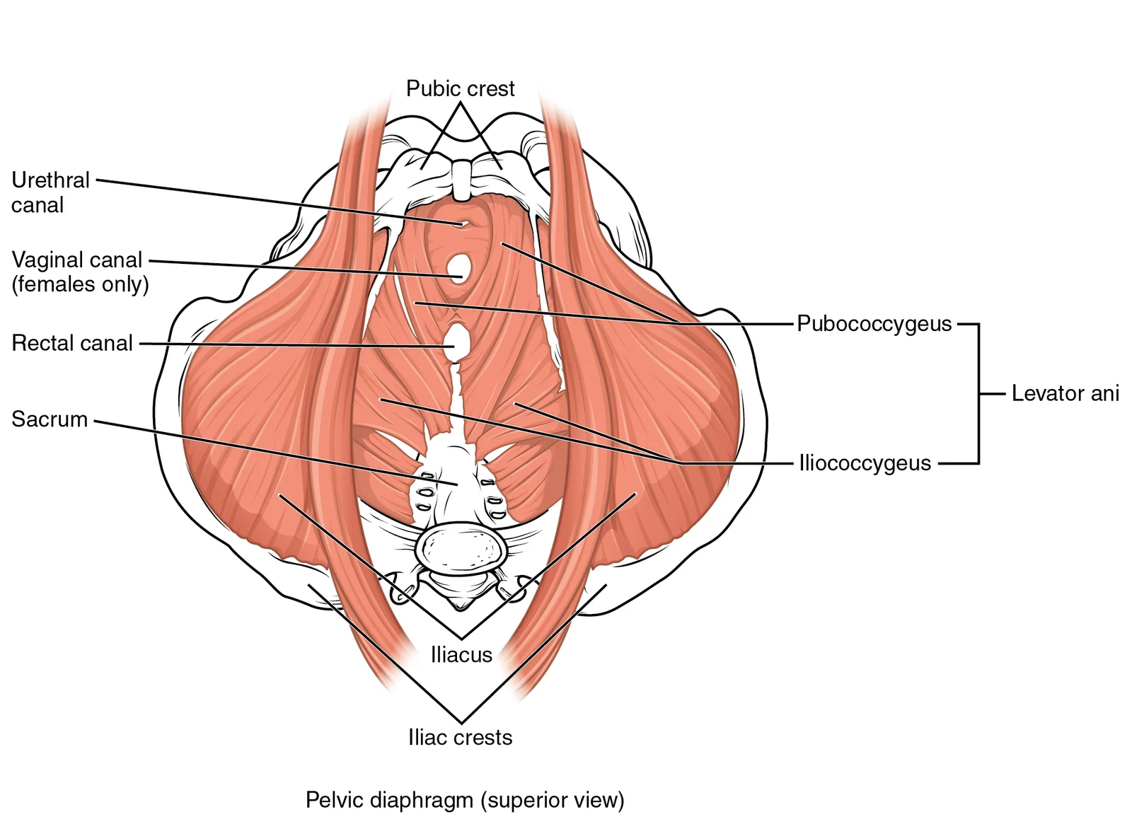
The pelvic floor is primarily composed of the levator ani and coccygeus muscles, collectively known as the pelvic diaphragm. These muscles originate from the inner surfaces of the pelvis and insert into the coccyx, anococcygeal raphe, and the walls of the pelvic viscera. Their primary action is to support the pelvic organs and maintain continence. The large levator ani consists of two skeletal muscles, the pubococcygeus and the iliococcygeus. The pubococcygeus arises from the pubis and insert [2]
| Muscle | Origin | Insertion | Action | Nerve Supply |
|---|---|---|---|---|
| Levator Ani (Pubococcygeus) | Pubis | Coccyx, anococcygeal raphe | Supports pelvic viscera | Nerve to levator ani |
| Levator Ani (Puborectalis) | Pubis | Anorectal junction | Maintains anorectal angle | Nerve to levator ani |
| Levator Ani (Iliococcygeus) | Ischial spine, pubic arch | Coccyx, anococcygeal raphe | Supports pelvic viscera | Nerve to levator ani |
| Coccygeus | Ischial spine | Sacrum, coccyx | Supports pelvic viscera | Nerve to coccygeus |
| Obturator Internus | Lateral wall of pelvis | Greater trochanter of femur | Lateral rotation of thigh | Nerve to obturator internus |
The sacral plexus provides the nerve supply to many of these muscles. The nerve to the levator ani arises from the ventral rami of S3 and S4. The nerve to the coccygeus arises from S4 and S5. These nerves emerge from the sacral plexus and travel to their respective muscles. It gives rise t
DefinitionPelvic Diaphragm
The levator ani and coccygeus muscles form the pelvic diaphragm, which supports the pelvic viscera and maintains continence.
Its action is lateral rotation of the thigh when the leg is extended. This muscle is innervated by the nerve to the obturator internus. It is important to distinguish the obturator internus from the obturator externus, which is located on the external surface of the hip bone.
Exam trapNerve Supply
The levator ani is innervated by the nerve to the levator ani (S3, S4), not the pudendal nerve, although they are closely related in the pudendal canal.
It is innervated by the nerve to the coccygeus. The coccygeus and levator ani together form the pelvic diaphragm. This diaphragm is crucial for maintaining intra-abdominal pressure during activities such as coughing and defecation. Damage to these muscles can lead to pelvic organ prolapse.
NoteClinical Relevance
Pelvic floor dysfunction can result from childbirth trauma or aging, leading to incontinence or prolapse. Rehabilitation often targets the levator ani muscles.
The pelvic walls are formed by the hip bones, which consist of the ilium, pubis, and ischium. These bones converge to form the acetabulum, which articulates with the head of the femur. This membrane serves for the attachment of muscles on both its internal and external surfaces. The hip bone provides the structural framework for the pelvic cavity. [2] [3]
DefinitionAcetabulum
The acetabulum is a deep, cup-shaped cavity formed by the convergence of the ilium, pubis, and ischium, articulating with the femoral head.
The pelvic floor muscles work in coordination with the abdominal muscles and diaphragm to regulate intra-abdominal pressure. This coordination is essential for functions such as defecation, urination, and childbirth. The Valsalva maneuver involves holding the breath while contracting the abdominal muscles, which increases pressure on the pelvic viscera. The pelvic diaphragm must relax to allow for the passage of stool or urine. Understanding the anatomy of the pelvic walls and floor is essential for clinical practice, particularly in obstetrics and urology. [2]
Figure 21115 Muscles of the Pelvic Floor
Long description
The pelvic floor muscles support the pelvic organs, resist intra-abdominal pressure, and work as sphincters for the urethra, rectum, and vagina.

Figure 2: The pelvic floor muscles support the pelvic organs, resist intra-abdominal pressure, and work as sphincters for the urethra, rectum, and vagina.
The arterial supply to the pelvic walls and floor is derived primarily from the internal iliac artery. This vessel arises from the common iliac artery. It descends into the lesser pelvis, running along the lateral pelvic wall. [1]
The anterior division supplies pelvic viscera. The posterior division supplies the gluteal region and pelvic walls.
The anterior division gives rise to visceral and parietal branches. The visceral branches supply the pelvic organs. The parietal branches supply the pelvic walls. The posterior division gives rise to branches that supply the gluteal region and lower back.
The superior gluteal artery arises from the posterior division. It exits the pelvis above the piriformis muscle. It supplies the gluteus medius and minimus muscles.
The middle rectal artery arises from the anterior division. It supplies the rectum. It anastomoses with the superior and inferior rectal arteries. These branches supply the external genitalia. [1][2]
The arterial supply is rich in anastomoses. This ensures collateral circulation. Ischaemia is rare unless major vessels are damaged. Knowledge of these vessels is crucial for trauma surgery. It is also important for oncological resections.
The venous drainage of the pelvis is complex, involving both deep and superficial systems that communicate extensively. Understanding this network requires examining the walls and floor separately, as their drainage patterns differ significantly. This diagram shows the pelvic veins and venous plexuses in a posterior view. The orientation is standard anatomical position. You can see the internal iliac veins forming the main outflow. The plexuses are situated around the pelvic organs. The walls drain into the lateral sacral and internal iliac veins. The floor drains via the pudendal system. The image labels the superior and inferior gluteal veins. These structures are crucial for understanding pelvic pathology. [2]
The pelvic walls are drained primarily by the internal iliac veins. These veins are formed by the union of several tributaries. The superior gluteal vein drains the gluteal region. It exits the pelvis through the greater sciatic foramen. The inferior gluteal vein drains the lower gluteal muscles. It also passes through the greater sciatic foramen. The middle rectal vein drains the rectum. It anastomoses with the superior rectal vein. It runs along the lateral pelvic wall. The internal iliac vein ascends to join the external iliac vein. This union forms the common iliac vein. The common iliac veins converge to form the inferior vena cava. This pathway is essential for venous return from the lower limbs. [3]
The pelvic floor has a distinct venous drainage pattern. Both plexuses drain into the internal iliac veins. It anastomoses with the ovarian veins. This connection allows for collateral circulation. The ovarian veins drain into the inferior vena cava on the right. On the left, they drain into the left renal vein. This asymmetry is clinically significant.
Varicose veins in the pelvis can result from valve incompetence. The internal iliac veins may become dilated. This condition is known as pelvic congestion syndrome. It causes chronic pelvic pain. The plexuses can also be sites of metastasis. Prostate cancer often spreads via the vertebral venous plexus. This plexus connects with the internal iliac veins. It provides a route for tumour cells to reach the spine. Understanding these connections is vital for staging cancer. Surgical procedures in the pelvis must respect these veins. Injury to the internal iliac vein can cause severe bleeding. Compression of the veins can lead to oedema. The lymphatic drainage often follows the venous paths. This is important for understanding infection spread.
The innervation of the pelvic walls and floor is complex, involving both somatic and autonomic components that control motor function and provide sensory feedback. Understanding the specific pathways is essential for localizing lesions and understanding clinical presentations such as incontinence or sexual dysfunction. The primary nerve supply arises from the lumbosacral plexus and the pelvic splanchnic nerves.
The motor supply to the pelvic floor muscles is primarily provided by the pudendal nerve and direct branches from the sacral plexus. The pudendal nerve supplies the external anal sphincter and the muscles of the urogenital triangle. This direct innervation allows for precise control of the pelvic diaphragm.
Sensory innervation is divided between the pudendal nerve for the perineum and the pelvic splanchnic nerves for the viscera. The pudendal nerve provides cutaneous sensation to the anal region and the external genitalia. Visceral sensation from the pelvic organs travels via the pelvic splanchnic nerves to the sacral spinal cord.
| Nerve | Motor Supply | Cutaneous/Sensory Area |
|---|---|---|
| Pudendal | External anal sphincter, bulbospongiosus, ischiocavernosus | Perineum, external genitalia, anal canal below pectinate line |
| Nerve to Levator Ani | Levator ani, coccygeus | None (proprioception only) |
| Pelvic Splanchnic | Smooth muscle of pelvic viscera | Visceral afferents from pelvic organs |
The pelvic nerves must navigate around these major vascular structures to reach their targets. [2]
Lymphatic drainage of the pelvic walls and floor follows a predictable anatomical pathway that mirrors the systemic venous return, converging primarily upon groups of nodes located along the major pelvic vessels. Understanding this architecture is vital when staging pelvic malignancies or assessing infectious spread from the perineum.
DefinitionPelvic Lymphatic System
A deeply situated network of interconnected channels and filtering nodes that primarily drains the pelvic viscera, pelvic walls, and perineal structures into the lumbar trunk.
Lymph from the superficial structures of the perineum and external genitalia drains primarily into the superficial inguinal lymph nodes, which lie in the subcutaneous tissue along the saphenous opening of the fascia lata. From these superficial stations, efferent vessels ascend through the cribriform fascia to reach the external iliac lymph nodes.
| Lymph Node Group | Anatomical Location | Primary Drainage Target |
|---|---|---|
| External iliac nodes | Along the external iliac vessels | Common iliac nodes |
| Internal iliac nodes | Around the internal iliac artery branches | Common iliac nodes |
| Obturator nodes | Within the obturator fossa near the nerve | Internal iliac nodes |
| Sacral nodes | Anterior to the sacrum and piriformis | Common iliac and lumbar nodes |
Within the deep pelvis, the external iliac lymph nodes form three chains (medial, intermediate, and lateral) situated along the external iliac artery and vein. They receive lymph from the lower abdominal wall, inguinal nodes, and pelvic viscera. Posteriorly, the internal iliac lymph nodes cluster around the branches of the internal iliac artery, draining the pelvic walls, deep perineum, and pelvic organs.
Further posterolaterally, the obturator lymph nodes sit in the obturator fossa adjacent to the obturator nerve and vessels.
The superficial inguinal lymph nodes appear in the subcutaneous fat inferior to the inguinal ligament, sending efferents superiorly through the lacunar ligament. Deep to this, the external iliac lymph nodes form a continuous chain along the medial and lateral borders of the external iliac vein. Centrally, the internal iliac lymph nodes cluster around the bifurcation of the internal iliac artery against the posterolateral pelvic wall. Posteriorly, the sacral lymph nodes lie in a vertical row in front of the middle sacral vessels on the anterior surface of the sacrum. All these nodal groups ultimately drain superiorly into the common iliac nodes situated along the common iliac vessels at the pelvic brim. [1]
The enlarged medial portion of the pubis is the pubic body. Located superiorly on the pubic body is a small bump called the pubic tubercle. This tubercle serves as a key landmark for identifying the superior pubic ramus, which is the segment of bone that passes laterally from the pubic body to join the ilium. The narrow ridge running along the superior margin of the superior pubic ramus is the pectineal line of the pubis. This line extends posteriorly to merge with the pectineal line of the ilium, forming the pectineal line of the pelvis, which marks the boundary between the false and true pelvis. [3]
The pelvic floor is formed by the levator ani and coccygeus muscles, which support the pelvic viscera. The levator ani consists of the pubococcygeus, puborectalis, and iliococcygeus parts. The pubococcygeus arises from the posterior surface of the pubis and the tendinous arch of the obturator fascia. It passes posteriorly and medially to insert into the anococcygeal raphe and the coccyx. The puborectalis forms a sling around the rectum, contributing to the anorectal angle. The iliococcygeus arises from the ischial spine and the tendinous arch, inserting into the coccyx and anococcygeal raphe.
The pelvic cavity contains the urinary bladder, rectum, and reproductive organs. In males, the prostate lies inferior to the bladder. In females, the uterus and ovaries are located within the true pelvis. The rectum lies posterior to the bladder in males and posterior to the vagina in females. The pelvic walls are formed by the hip bones, sacrum, and coccyx. The obturator foramen is bounded by the pubis and ischium, and is largely closed by the obturator membrane. These foramina allow passage of nerves and vessels to and from the lower limb and perineum. [3]
| Muscle | Origin | Insertion | Action | Nerve Supply |
|---|---|---|---|---|
| Pubococcygeus | Posterior pubis, obturator fascia | Anococcygeal raphe, coccyx | Supports viscera, constricts vagina/urethra | Nerve to levator ani |
| Puborectalis | Posterior pubis | Anococcygeal raphe | Maintains anorectal angle | Nerve to levator ani |
| Iliococcygeus | Ischial spine, tendinous arch | Coccyx, anococcygeal raphe | Supports viscera | Nerve to levator ani |
| Coccygeus | Ischial spine | Coccyx, lower sacrum | Supports viscera | Nerve to coccygeus |
Radiological assessment of the pelvic walls and floor relies on plain radiography, computed tomography, magnetic resonance imaging, and ultrasound. Each modality provides specific tissue contrast to evaluate bony architecture, muscular slings, and fascial supports.
Plain abdominal and pelvic radiographs demonstrate the bony pelvis in coronal projection. The ilium, ischium, and pubis form a rigid ring, while the sacroiliac joints and pubic symphysis maintain pelvic stability. Computed tomography provides superior spatial resolution for complex fractures and bony landmarks of the pelvic walls. Axial computed tomography sections trace the piriformis muscle exiting through the greater sciatic foramen and the obturator internus lining the lateral pelvic wall. Intravenous and oral contrast agents help delineate the pelvic viscera from the surrounding musculature and vascular bundles.
Magnetic resonance imaging is the gold standard for soft tissue evaluation of the pelvic floor due to its exceptional multiplanar soft tissue contrast. T2-weighted sequences clearly distinguish the puborectalis muscle and the levator ani plate from surrounding fat and connective tissue. Sagittal magnetic resonance imaging demonstrates the hammock-like support of the levator ani running from the pubis to the coccyx. Ultrasound offers dynamic, real-time assessment of pelvic floor descent and urethral mobility. Transperineal and endoanal ultrasound visualize the integrity of the anal sphincter complex and the resting tone of the puborectalis sling during voluntary contraction and Valsalva manoeuvres.
| Muscle | Origin | Insertion | Action | Nerve Supply |
|---|---|---|---|---|
| Levator ani | Pubis, arcus tendineus, ischial spine | Anococcygeal body, perineal body, walls of viscera | Supports pelvic viscera, aids continence | Nerve to levator ani (S4), pudendal nerve |
| Piriformis | Anterior surface of sacrum (S2-S4) | Greater trochanter of femur | Lateral rotator of extended hip | Ventral rami of S1-S2 |
| Obturator internus | Inner surface of obturator membrane and bony pelvis | Greater trochanter of femur | Lateral rotator of extended hip | Nerve to obturator internus (L5-S1) |
| Coccygeus | Ischial spine and sacrospinous ligament | Inferior sacrum and coccyx | Supports pelvic floor, flexes coccyx | Ventral rami of S4-S5 |
The symphysis pubis lies most anteriorly, serving as the midline landmark for the anterior pelvic wall. Deep to the obturator internus, the obturator canal transmits the obturator nerve and vessels. Posteriorly, the piriformis muscle arises from the anterior surface of the sacrum and passes laterally through the greater sciatic foramen, separating the greater sciatic notch into supra-piriform and infra-piriform compartments. Lateral to the rectum, the iliococcygeus and pubococcygeus muscles extend from the arcus tendineus levator ani towards the midline anococcygeal raphe, forming a continuous muscular floor that supports the pelvic viscera against intra-abdominal pressure. [2]
Clinical pearlLevator ani assessment
Magnetic resonance imaging in the sagittal plane best demonstrates the normal angulation and resting tone of the levator plate.
The bony pelvis forms a rigid ring that is vulnerable to high-energy trauma such as road traffic collisions. When a pelvic fracture occurs, the disruption of the posterior ligamentous complex and the bony ring can lead to severe retroperitoneal bleeding. Understanding the applied anatomy of pelvic haemorrhage is vital because the posterior venous plexus and the branches of the internal iliac artery, particularly the superior gluteal and internal pudendal vessels, lie directly against the pelvic
The pubic symphysis lies anteriorly, articulating the two pubic bones, while the sacrum and sacroiliac joints lie posteriorly.
| Muscle | Origin | Insertion | Action | Nerve Supply |
|---|---|---|---|---|
| Levator ani | Posterior body of pubis, arcus tendineus, spine of ischium | Coccyx, anococcygeal body, walls of vagina and anal canal | Supports pelvic viscera, resists increases in intra-abdominal pressure | Nerve to levator ani (S4), pudendal nerve |
| Coccygeus | Ischial spine and sacrospinous ligament | Lateral margin of coccyx and lower sacrum | Flexes coccyx and supports pelvic floor | S4 and S5 spinal nerves |
Surgical and obstetric procedures frequently interact with the pelvic floor. Episiotomy or childbirth can stretch or tear the levator ani muscle and the pudendal nerve as it crosses the sacrospinous ligament, leading to subsequent pelvic organ prolapse or stress urinary incontinence. Nerve palsies may also arise from compression during prolonged lithotomy positioning or regional trauma. Surface landmarks used clinically include the ischial spines, which serve as the target for pudendal nerve block during second-stage labour, and the coccyx, which is palpated externally during digital rectal examination to assess pelvic floor tone.
Clinical pearlPudendal nerve block landmark
The ischial spine is palpated through the vaginal wall to guide the needle for local anaesthesia of the pudendal nerve.
Clinical pearlLevator ani components
The levator ani muscle is composed of the pubococcygeus, iliococcygeus, and puborectalis muscles, which together form the primary structural support of the pelvic floor.
Clinical pearlPerineal body support
The perineal body serves as a vital central tendinous attachment point for multiple pelvic floor and perineal muscles.
Exam trapUrogenital hiatus trap
Mistaking the urogenital hiatus for a bony opening is a frequent error; it is a muscular and fascial cleft transmitting the urethra and vagina.
Exam trapNerve supply confusion
Remember that the levator ani receives direct motor branches from the sacral plexus (S4) and the pudendal nerve, not solely the pudendal nerve.
Clinical pearlEndopelvic fascia
The endopelvic fascia suspends the pelvic viscera and contributes to the formation of important condensations like the cardinal and uterosacral ligaments.
Clinical pearlArcus tendineus
The arcus tendineus levator ani is a thickened condensation of the obturator fascia that provides origin to parts of the levator ani muscle.
Exam trapGreater sciatic foramen boundaries
Do not confuse the greater sciatic notch with the greater sciatic foramen; the latter is converted into an actual foramen by the sacroligamentous structures.
Clinical pearlPiriformis nerve relation
The sciatic nerve typically emerges inferior to the piriformis muscle, though variations involving nerve splitting can occur.
A common mistake is confusing the boundaries of the pelvic inlet and outlet. The pelvic inlet is bounded anteriorly by the pubic symphysis, laterally by the linea terminalis (pecten pubis, arcuate line, and iliopectineal line), and posteriorly by the sacral promontory. The pelvic outlet is bounded anteriorly by the pubic arch, posteriorly by the coccyx, and laterally by the ischial spines and sacrotuberous ligaments.
Students often misidentify the bony landmarks of the hip bone. The ilium is the fan-like, superior region that forms the largest part of the hip bone. The ischium forms the posteroinferior region and has the expanded ischial tuberosity that supports body weight when sitting. The pubis forms the anterior portion and curves medially to join the opposite pubis at the pubic symphysis. [3]
Another error is misunderstanding the sacroiliac joint anatomy. The sacroiliac joint is a largely immobile joint where the ilium is firmly united to the sacrum. Students sometimes confuse this with the sacrospinous and sacrotuberous ligaments, which are important for pelvic outlet stability. [3]
The lesser sciatic notch is often mislocated. It is found on the posterior margin of the ischium, between the ischial spine and the ischial tuberosity. The ischial spine separates the greater and lesser sciatic notches. [3]
Exam trapCommon Misconception
The pelvic floor muscles do not attach directly to the sacrum but to the pelvic walls via the levator ani and coccygeus muscles.
At the bedside, knowledge of pelvic walls and floor is essential for performing a digital rectal examination. The examiner palpates the ischial spines, which are important landmarks for assessing pelvic outlet dimensions. The anorectal junction is located approximately 4-5 cm from the anal verge, where the puborectalis muscle forms a sling around the rectum.
In the operating theatre, understanding pelvic anatomy is crucial for procedures like hysterectomy and prostatectomy. The surgeon must identify the pelvic diaphragm formed by the levator ani and coccygeus muscles. The ischial spines serve as important surgical landmarks when approaching the rectum and pelvic organs.
On imaging, radiologists evaluate pelvic fractures by recognizing the bony landmarks. The sacroiliac joint is a common site for injury in pelvic trauma. Computed tomography (CT) scans clearly show the ischial tuberosity and spines, which help assess pelvic ring stability.
In examinations, students are tested on their ability to identify pelvic structures on diagrams and models. Common questions involve tracing the course of the pudendal nerve and vessels through the greater and lesser sciatic foramina. The relationship between the pelvic inlet and outlet dimensions is frequently tested in obstetrics questions.
Pelvic Floor Assessment
- Patient PositionLithotomy or left lateral position
- Question: Digital Rectal Exam?
Yes
- Palpate Ischial SpinesAssess pelvic outlet dimensions
- Check Puborectalis SlingEvaluate anorectal junction
No
- Imaging AssessmentX-ray, CT, or MRI for bony landmarks
- Document FindingsRecord any abnormalities or measurements
The ilium forms the largest part of the hip bone and is united to the sacrum at the sacroiliac joint. The ischium forms the posteroinferior region and has the ischial tuberosity that supports body weight when sitting. The pubis forms the anterior portion and joins the opposite pubis at the pubic symphysis. The ischial spine separates the greater and lesser sciatic notches. Knowledge of pelvic walls and floor is essential for digital rectal examination and surgical procedures. The pelvic inlet and outlet have specific boundaries important for obstetric assessment.
Answer each one in your head first, then open the answer.
Which bone forms the largest part of the hip bone?
Show answerHide answer
IliumWhat structure supports body weight when sitting?
Show answerHide answer
Ischial tuberosityWhat joint joins the pubis of opposite hip bones?
Show answerHide answer
Pubic symphysisWhat separates the greater and lesser sciatic notches?
Show answerHide answer
Ischial spineWhich joint is largely immobile and unites the ilium to the sacrum?
Show answerHide answer
Sacroiliac joint
- Betts JG, Young KA, Wise JA, et al. Appendicular Muscles of the Pelvic Girdle and Lower Limbs. OpenStax, Anatomy and Physiology 2e (CC BY-NC-SA 4.0). Accessed .
- Betts JG, Young KA, Wise JA, et al. Axial Muscles of the Abdominal Wall, and Thorax. OpenStax, Anatomy and Physiology 2e (CC BY-NC-SA 4.0). Accessed .
- Betts JG, Young KA, Wise JA, et al. The Pelvic Girdle and Pelvis. OpenStax, Anatomy and Physiology 2e (CC BY-NC-SA 4.0). Accessed .
Figure credits
Educational content for medical students and doctors. It is not a substitute for clinical judgement or local guidelines.