Pelvic fascia
Anatomical overview of the pelvic fascia, supports, and boundaries for medical students.
The space enclosed by the bony pelvis is divided into the greater pelvis, defined laterally by the large fan-like portion of the upper hip bone, and the lesser pelvis, which contains the bladder and other pelvic organs. The pelvic brim forms the superior margin of the lesser pelvis, separating it from the greater pelvis.
The region features specialized connective tissue membranes divided into parietal and visceral layers. The visceral pelvic fascia is a fibrous envelope that surrounds the mesorectum, also known as the fascia propria of the rectum. Posterior pelvic dissection is performed along this rectal proper fascia, leaving the hypogastric nerves along the pelvic sidewall. Waldeyer's fascia is defined as the presacral parietal fascia or its most caudal part at the junction with the visceral fascia at the lev
The pelvic diaphragm is formed by the levator ani and coccygeus muscles, which span the floor of the lesser pelvis.
| Muscle | Origin | Insertion | Action | Nerve Supply |
|---|---|---|---|---|
| Levator ani | Pubis, obturator fascia, ischial spine | Anococcygeal body, perineal body, wall of anal canal | Supports pelvic viscera, aids continence | S4 nerve, pudendal nerve |
| Coccygeus | Ischial spine, sacrospinous ligament | Coccyx, lower sacrum | Supports pelvic floor | S4-S5 spinal nerves |
The bladder, vagina, and rectum are held in place by supporting ligaments, including the pubo-urethral and pubo-vesical ligaments, the utero-sacral ligaments, the cardinal ligaments, and the arcus tendineus fascia pelvis. The pelvic floor fascia joins these ligaments and the perineal body, with collagen serving as the main component of both the fascia and the supporting ligaments. The pelvic floor muscles pull back the pelvic organs and the pelvic floor fascia in three different directions, providing support and maintaining their form and strength. [5]
Pelvic organ prolapse is defined as a protrusion or herniation of the pelvic organs through the vaginal walls and pelvic floor. Weakening of the pelvic floor connective tissue and collagen allows the pelvic organs to prolapse through the vaginal walls. Prolapse is categorized by descent of the anterior, posterior, or apical vaginal compartments. Prolapse of the anterior compartment occurs most frequently, reported to be twice as prevalent as posterior compartment prolapse and three times more prevalent than apical prolapse. Additionally, two thirds of women have a combined prolapse of all three compartments. The prevalence of vaginal cuff prolapse following hysterectomy is reported to be as high as 6% to 12%. Damage to the supporting ligaments and fascia brings about the collapse of this suspension system, contributing directly to urinary incontinence and pelvic organ prolapse. [5]
Clinical pearlFascial planes in surgery
Identification of the avascular planes between the visceral pelvic fascia and parietal pelvic fascia is essential during total mesorectal excision to protect the pelvic plexus and hypogastric nerves.
Pelvic Organ Support Architecture
- Pelvic BrimSeparates greater and lesser pelvis.
- Question: Component Intact?
Yes
- Suspension bridge maintained; organs supported.
No
- Collagen and ligament injury leads to pelvic organ prolapse.
The pelvic fascia does not form joints in the conventional synovial sense, but it provides the critical connective tissue architecture that stabilizes the sacroiliac and pubic symphysis joints, while also defining the fascial compartments that constrain pelvic organ movement. Understanding these fascial relationships is essential for appreciating how mechanical stress is distributed across the pelvic floor during activities such as coughing, lifting, or childbirth. The integrity of the pelvic di
In the posterior compartment, the rectosacral fascia plays a pivotal role in stabilizing the rectum relative to the sacrum. Dissection studies involving 35 female cadavers have demonstrated that this fascia originates from the presacral fascia at the level of S2, S3, or S4 and joins the fascia propria of the rectum 3–5 cm above the anorectal junction. This specific anatomical arrangement allows the rectosacral fascia to clearly divide the retrorectal space into inferior and superior portions. This division is clinically significant because it creates distinct potential spaces that can be involved in the spread of infection or neoplastic disease. The precise origin points at S2, S3, or S4 mean that surgical dissection in this region must account for individual anatomical variations to avoid inadvertent injury to the presacral venous plexus or the hypogastric nerves. [6]
The inferior fascia of the pelvic diaphragm, also known as Waldeyer’s fascia, originates from the presacral fascia but differs in its functional role. It forms the floor of the inferior portion of the retrorectal space. This distinction is crucial for surgeons performing posterior pelvic dissections, such as abdominoperineal resections. The visceral pelvic fascia, or fascia propria of the rectum, envelops the mesorectum, and dissection should continue along this plane to the rectosacral fascia. By staying within this fascial plane, surgeons can leave the hypogastric nerves along the pelvic sidewall intact, preserving autonomic function. The rectosacral fascia thus acts as a key landmark, separating the visceral compartment from the parietal presacral fascia. [6]
Controversies persist regarding the exact definition and distinction between the inferior fascia of the pelvic diaphragm and the rectosacral fascia, with some literature treating them as synonymous. However, according to Garcia-Armengol et al., the rectosacral fascia is a distinct structure that generally arises at the level of S4 to separate the retrorectal space into a superior and an inferior portion. This anatomical clarity is vital for understanding the limits of total mesorectal excision.
The tendinous arch of pelvic fascia (arcus tendineus fasciae pelvis) provides essential attachment points for the pelvic floor muscles, contributing significantly to the stability of the pelvic joints and organs. This structure, along with components like the pubocervical fascia, rectovaginal fascia, and paracolpium, forms a complex network that supports the pelvic organs. In pregnant women, the increased load on the pelvic floor places additional stress on these fascial attachments. Weakness or tearing of the endopelvic fascia can lead to pelvic organ prolapse, highlighting the importance of maintaining fascial integrity. Similarly, in older adults, degenerative changes in the collagen content of these fascial layers can compromise pelvic support, leading to symptoms of pelvic floor dysfunction. Understanding these fascial mechanics is therefore not just an anatomical exercise but a clinical imperative for managing pelvic health across different life stages.
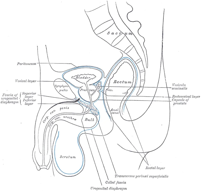
Figure 1Gray403
Long description
Median sagittal section of pelvis, showing arrangement of fasciæ.

Figure 1: Median sagittal section of pelvis, showing arrangement of fasciæ.
The surface anatomy of the pelvic fascia is best understood by correlating deep fascial layers with external bony landmarks and the overlying skin. The posterior boundary of the pelvic cavity is defined by the sacrum. At the inferior end of a large, roughened area called the auricular surface of the ilium, the sacroiliac joint is formed. This joint is stabilized by the posterior superior and posterior inferior iliac spines, which serve as attachment points for the muscles and very strong ligaments that support the sacroiliac joint. These bony prominences are palpable on the posterior aspect of the pelvis and serve as key reference points for locating the deeper presacral fascia. [2]
At the S4 level, relatively dense connective tissue links the presacral fascia and the MRF. The levator ani forms the pelvic floor, consisting of the pubocococcygeus, iliococcygeus, and puborectalis muscles. The U-shaped puborectalis muscle creates a sharp anorectal angle, preventing fecal incontinence. The mesorectum is usually tapered out 2 or 3 cm proximally from the pelvic floor. [3]
The anterior aspect of the pelvic fascia involves Denonvilliers' fascia (DVF). Although DVF has been frequently studied, there is no consensus on its embryological origin and topological anatomy. It separates the rectum from the prostate in males or the vagina in females. The mesorectum is in contact with the fascia of the levator ani muscle. The shape and angle of the funnel-shaped pelvic floor differ with patient characteristics and sex. The rectum and mesorectum should be usually mobilized completely from the pelvic floor. [3]
The lateral attachments include the middle rectal artery and unnamed attachments at the lateral wall of the mesorectum at deep pelvis. The pelvic autonomic nerves run in close proximity to these fascial planes. A proper understanding of the anatomy of the rectum, especially the fascia and autonomic nerve, is necessary to perform autonomic-nerve–preserving TME. The presacral venous plexus exists and is covered with the parietal pelvic fascia. Blunt dissection should be avoided since this maneuver may cause a partial tear and hemorrhage due to injury of the presacral venous plexus. [3]
DefinitionMesorectal Fascia
The MRF corresponds to the visceral fascia and envelops the mesorectum, containing possible metastatic lymph nodes and blood vessels. [3]
Clinical pearlS4 Landmark
At the S4 level, relatively dense connective tissue links the presacral fascia and the MRF.
| Structure | Location | Relation |
|---|---|---|
| Presacral Fascia | Posterior | Adheres to sacral periosteum |
| Mesorectal Fascia (MRF) | Anterior to presacral | Envelops mesorectum |
| Denonvilliers' Fascia | Anterior | Separates rectum from prostate/vagina |
| Levator Ani Fascia | Inferior | Forms pelvic floor boundary |
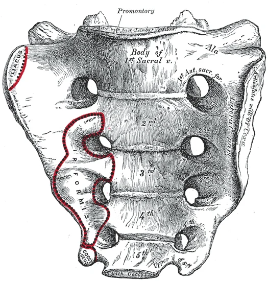
Figure 2Gray95
Long description
Sacrum, pelvic surface. Labelled illustration from Wikimedia Commons, used under its open licence.

Figure 2: Sacrum, pelvic surface.
pelvic girdle consists of the hip bones, sacrum, and coccyx [P35]. [2]
DefinitionPelvic Brim
The pelvic brim forms the boundary between the greater and lesser pelvis.
The sacral promontory marks the anterior margin of the superior sacrum in the posterior midline. Traced laterally from the sacral promontory, the arcuate line of the ilium meets the pectineal line of the pubis and the upper margin of the pubic symphysis anteriorly to define the pelvic brim, also known as the pelvic inlet. The broad space of the upper pelvis is the greater pelvis, while the narrow, inferior space below the pelvic brim is the lesser pelvis. The inferior limit of the lesser pelvic cavity is the pelvic outlet, a large opening defined by the inferior margin of the pubic symphysis anteriorly, and posteriorly by the ischiopubic ramus, the ischial tuberosity, the sacrotuberous ligament, and the inferior tip of the coccyx. [2]
| Feature | Greater Pelvis | Lesser Pelvis |
|---|---|---|
| Location | Upper expanded space | Narrow inferior space |
| Boundary | Above pelvic brim | Below pelvic brim |
| Sex Differences | Wider in females | Narrower and deeper in males |
The female sacrum is wider, shorter, and less curved, with a sacral promontory that projects less into the pelvic cavity, giving the female pelvic inlet a more rounded or oval shape. Furthermore, the female pelvis is wider to accommodate childbirth, possesses a larger subpubic angle, and displays a broader greater sciatic notch compared to the male. [2]
The pelvic fascia lines the pelvic cavity and forms specialized condensations that support the pelvic viscera. Weakness of the endopelvic fascia is the main factor in the etiology of pelvic organ prolapse, leading to herniation of the pelvic organs and prolapse. Urogenital prolapse affects about 45% of post-menopausal women, with a 30% to 50% prevalence in a lifespan. Models by Petros and Delancey enable clinicians to locate defects, diagnose, and treat pelvic organ prolapse. In clinical practice, aging, pregnancy, delivery, and a history of pelvic surgery are identified as some of the main causes of pelvic organ prolapse. Furthermore, cervical elongation is reported to affect approximately 40% of women with uterine prolapse, and measurements demonstrate that the cervical length in women with uterine prolapse is about 36% longer than in women without uterine prolapse. [4] [5]
The pararectal and paravesical spaces lie lateral to the rectum and bladder respectively, bounded medially by visceral fascia and laterally by the parietal pelvic fascia covering the obturator internus muscle.
DefinitionEndopelvic fascia
Continuous sheet of connective tissue lining the pelvic floor that suspends pelvic organs.
| Level | Location | Supporting Structures | Prolapse Result |
|---|---|---|---|
| Level I | Superior | Uterosacral-cardinal ligament complex | Uterine or apical prolapse |
| Level II | Intermediate | Arcus tendineus fasciae pelvis | Cystocele or rectocele |
| Level III | Inferior | Perineal membrane and body | Distal vaginal laxity |
Pelvic Organ Prolapse Evaluation
- Identify symptomsCheck for bulge or pressure.
- Question: Bothersome symptoms?
Yes
- Clinical examinationAssess prolapse stage.
No
- Conservative careObserve or lifestyle advice.
The pelvic girdle serves as the critical attachment point that connects the lower limbs to the axial skeleton, forming the foundation for the hip joint where the pelvic girdle and leg converge. The musculature originating from this region is extensive, with most muscles that insert on the femur and facilitate its movement beginning their course on the pelvic girdle. These muscles are broadly categorised into groups based on their functional compartments and anatomical relationships. [1]
Deep to the gluteal region, the gluteus maximus is the largest and most superficial muscle, with the gluteus medius lying deep to it, and the gluteus minimus positioned deepest as the smallest of the trio. The lateral rotators of the femur at the hip include the piriformis, obturator internus, obturator externus, superior gemellus, inferior gemellus, and quadratus femoris. On the medial aspect of the thigh, the adductor longus, adductor brevis, and adductor magnus are responsible for adducting the thigh and medially rotating it. The pectineus muscle also contributes by adducting and flexing the femur at the hip. Additionally, the tensor fascia lata muscle works alongside the gluteus maximus to form the iliotibial tract. [1]
The following table details the key muscles of the pelvic and proximal lower limb region, grouped by compartment, with their origins, insertions, actions, and nerve supplies.
| Compartment | Muscle | Origin | Insertion | Action | Nerve Supply |
|---|---|---|---|---|---|
| Anterior | Iliopsoas (Psoas major & Iliacus) | Pelvic girdle | Femur | Flexes thigh | Lumbar plexus |
| Posterior (Gluteal) | Gluteus maximus | Pelvic girdle | Femur & Iliotibial tract | Extends and abducts femur | Inferior gluteal nerve |
| Posterior (Gluteal) | Gluteus medius | Pelvic girdle | Femur | Extends and abducts femur | Superior gluteal nerve |
| Posterior (Gluteal) | Gluteus minimus | Pelvic girdle | Femur | Extends and abducts femur | Superior gluteal nerve |
| Posterior (Lateral Rotators) | Piriformis, Obturator internus/externus, Gemelli, Quadratus femoris | Pelvic girdle | Femur | Lateral rotation of femur | Sacral plexus branches |
| Medial | Adductor longus, brevis, magnus | Pelvic girdle | Femur | Adducts and medially rotates thigh | Obturator nerve |
| Medial | Pectineus | Pelvic girdle | Femur | Adducts and flexes femur | Femoral/Obturator nerve |
| Anterior (Thigh) | Tensor fascia lata | Pelvic girdle | Iliotibial tract | Forms iliotibial tract with gluteus maximus | Superior gluteal nerve |
DefinitionIliopsoas Group
The psoas major and iliacus combine to form the iliopsoas, a primary flexor of the thigh originating from the pelvic girdle.
DefinitionGluteal Group
The gluteal muscles are among the largest and most powerful in the body, with the gluteus maximus being the largest, followed by the medius and minimus.
NoteClinical Relevance
The iliotibial tract is formed by the tensor fascia lata and the gluteus maximus, providing lateral stability to the knee and hip.
The posterior compartment of the thigh, while distinct from the pelvic muscles, includes the hamstring group (biceps femoris, semitendinosus, and semimembranosus) which flexes the knee and extends the thigh. These muscles originate from the ischial tuberosity, a part of the pelvic girdle, and their tendons contribute to the boundaries of the popliteal fossa. Understanding the origin of these muscles on the pelvic girdle is essential for comprehending their role in lower limb movement. [1]
Disruption of this supply, such as during hysterectomy, can compromise tissue viability and contribute to pelvic organ prolapse [P141]. [5]
The internal iliac artery lies against the lateral pelvic wall, lateral to the ureter and deep to the peritoneum. The internal pudendal artery exits the greater sciatic foramen to re-enter via the lesser sciatic foramen, supplying the perineal fascia. [2]
| Vessel | Origin | Course | Branches |
|---|---|---|---|
| Internal iliac artery | Common iliac artery | Descends over pelvic brim into lesser pelvis | Parietal and visceral branches |
| Uterine artery | Internal iliac artery | Runs medially in cardinal ligament above ureter | Vaginal and cervical branches |
| Middle rectal artery | Internal iliac artery | Travels medially in lateral attachments to rectum | Rectal wall and fascial branches |
| Internal pudendal artery | Internal iliac artery | Exits via greater sciatic foramen, curves around pudendal spine | Perineal and labial/scrotal branches |
Precise identification of these vessels during fascial dissection is essential to prevent intraoperative bleeding [P76]. [3]
The venous drainage of the pelvic region is intimately associated with the fascial planes that compartmentalise the viscera. Understanding this drainage requires first recognising that the pelvic fascia provides the structural scaffolding within which these vascular networks reside. The venous return from the pelvic organs and walls does not follow a single linear path but rather forms extensive, interconnected plexuses embedded within the endopelvic fascia. These plexuses drain into the internal iliac veins, which serve as the primary outflow tract for the pelvis.
The internal iliac vein is situated along the pelvic sidewall, receiving tributaries from the vesical, uterine, and rectal venous plexuses. These plexuses are embedded within the endopelvic fascia, which surrounds the pelvic organs. The obturator vein courses superiorly across the lateral pelvic wall from the obturator foramen to empty into the internal iliac vein, running inferior to the obturator nerve. The middle rectal vein emerges from the fascial envelopes surrounding the rectum and passes laterally along the pelvic floor to join the internal iliac vein. [2][3]
The female pelvis is adapted for childbirth, being wider than the male pelvis, as evidenced by the distance between the anterior superior iliac spines. This increased pelvic width influences the course of the venous structures, which must traverse a broader cavity. The subpubic angle is larger in females (greater than 80 degrees) than it is in males (less than 70 degrees). This anatomical difference affects the orientation of the pelvic floor muscles and the associated fascial supports, thereby altering the pathway of the venous drainage from the lower urinary tract and genital organs. [2]
DefinitionEndopelvic Fascia
The connective tissue framework that suspends pelvic viscera and contains the neurovascular bundles, providing the pathway for venous drainage into the internal iliac system.
The venous plexuses within the pelvic fascia communicate freely with the vertebral venous system, providing a route for metastatic spread without filtering through the hepatic capillary bed. This connection is clinically significant, particularly in the context of pelvic organ prolapse, where surgical repair may involve synthetic mesh. The presence of such mesh can alter local haemodynamics and fascial tension, potentially affecting venous return. Furthermore, functional issues such as urinary incontinence can occur due to nerve injury during dissection, which may also compromise the integrity of the fascial planes and associated venous channels. [4] [3]
In the context of rectal surgery, the middle rectal artery may appear occasionally during the procedure. Its accompanying vein follows a similar course, draining into the internal iliac system. Surgeons must be aware of these structures to avoid excessive bleeding and to maintain the integrity of the mesorectal fascia. The lack of proper space in the deep pelvis can induce coning down and breaching of the mesorectum. This breach can disrupt the normal fascial planes and lead to uncontrolled venous bleeding from the plexuses embedded within them. [3]
| Structure | Origin | Course | Termination |
|---|---|---|---|
| Internal Iliac Vein | Common iliac vein bifurcation | Descends along pelvic sidewall | Common iliac vein |
| Obturator Vein | Obturator foramen | Superiorly across lateral wall | Internal iliac vein |
| Middle Rectal Vein | Rectal plexus | Laterally along pelvic floor | Internal iliac vein |
The autonomic innervation of the pelvic viscera is formed by sympathetic fibers from the inferior hypogastric plexus and parasympathetic fibers from the pelvic splanchnic nerves. These nerves run in close proximity to the venous plexuses within the pelvic fascia. Injury to these nerves during dissection can lead to functional issues, such as urinary incontinence and sexual dysfunction. The superior hypogastric plexus around the inferior mesenteric artery descends along the sacral promontory and bifurcates into hypogastric nerves. The paired hypogastric nerves enter the pelvis at the level of the first sacrum and then run along the posterolateral wall of the pelvis. This anatomical relationship underscores the importance of precise dissection to preserve both neural and vascular integrity. [3]
The pelvic fascia is intimately associated with the autonomic nervous system, which governs critical functional outcomes. With the improvement in survival outcomes after adopting TME, functional outcomes, such as sexual and urinary dysfunction, have also been highlighted. In rectal cancer surgery, surgeons should have complete knowledge about the anatomy of the autonomic nerve, because functional outcomes are related to nerve damage during surgery. In open surgery, autonomic nerves and nerve plexuses are difficult to recognize, making it very difficult to avoid nerve injury. However, the nerve structure can be identified in MIS. [3]
The plexus is embedded in the connective tissue of the pelvic fascia. Branches from this plexus supply the pelvic viscera. The nerves are surrounded by fatty tissue and fascial layers. This arrangement protects the nerves but also makes them vulnerable during dissection.
DefinitionAutonomic Nerves
Nerves that control involuntary physiological processes, including those regulating bladder and sexual function, located within the pelvic fascia.
The motor supply to the pelvic floor muscles is derived from the sacral spinal nerves. The pudendal nerve provides motor innervation to the external anal sphincter and the muscles of the perineum. It exits the pelvis through the greater sciatic foramen and re-enters via the lesser sciatic foramen. The nerve runs along the lateral wall of the ischiorectal fossa within the pudendal canal. Cutaneous supply to the perineum is also provided by the pudendal nerve. Its branches include the inferior rectal nerve, the perineal nerve, and the dorsal nerve of the penis or clitoris. The anterior cutaneous branches of the femoral nerve supply the skin of the upper medial thigh. The posterior cutaneous nerve of the thigh supplies the skin of the posterior thigh and perineum.
| Nerve | Muscles Supplied | Skin Area Supplied |
|---|---|---|
| Pudendal | External anal sphincter, perineal muscles | Perineum, distal vagina, scrotum/labia |
| Inferior Rectal | External anal sphincter | Anal canal skin |
| Perineal | Bulbospongiosus, ischiocavernosus | Posterior scrotum/labia majora |
| Dorsal Nerve of Penis/Clitoris | Corpus cavernosum | Glans penis/clitoris |
Exam trapNerve Identification
Autonomic nerves and nerve plexuses are difficult to recognize in open surgery, making it very difficult to avoid nerve injury.
The parasympathetic supply to the pelvic viscera arises from the pelvic splanchnic nerves. These nerves arise from the ventral rami of S2, S3, and S4. They join the inferior hypogastric plexus. The sympathetic supply comes from the lumbar splanchnic nerves and the superior hypogastric plexus. These nerves travel through the pelvic fascia to reach the organs. Damage to these nerves during pelvic surgery can lead to significant morbidity. Understanding the fascial planes is essential for preserving nerve integrity. The fascia provides a pathway for these nerves, and dissection must respect these anatomical boundaries.
Laterally, the parietal pelvic fascia lines the internal obturator and piriformis muscles, guiding drainage toward the internal iliac nodes. The mesorectum serves as the primary container for the draining lymph nodes associated with the rectum[1]. Understanding this anatomical arrangement is essential for oncological surgery, as complete removal of the mesorectum within its fascial envelope reduces local recurrence rates significantly[1]. [3]
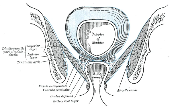
Figure 3Gray402
Long description
Coronal section of pelvis, showing arrangement of fasciæ. Viewed from behind. (Diagrammatic.)

Figure 3: Coronal section of pelvis, showing arrangement of fasciæ (viewed from behind).
The pelvic fascia provides structural support and defines the compartments through which lymphatic fluid travels. The pelvic girdle, consisting of the hip bones, sacrum, and coccyx, supports the upper body and transfers weight to the lower limbs. The hip bone articulates posteriorly with the sacrum at the sacroiliac joint and anteriorly with the opposite hip bone at the pubic symphysis. This bony framework anchors the pelvic floor muscles and their overlying fascia. Deep fascia in the thigh separates it into medial, anterior, and posterior compartments. While these thigh muscles are not part of the pelvic fascia, their fascial continuities influence lower limb lymphatic return. [1] [2]
Dissection within the pelvis must respect these fascial boundaries to prevent injury to the lymphatic channels and autonomic nerves. The total mesorectal excision (TME) technique involves sharp pelvic dissection of the mesorectum[1]. This procedure is performed in the holy plane to ensure complete removal of the lymphatic container[1]. After adoption of the TME technique, local recurrence rates decreased to 6–12% and the 5-year survival rate increased up to 53–87%[1]. Damage to the inferior hypogastric nerves can occur if the correct plane is missed, resulting in sympathetic damage[1]. Full mobilization of the rectum requires complete division of the recto-sacral fascia[1]. Major complications like bleeding from the presacral plexus of veins can occur during this step[1]. Therefore, precise identification of the fascial planes is paramount for both oncological clearance and functional preservation. [3]
Unlike the pectoral girdle, the bones of the pelvis are strongly united to form a largely immobile, weight-bearing structure.
The pubis forms the anterior portion of the hip bone. The enlarged medial portion is the pubic body. Located superiorly on the pubic body is a small bump called the pubic tubercle. The superior pubic ramus is the segment of bone that passes laterally from the pubic body to join the ilium. The narrow ridge running along the superior margin of the superior pubic ramus is the pectineal line of the pubis. When standing in the anatomical position, the pelvis is tilted anteriorly. In this position, th [2]
The smaller, more inferior lesser sciatic foramen is formed by the lesser sciatic notch of the hip bone, together with the sacrospinous and sacrotuberous ligaments. [2]
The primary role of the pelvis is to support the weight of the upper body when sitting and to transfer this weight to the lower limbs when standing. It serves as an attachment point for trunk and lower limb muscles, and also protects the internal pelvic organs. The presence of lateral ligaments has been a topic of much controversy. They are thought to be a fascial condensation on both anterolateral aspects of the extraperitoneal rectum. In past studies, there was some concern about the bleeding from the middle rectal artery when the rectum was mobilized from the lateral pelvic wall. [2] [3]
Clinical pearlStability
The immobility of the pelvis provides a strong foundation for the upper body as it rests on top of the mobile lower limbs.
The superior hypogastric plexus is carefully preserved at the root of the inferior mesenteric artery from the abdominal aorta. It is difficult to grossly differentiate lymphatics from the nerve structure. The magnified view through a camera could be helpful in distinguishing the inferior mesenteric artery pedicle from the underlying superior hypogastric plexus covered with [3]
| Opening | Formation |
|---|---|
| Greater sciatic foramen | Greater sciatic notch, sacrum, sacrospinous ligament |
| Lesser sciatic foramen | Lesser sciatic notch, sacrospinous ligament, sacrotuberous ligament |
X-ray imaging of the pelvis offers limited direct visualisation of the soft tissues of the pelvic fascia because plain radiographs lack the necessary soft tissue contrast resolution. Instead, conventional radiography demonstrates only the bony pelvis, outlining the pelvic brim, ischial spines, and pubic symphysis, which serve as crucial bony landmarks when inferring the spatial planes of the endopelvic fascia and pelvic floor.
Computed tomography provides superior spatial resolution compared to plain radiography and allows visualization of the pelvic wall muscles, fat-filled pelvirectal spaces, and thickened fascial condensations such as the levator ani sling. On standard contrast-enhanced CT scans, the pelvic fascia appears as linear bands or partitions dividing the extraperitoneal spaces, though differentiation between distinct fascial layers remains constrained by soft tissue contrast limits.
Magnetic resonance imaging is the gold standard for evaluating pelvic fascial anatomy due to its exceptional soft tissue contrast. On T2-weighted MRI sequences, the endopelvic fascia, levator ani muscle, and fascial ligaments appear as low-signal intensity dark structures contrasting sharply against the bright signal of high water content fat in the ischioanal and pararectal spaces.
Ultrasound, particularly transperineal, transrectal, and endoanal modalities, permits dynamic real-time assessment of the pelvic fascia and pelvic floor structures. On ultrasound imaging, collagen-rich fascial condensations appear hyperechoic relative to the surrounding muscle bellies. Transperineal ultrasound clearly depicts the integrity of the puborectalis sling and the displacement of fascial supports during a Valsalva manoeuvre, assisting in the diagnosis of pelvic organ prolapse.
DefinitionEndopelvic Fascia
A continuous sheet of connective tissue lining the pelvic cavity that invests the pelvic organs and forms supportive ligaments.
| Structure | Location | Function |
|---|---|---|
| Denonvilliers fascia | Anterior to rectum | Separates rectum from seminal vesicles |
| Pubocervical fascia | Between bladder and vagina | Supports the bladder base |
| Arcus tendineus | Pubic bone to ischial spine | Attachment for levator ani |
Complete total mesorectal excision aims to preserve the neurovascular bundle and maintain an intact mesorectal fascia [P50]. [3]
The levator ani muscle lies centrally forming the muscular pelvic floor, suspended from the thickened obturator fascia, and relates laterally to the bony pelvic sidewall and medially to the pelvic viscera. The endopelvic fascia bridges the pelvic floor structures and provides critical suspensory support to the bladder, vagina, and prostate. The Denonvilliers fascia spans transversely between the rectogenital spaces, separating the seminal vesicles or prostate from the rectum posteriorly. The neurovascular bundle runs posterolateral to the rectum and prostate within these fascial reflections, where it remains vulnerable to traction or thermal injury during radical pelvic dissections unless precise anatomical cleavage planes are strictly maintained. [3]
Clinical pearlFascial layers in surgery
Recognising whether fascial layers such as the Denonvilliers fascia are fused or distinct dictates the correct plane of surgical dissection around the prostate and rectum.
| Structure | Location | Clinical Significance |
|---|---|---|
| Denonvilliers fascia | Rectovesical space | Separates seminal vesicles and prostate from rectum; multilayered superiorly, single layer inferiorly |
| Endopelvic fascia | Pelvic floor and sidewalls | Provides structural support to pelvic viscera and anchors pelvic organs |
| Mesorectal fascia | Surrounding the rectum | Encloses the rectum and perirectal fat for safe total mesorectal excision |
The Denonvilliers fascia, also known as the rectovesical septum, exhibits a multilayer structure at the level of the seminal vesicle [P88]. [3]
The sacroiliac joint is largely immobile and supported by strong ligaments attaching the sacrum to the ilium. These include the anterior sacroiliac ligament on the anterior side and the posterior sacroiliac ligament on the posterior side. [2]
Two additional ligaments span the sacrum and hip bone to support and immobilize the sacrum. The sacrospinous ligament runs from the sacrum to the ischial spine. [2]
The sacrotuberous ligament runs from the sacrum to the ischial tuberosity, helping to carry the weight of the body. [2]
The pubis forms the anterior portion of the hip bone and articulates with the opposite pubis at the pubic symphysis.
The superior margin of the pubic body has the pubic tubercle, while the superior pubic ramus forms the pectineal line. [2]
The inferior pubic ramus joins the ischial ramus to form the ischiopubic ramus, which contributes to the subpubic angle. [2]
Stress urinary incontinence is defined as a complaint of involuntary loss of urine on effort or physical exertion including sporting activities etc. [4]
Its incidence is estimated to be about 15-20% of adult women, making it a common subtype of urinary incontinence. [4]
The prevalence of urinary incontinence ranges between 11.4 and 73.0%, and it is a major public health issue for women. [4]
By the age of 80 years, the lifetime risk of a woman in the USA undergoing at least one surgery for POP is 6.3%. [5]
Women with two children have an 8.4 times greater likelihood of experiencing POP requiring hospital attention compared with nulliparous women. [5]
DefinitionSacrospinous Ligament
The sacrospinous ligament runs from the sacrum to the ischial spine, helping to support and immobilize the sacrum.
| Structure | Attachment | Function |
|---|---|---|
| Sacrospinous | Sacrum to ischial spine | Support sacrum |
| Sacrotuberous | Sacrum to ischial tuberosity | Support sacrum |
| Anterior SI | Sacrum to ilium | Stabilize joint |
| Posterior SI | Sacrum to ilium | Stabilize joint |
Students often confuse the exact layers of the pelvic fascia, particularly mistaking the parietal pelvic fascia for visceral pelvic fascia. Remember that parietal pelvic fascia lines the pelvic walls, whereas visceral pelvic fascia invests individual pelvic organs. Another frequent error is assuming the Denonvilliers fascia is a single embryonic fusion plane without clinical significance; in reality, it serves as a critical surgical landmark during rectal cancer surgery.
Learners also misidentify the plane of dissection during total mesorectal excision. Dissection must be directed below the Denonvilliers fascia unless the tumor directly invades the seminal vesicle. Confusing the anterior and posterior planes around the Denonvilliers fascia is a major pitfall, as some authors argue that dissection posterior to it is oncologically inadequate while others consider the posterior plane optimal. [3]
Students frequently overlook the intimate relationship between the Denonvilliers fascia and the mesorectal fascia, noting incorrectly that they are entirely separate structures without continuity. Another mistake is ignoring the genetic risk factors for pelvic floor weakness, assuming pelvic organ prolapse is solely obstetric in origin, even though a family history confers a 2.5-fold increased incidence of pelvic organ prolapse compared with the general population. Finally, candidates forget that validated instruments assessing quality of life and female sexual function must form an integral part of the therapeutic process to limit undesirable consequences. [4] [5]
At the bedside, clinicians evaluate pelvic fascia integrity when patients present with pelvic organ prolapse or stress urinary incontinence, noting that a woman lifetime risk of undergoing surgery for these conditions is 11% to 20%. Counselors use standardized assessments to guide patients toward the best therapeutic option among conservative and surgical choices. [5]
In the operating theatre, surgeons rely on the Denonvilliers fascia as a landmark structure during anterior dissection. The optimal surgical dissection during total mesorectal excision is conducted anterior to the Denonvilliers fascia to ensure radical removal, or posterior to it depending on the mobilization strategy chosen by the operating team. If the middle rectal artery is encountered, it is easily controlled by cauterization with monopolar or bipolar diathermy. [3]
On imaging, magnetic resonance imaging delineates the fascial planes of the pelvis, helping radiologists and surgeons stage rectal tumors and assess whether a tumor directly invades the seminal vesicle. In examinations, students are tested on the precise anatomical boundaries of the endopelvic fascia, the nerve-sparing planes of the pelvis, and the genetic associations such as the collagen type 3 alpha 1 genotype linked to pelvic weakness.
Surgical Dissection Planes around Denonvilliers Fascia
- Identify seminal vesicle in men or vaginal wall in womenAnatomical landmarks for anterior dissection
- Question: Does tumor directly invade seminal vesicle?
Yes
- Perform anteriorly directed dissectionRequired for anterior tumors or threatened margin
No
- Direct dissection below Denonvilliers fasciaOptimal plane for nerve preservation
- Parietal pelvic fascia lines the inner walls of the pelvis while visceral fascia invests the pelvic organs.
- Denonvilliers fascia acts as a key landmark structure during anterior rectal and pelvic dissection.
- Dissection below the Denonvilliers fascia is the optimal plane for nerve preservation unless the tumor invades the seminal vesicle.
- Debate exists regarding whether the optimal anterior rectal mobilization plane lies anterior or posterior to the Denonvilliers fascia. [3]
- Middle rectal bleeding during dissection can be controlled with monopolar or bipolar diathermy.
- A woman lifetime risk of undergoing surgery for pelvic organ prolapse or stress urinary incontinence is 11% to 20%.
- A family history of prolapse results in a 2.5-fold increased incidence of pelvic organ prolapse compared with the general population.
- Validated instruments assessing quality of life are crucial for proper patient counseling and therapeutic choices.
Answer each one in your head first, then open the answer.
What structure is identified in men after the peritoneal reflection is divided during anterior dissection?
Show answerHide answer
seminal vesicleWhat is the recommended dissection direction if a tumor directly invades the seminal vesicle?
Show answerHide answer
anteriorly directed dissectionHow is an identified middle rectal artery easily controlled during surgery?
Show answerHide answer
cauterization with monopolar or bipolar diathermyWhat is a woman lifetime risk of undergoing surgery for pelvic organ prolapse or stress urinary incontinence?
Show answerHide answer
11% to 20%What is the fold increase in pelvic organ prolapse incidence for women with a positive family history?
Show answerHide answer
2.5-fold
- Betts JG, Young KA, Wise JA, et al. Appendicular Muscles of the Pelvic Girdle and Lower Limbs. OpenStax, Anatomy and Physiology 2e (CC BY-NC-SA 4.0). Accessed .
- Betts JG, Young KA, Wise JA, et al. The Pelvic Girdle and Pelvis. OpenStax, Anatomy and Physiology 2e (CC BY-NC-SA 4.0). Accessed .
- Kim NK, Kim HS, Alessa M, Torky R. Optimal Complete Rectum Mobilization Focused on the Anatomy of the Pelvic Fascia and Autonomic Nerves: 30 Years of Experience at Severance Hospital. Yonsei medical journal 2021;62(3):187-199. doi:10.3349/ymj.2021.62.3.187. Accessed .
- La Rosa VL, Ciebiera M, Lin LT, et al. Multidisciplinary management of women with pelvic organ prolapse, urinary incontinence and lower urinary tract symptoms.A clinical and psychological overview. Przeglad menopauzalny = Menopause review 2019;18(3):184-190. doi:10.5114/pm.2019.89496. Accessed .
- Weintraub AY, Glinter H, Marcus-Braun N. Narrative review of the epidemiology, diagnosis and pathophysiology of pelvic organ prolapse. International braz j urol : official journal of the Brazilian Society of Urology 2020;46(1):5-14. doi:10.1590/s1677-5538.ibju.2018.0581. Accessed .
- Roch M, Gaudreault N, Cyr MP, et al. The Female Pelvic Floor Fascia Anatomy: A Systematic Search and Review. Life (Basel, Switzerland) 2021;11(9):900. doi:10.3390/life11090900. Accessed .
Figure credits
Educational content for medical students and doctors. It is not a substitute for clinical judgement or local guidelines.