Urinary bladder
A hollow muscular pelvic organ that stores urine before voiding via the urethra.
The urinary bladder is a retroperitoneal organ located posterior to the pubic bone. In females, the bladder lies anterior to the uterus, whereas in males, the prostate is situated immediately inferior to the bladder. The bladder collects urine from both ureters. The ureters lead from the renal pelvis of the kidney to the trigone area at the base of the bladder. As the ureters approach the bladder, they turn medially and pierce the bladder wall obliquely, creating a physiological one-way valve that allows urine entry while preventing reflux. [1]
DefinitionTrigone
A smooth triangular region at the base of the bladder defined by the two ureteric orifices superiorly and the internal urethral orifice inferiorly.
The bladder wall consists of a thick muscular layer containing smooth muscle that helps move urine. The dome of the bladder distends superiorly when the organ is filling with urine, and it possesses a peritoneal covering only over this dome. Micturition results from an interplay of involuntary and voluntary actions by the internal and external urethral sphincters. When bladder volume reaches about 150 mL, an urge to void is sensed. Incontinence occurs as bladder volume approaches 300 to 400 mL, or when involuntary voiding happens around 250 mL. [1]
| Sex | Anterior relation | Posterior relation | Inferior relation |
|---|---|---|---|
| Female | Pubic bone | Uterus | Pelvic floor |
| Male | Pubic bone | Rectum | Prostate |
Catheterisation involves passing a tube through the urethra into the bladder lumen to drain urine. Bladder rupture can occur due to severe pelvic trauma or overdistension, potentially leading to the leakage of urine into surrounding retroperitoneal or peritoneal spaces depending on the anatomical site of the tear.
Bladder filling and micturition milestones
- Bladder filling beginsUrine propelled by ureteric peristalsis enters via the oblique ureteric orifices into the trigone.
- Question: Volume reaches 150 mL?
Yes
- Initial urge to voidSensed but easily overridden by voluntary control of the external urethral sphincter.
No
- Continued fillingDome distends superiorly into the retroperitoneal space.
- Question: Volume approaches 300 to 400 mL?
Yes
- Voluntary constraint failsResults in involuntary micturition or incontinence.
No
- Maintain continenceConsciously prevent relaxation of the external urethral sphincter.
Figure 1Female and Male Urethra
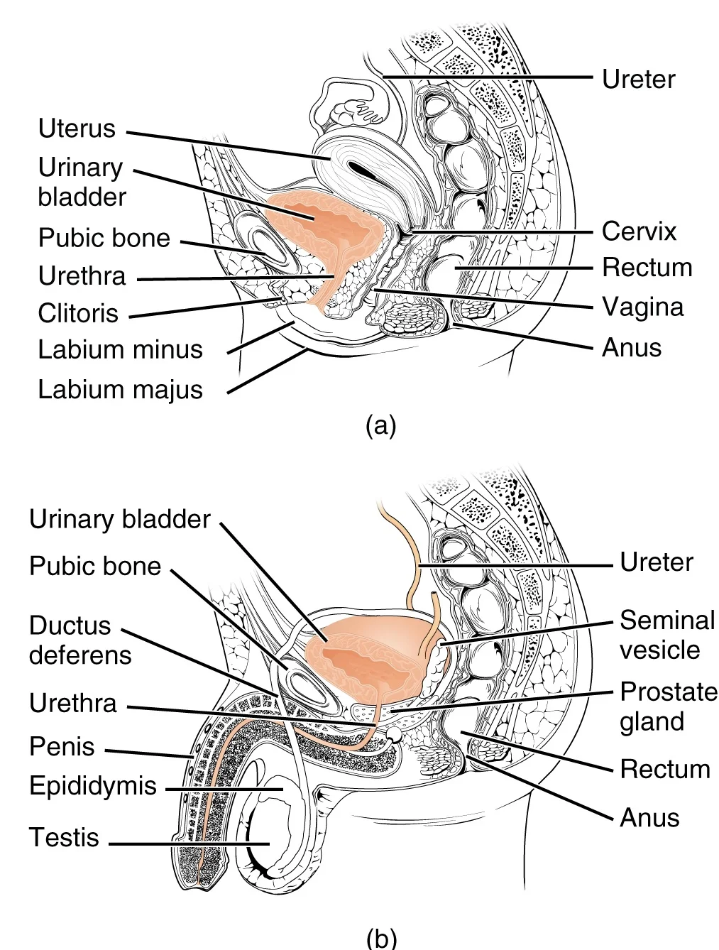
Long description
The urethra transports urine from the bladder to the outside of the body. This image shows (a) a female urethra and (b) a male urethra.

Figure 1: The urethra transports urine from the bladder to the outside of the body.
The urethra begins inferior and central to the two ureteral openings at the base of the bladder, forming the trigone[1]. The proximal urethra is lined by transitional epithelium, whereas the terminal portion is a nonkeratinized, stratified squamous epithelium[1]. In the male, pseudostratified columnar epithelium lines the urethra between these two cell types[1]. Voiding is regulated by an involuntary internal urinary sphincter consisting of smooth muscle, and a voluntary external urinary sphincter of skeletal muscle located below it[1].
DefinitionTrigone
A triangular-shaped area at the base of the bladder formed by the two ureteral openings and the urethral opening[1].
In the female, the external urethral orifice is embedded in the anterior vaginal wall inferior to the clitoris, superior to the vaginal opening, and medial to the labia minora[1]. The female urethra has a short length, about 4 cm, which is less of a barrier to fecal bacteria than the longer male urethra and provides the best explanation for the greater incidence of urinary tract infections in females[1]. Voluntary control of the female external urethral sphincter is a function of the pudendal nerve, arising in the sacral region of the spinal cord and traveling via the S2 to S4 nerves of the sacral plexus[1].
In the male, the urethra passes through the prostate gland immediately inferior to the bladder before passing below the pubic symphysis[1]. The male urethra averages 20 cm in length and is divided into four regions: the preprostatic urethra, the prostatic urethra, the membranous urethra, and the spongy or penile urethra[1]. The preprostatic urethra is very short and incorporated into the bladder wall[1]. The prostatic urethra passes through the prostate gland, where it receives sperm via the ejaculatory ducts and secretions from the seminal vesicles during sexual intercourse[1]. Paired Cowper glands, also known as bulbourethral glands, produce and secrete mucus into the prostatic urethra to buffer urethral pH and lubricate the passage during sexual stimulation[1].
| Region | Anatomical location and relations | Key secretions or structures received |
|---|---|---|
| Preprostatic urethra | Incorporated into the bladder wall[1] | None |
| Prostatic urethra | Passes through the prostate gland[1] | Sperm, seminal vesicle secretions, Cowper gland mucus[1] |
| Membranous urethra | Passes through the deep muscles of the perineum[1] | Urethral sphincters investment[1] |
| Spongy urethra | Passes through the corpus spongiosum to the external urethral orifice[1] | Mucous gland secretions[1] |
The membranous urethra passes through the deep muscles of the perineum, where it is invested by the overlying urethral sphincters[1]. The spongy urethra exits at the external urethral orifice at the tip of the penis after passing through the corpus spongiosum[1]. Mucous glands are found along much of the length of the urethra to protect the tissue from extremes of urine pH[1].
Figure 2Position of the urinary bladder 1
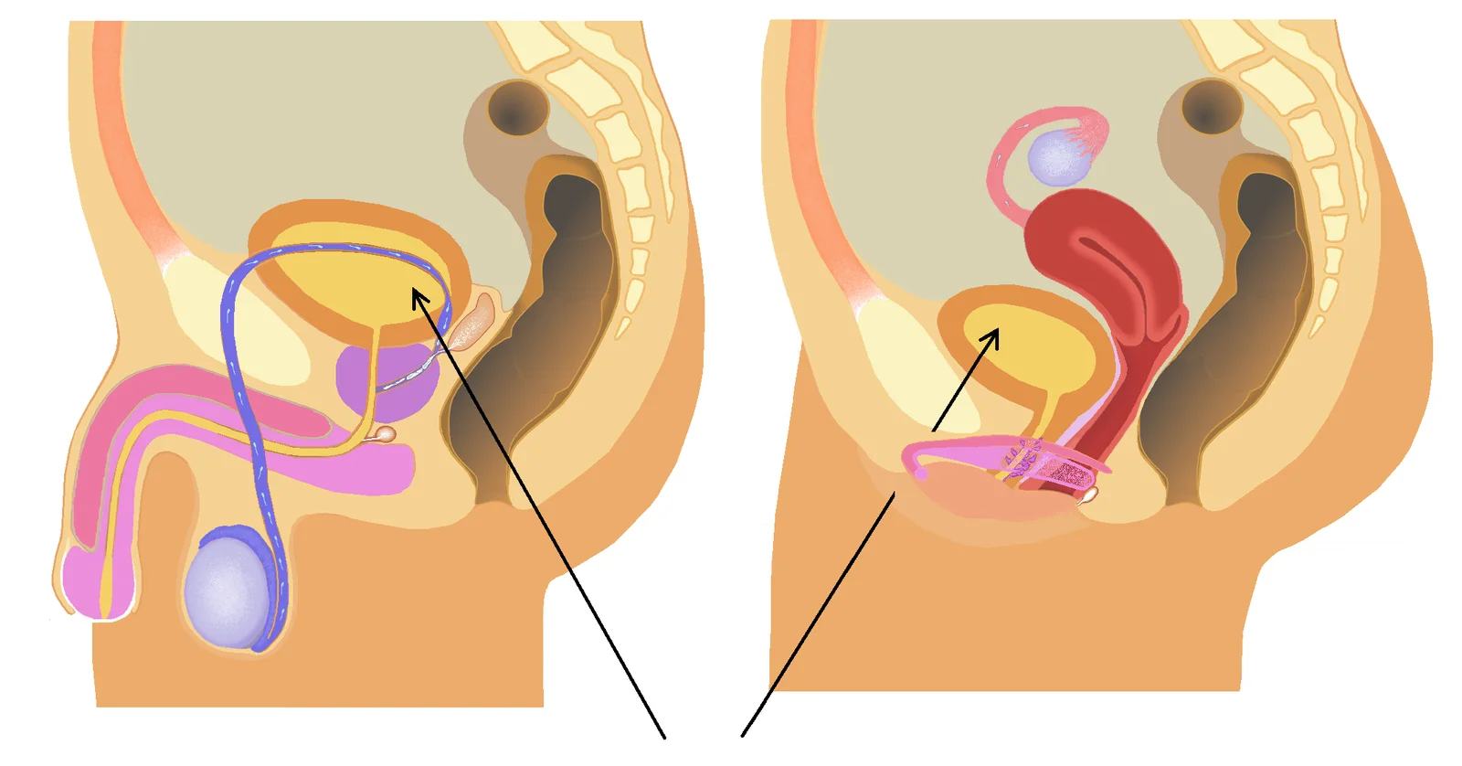
Long description
Position of the urinary bladder in man and woman.

Figure 2: Position of the urinary bladder in man and woman.
DefinitionBladder function
The urinary bladder stores urine for most of the time and expels it several times a day. In healthy humans, the number of voiding events is less than eight times per day. [2]
The apex of the bladder points anterosuperiorly toward the superior border of the pubic symphysis. The base or posterior wall lies directly against the anterior wall of the vagina and cervix in females, and against the seminal vesicles, terminal parts of the ureters, and rectum in males. The inferolateral surfaces are related externally to the retropubic fat pad and the pubic bones, as well as the obturator internus and levator ani muscles. The neck of the bladder is anchored inferiorly at the base of the prostate gland in males or directly onto the superior fascia of the urogenital diaphragm in females. [1]
Figure 3Gray1135
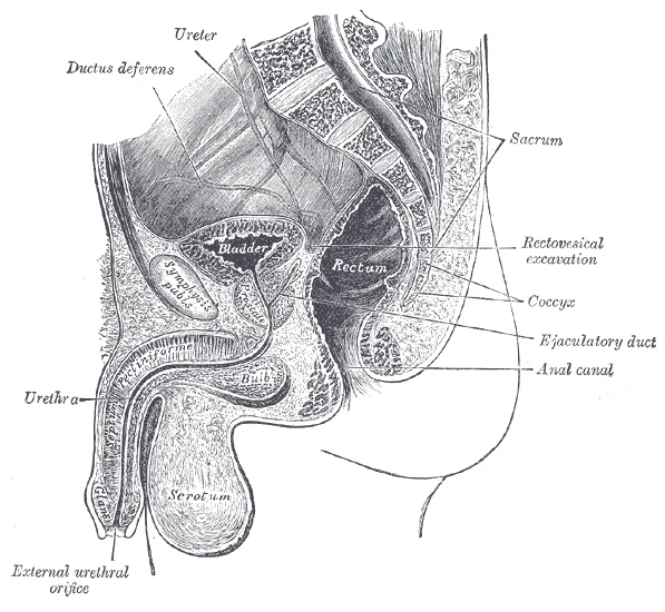
Long description
Median sagitta section of male pelvis. Labelled illustration from Wikimedia Commons, used under its open licence.

Figure 3: Median sagitta section of male pelvis.
Storage of urine requires the bladder body to distend, which is mediated by smooth muscle relaxation of the detrusor muscle. A key physiological mechanism to induce detrusor relaxation is beta-adrenoceptor stimulation, which in most species involves a strong beta-3 component, and in humans predominantly if not exclusively occurs via this beta-3 receptor. Although the prototypical signaling pathway of the beta-adrenoceptor is the activation of adenylyl cyclase with subsequent formation of cyclic AMP, cyclic AMP apparently contributes to beta-adrenoceptor-mediated bladder relaxation only in a minor way. Instead, large-conductance calcium-activated potassium channels, known as BKCa channels, may play a greater role in beta-adrenoceptor-mediated bladder relaxation, and activators of these channels are currently under investigation to treat an overactive bladder. The strength of coupling of calcium sparks to BKCa channels is regulated by membrane potential and stimuli that modulate calcium sensitivity. Consequently, activators of BKCa channels have been synthesized as possible novel drugs for the treatment of overactive bladder. [1][2]
| Region | Male Relations | Female Relations |
|---|---|---|
| Superior surface | Covered by peritoneum, sigmoid colon loops | Covered by peritoneum, body of uterus |
| Posterior base | Seminal vesicles, ampulla of vas deferens, rectum | Anterior wall of vagina, supravaginal cervix |
| Inferolateral | Retropubic space, levator ani, obturator internus | Retropubic space, levator ani, pubocervical fascia |
| Neck | Superior surface of prostate gland | Superior fascia of urogenital diaphragm |
Bladder relations and autonomic control
- Bladder BodyDistends during urine storage via detrusor smooth muscle relaxation
- Question: Mechanism involved?
Beta-AR stimulation
- Beta-3 adrenoceptor dominance in humans
BKCa channels
- Contribute to relaxation pathways
Figure 4Anatomytool Male and female urinary tract English
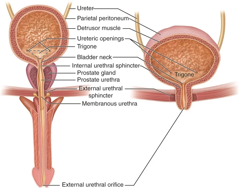
Long description
Anatomical structures in item: Systema urinarium Ureter Musculus detrusor vesicae Ostium ureteris Trigonum vesicae Cervix vesicae; Collum vesicae Sphincter urethrae internus Prostata (Glandula prostatica) Pars prostatica

Figure 4: Anatomical structures in item: Systema urinarium Ureter Musculus detrusor vesicae Ostium ureteris Trigonum vesicae Cervix vesicae; Collum vesicae Sphincter urethrae internus Prostata (Glandula prostatica) Pars prostatica.
The urinary bladder is a highly distensible hollow organ designed to accommodate large volume fluctuations, with adult capacities ranging from nearly zero to 500 to 600 mL. [1]
The organ wall is composed of irregular crisscrossing bands of smooth muscle known collectively as the detrusor muscle. Contraction of this muscular network is required for bladder emptying and is coordinated by autonomic and somatic neural inputs. [1]
The interior surface of the urinary bladder is lined by a specialized transitional cellular epithelium, which is structurally suited for accommodating significant organ stretching. When the bladder is empty, this mucosa resembles columnar epithelia. As the bladder fills and stretches, the cellular layer transitions in appearance to a flattened, squamous configuration. [1]
Figure 5Gray1140
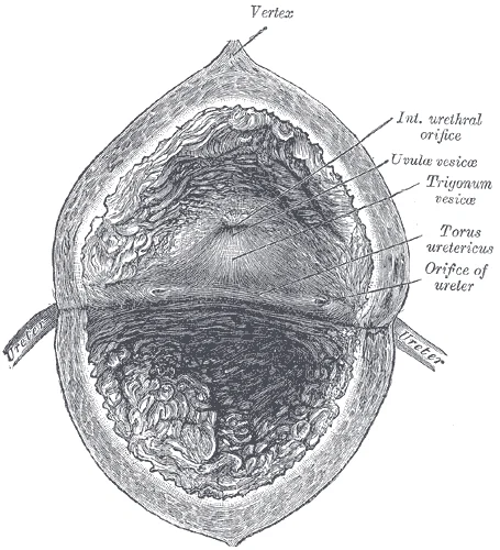
Long description
The interior of bladder. Labelled illustration from Wikimedia Commons, used under its open licence.

Figure 5: The interior of bladder.
DefinitionTransitional Epithelium
A specialized stratified epithelium lining the urinary tract that changes its morphological appearance from columnar to squamous upon tissue stretching.
The superior dome of the organ is formed by the bladder body, leading inferiorly toward the funnel-shaped trigone region at the base. Descending further inferiorly, the internal urethral orifice marks the transition from the bladder cavity into the proximal urethra. Flanking this outlet, the muscular wall thickens to form the circular internal urethral sphincter.
| Structure | Cell Type | Primary Action | Innervation |
|---|---|---|---|
| Detrusor muscle | Smooth muscle | Contracts to empty the bladder | Parasympathetic via pelvic nerves |
| External urinary sphincter | Skeletal muscle | Maintains voluntary urinary continence | Somatic via pudendal nerve |
The arterial supply of the urinary bladder is derived primarily from the superior and inferior vesical arteries, which are branches of the internal iliac artery. These vessels enter the bladder at the lateral aspects of its base, specifically near the ureteric openings and the neck. The superior vesical artery typically arises from the patent portion of the umbilical artery, while the inferior vesical artery may arise directly from the internal iliac or share a common trunk with the middle rectal artery. Upon entering the bladder wall, these arteries anastomose extensively with branches from the opposite side and with the ureteric arteries, forming a rich submucosal and muscular plexus. This vascular network ensures robust perfusion to the detrusor muscle and the mucosal lining. The venous drainage follows a similar pattern, with the vesical venous plexus draining into the internal iliac veins. Lymphatic drainage from the bladder body and fundus flows to the external and internal iliac lymph nodes, while drainage from the bladder neck and trigone may also reach the presacral nodes.
The detrusor muscle contracts with significant force in the young. The bladder’s strength diminishes with age, but voluntary contractions of abdominal skeletal muscles can increase intra-abdominal pressure to promote more forceful bladder emptying. Such voluntary contraction is also used in forceful defecation and childbirth. [1]
Noradrenaline released from the hypogastric nerves activates β-AR and induces detrusor smooth muscle relaxation. All three cloned β-AR subtypes are present in the urinary bladder of rat and humans at the mRNA level. At least in humans, the β3-AR is by far the most abundantly expressed subtype. Functionally, it appears that detrusor relaxation involves a strong β3-AR component in most species. [2]
DefinitionDetrusor Muscle
The smooth muscle layer of the bladder wall responsible for contraction during micturition.
| Structure | Nerve Supply | Primary Effect |
|---|---|---|
| Detrusor Muscle | Parasympathetic (Pelvic Splanchnic) | Contraction |
| Detrusor Muscle | Sympathetic (Hypogastric) | Relaxation |
| Internal Sphincter | Sympathetic (Hypogastric) | Contraction |
The venous drainage of the urinary bladder follows a systematic pattern, emptying into the internal iliac veins. This pathway is critical for understanding the spread of pathology and the anatomical relationships within the pelvis.
The middle vesical veins are located on the lateral aspects. They also terminate in the internal iliac veins. The inferior vesical veins are found at the base. They drain into the prostatic venous plexus in males or the vaginal venous plexus in females. These plexuses then connect to the internal iliac veins. The veins run alongside the arteries. They are embedded in the connective tissue of the bladder wall. The arrangement ensures efficient return of deoxygenated blood to the systemic circulation. The internal iliac veins are the primary recipients. They carry blood towards the common iliac veins. This anatomical layout is consistent across adults. It provides a reliable route for venous return. Understanding this drainage is essential for surgical procedures. It helps in controlling bleeding during cystectomy. It also aids in identifying metastatic spread. The veins are thin-walled and valvular. This structure allows for pressure regulation. The connections to the prostatic or vaginal plexuses are significant. They provide alternative pathways for drainage. This collateral circulation is important in cases of obstruction. The internal iliac veins are large and robust. They can accommodate increased blood flow. The bladder wall is highly vascular. This vascularity supports its function as a reservoir. The venous drainage mirrors the arterial supply. The arteries and veins run in parallel. They are accompanied by nerves. This neurovascular bundle is protected by surrounding tissues. The anatomical relationships are fixed. The bladder lies anterior to the rectum in males. In females, it lies anterior to the vagina. These relations influence the venous drainage patterns. The internal iliac veins are located posteriorly. They receive blood from multiple pelvic organs. The bladder is just one of these organs. The drainage is unidirectional. Blood flows from the bladder to the internal iliac veins. This flow is maintained by muscle contractions. The pelvic floor muscles assist in venous return. The anatomy is designed for efficiency. This area is a common site for pathology. The venous drainage here is particularly important. It connects to the systemic circulation via the internal iliac veins. This connection is vital for clinical assessment. It allows for the detection of venous congestion. The anatomy is consistent in healthy individuals. Variations are rare but possible. The internal iliac veins are the main outflow tract. They are located in the pelvis. The bladder drains into them directly or indirectly. The prostatic or vaginal plexuses are intermediate stations. They receive blood from the inferior vesical veins. These plexuses are extensive. They connect with other pelvic veins. This network provides redundancy. It ensures drainage even if one pathway is compromised. The internal iliac veins are large. They can handle significant blood volume. The bladder is a muscular organ. Its contraction aids venous return. The anatomy supports its physiological function. The venous drainage is a key feature. It is studied in detail for medical students. Understanding it is essential for clinical practice. The internal iliac veins are the final destination. They carry blood to the heart. The bladder is thus connected to the systemic circulation. This connection is fundamental to its physiology. The venous drainage is a critical aspect of bladder anatomy. It is studied alongside arterial supply and innervation. The three systems work together. They ensure proper bladder function. The venous drainage is often overlooked. It is nonetheless vital. It provides a route for waste removal. The veins are thin-walled. This allows for easy expansion. The bladder can hold large volumes. The veins accommodate this change. They expand and contract with the bladder. This dynamic nature is important. It reflects the bladder's function. The venous drainage is a complex system. It involves multiple veins and plexuses. The internal iliac veins are the main collectors. They are located in the pelvis. The bladder drains into them. This drainage is efficient. It ensures timely removal of blood. The anatomy is well-defined. It is taught in medical schools. It is essential for future doctors. Understanding it helps in diagnosis and treatment. The venous drainage is a key topic. It is part of the broader study of anatomy. It is linked to physiology and pathology. It is a vital component of medical knowledge. The internal iliac veins are the main outflow. They are located in the pelvis. The bladder drains into them. This drainage is crucial. It supports the bladder's function. The anatomy is fixed. It is consistent in humans. It is studied for clinical relevance. The venous drainage is a critical aspect. It is often tested in exams. It is important for practical skills. It aids in surgical planning. It helps in understanding disease spread. The internal iliac veins are the main route. They are large and robust. The bladder drains into them. This drainage is efficient. It ensures proper function. The anatomy is well-understood. It is taught clearly. It is essential for medical students. The venous drainage is a key feature. It is part of the bladder's structure. It is linked to its function. It is vital for health. The internal iliac veins are the main collectors. They are located in the pelvis. The bladder drains into them. This drainage is important. It supports the bladder's role. The anatomy is fixed. It is consistent. It is studied for clinical use. The venous drainage is a critical topic. It is part of medical education. It is essential for practice. The internal iliac veins are the main outflow. They are located in the pelvis. The bladder drains into them. This drainage is vital. It supports the bladder's function. The anatomy is well-defined. It is taught in detail. It is essential for students. The venous drainage is a key aspect. It is part of the bladder's anatomy. It is linked to its physiology. It is vital for understanding disease. The internal iliac veins are the main route. They are large. The bladder drains into them. This drainage is efficient. It ensures proper function. The anatomy is fixed. It is consistent. It is studied for clinical relevance. The venous drainage is a critical feature. It is part of medical knowledge. It is essential for practice. The internal iliac veins are the main collectors. They are located in the pelvis. The bladder drains into them. This drainage is important. It supports the bladder's role. The anatomy is well-understood. It is taught clearly. It is essential for medical students. The venous drainage is a key topic. It is part of the bladder's structure. It is linked to its function. It is vital for health. The internal iliac veins are the main outflow. They are located in the pelvis. The bladder drains into them. This drainage is vital. It supports the bladder's function. The anatomy is well-defined. It is taught in detail. It is essential for students. The venous drainage is a key aspect. It is part of the bladder's anatomy. It is linked to its physiology. It is vital for understanding disease. The internal iliac veins are the main route. They are large. The bladder drains into them. This drainage is efficient. It ensures proper function. The anatomy is fixed. It is consistent. It is studied for clinical relevance. The venous drainage is a critical feature. It is part of medical knowledge. It is essential for practice. [1]
Figure 6Gray1141
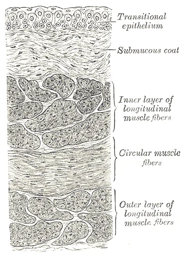
Long description
Vertical section of bladder wall. Labelled illustration from Wikimedia Commons, used under its open licence.

Figure 6: Vertical section of bladder wall.
Figure 7Gray1139
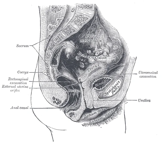
Long description
Median sagittal section of female pelvis.

Figure 7: Median sagittal section of female pelvis.
Figure 8Gray1136
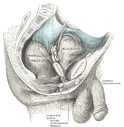
Long description
Male pelvic organs seen from right side. Bladder and rectum distended; relations of peritoneum to the bladder and rectum shown in blue. The arrow points to the rectovesical pouch.

Figure 8: Male pelvic organs seen from right side.
DefinitionAnatomical principle
Lymphatic outflow from the urinary bladder follows regional vascular pathways into pelvic and para-aortic nodes.
The lymphatic network originates within the mucosal and muscular layers of the urinary bladder wall, converging to form collecting channels that pierce the detrusor muscle. These initial efferent vessels course outwards towards the pelvic sidewall to reach the primary nodal groups.
The primary drainage stations comprise the external iliac, internal iliac, and obturator lymph nodes. The external iliac nodes lie along the medial and lateral aspects of the external iliac vein, receiving superior bladder surface drainage. The internal iliac nodes are situated along the internal iliac vessels within the posterior pelvis, draining the posterolateral walls and base.
From these primary stations, secondary efferent vessels ascend to drain into the common iliac lymph nodes, which lie along the common iliac vessels from the aortic bifurcation to the pelvic brim. Ultimately, lymph ascends further to reach the lateral aortic and preaortic lymph nodes surrounding the abdominal aorta.
In clinical scenarios involving malignancies, lymphatic mapping demonstrates that tumor cell dissemination follows these predictable anatomical pathways. Special populations such as older adults exhibit similar anatomical routes, though age-related sclerosis of lymphatic channels can occasionally alter flow dynamics. In low-resource settings without advanced imaging, knowledge of these stations guides traditional surgical staging.
| Nodal Station | Anatomical Location | Primary Drainage Area |
|---|---|---|
| External iliac | Along external iliac vein | Superior bladder surface |
| Internal iliac | Along internal iliac vessels | Bladder base and posterolateral walls |
| Obturator | Obturator fossa | Inferolateral surfaces |
| Common iliac | Along common iliac vessels | Primary pelvic nodes |
| Aortic | Surrounding abdominal aorta | Common iliac nodes |
DefinitionAutonomic and somatic innervation
The urinary bladder receives both autonomic and somatic nerve supplies. Autonomic control coordinates visceral smooth muscle activity, whereas somatic innervation governs voluntary control of skeletal muscle elements.
Normal micturition is a result of stretch receptors in the bladder wall that transmit nerve impulses to the sacral region of the spinal cord to generate a spinal reflex. The resulting parasympathetic neural outflow causes contraction of the detrusor muscle and relaxation of the involuntary internal urethral sphincter. At the same time, the spinal cord inhibits somatic motor neurons, resulting in the relaxation of the skeletal muscle of the external urethral sphincter. The micturition reflex is active in infants but with maturity, children learn to override the reflex by asserting external sphincter control, thereby delaying voiding. This reflex may be preserved even in the face of spinal cord injury that results in paraplegia or quadriplegia. [1]
However, various other signalling pathways have been demonstrated to be activated after stimulation of M3 receptors. [2]
One family is the small molecular weight secretory phospholipase A2 which consists of mainly calcium-dependent enzymes. Moreover, several cyclooxygenase products can also generate detrusor smooth muscle contraction. [2]
The urinary bladder is lined by a specialized transitional epithelium known as urothelium, which accommodates changes in organ volume. Link to the histology page.
DefinitionUrothelial adaptation
Specialized umbrella cells possess specialized plaques and fusiform vesicles to maintain a permeability barrier under high stretch.
| Marker Test | Reported Sensitivity | Reported Specificity | Clinical Utility |
|---|---|---|---|
| Cxbladder Monitor | 93% | Low | Postpone or avoid cystoscopy |
| Survivin assay | 64% | 93% | Superior to cytology |
| DNA Methylation | 89% | 97% | Surveillance setting |
Biomarker Surveillance Pathway
- Assess clinical riskEvaluate patient risk and prior history.
- Question: Low risk of recurrence?
Yes
- Consider biomarker test like Cxbladder Monitor to postpone cystoscopy
No
- Perform standard surveillance cystoscopy and cytology
Link to the embryology page.
The urinary bladder is a hollow, distensible muscular organ situated in the true pelvis, serving as the primary reservoir for urine before micturition. Its development is a complex process involving the integration of structures from the urogenital sinus and the mesoderm. Understanding its anatomy requires a clear distinction between its structural layers and its functional innervation, which governs both storage and voiding.
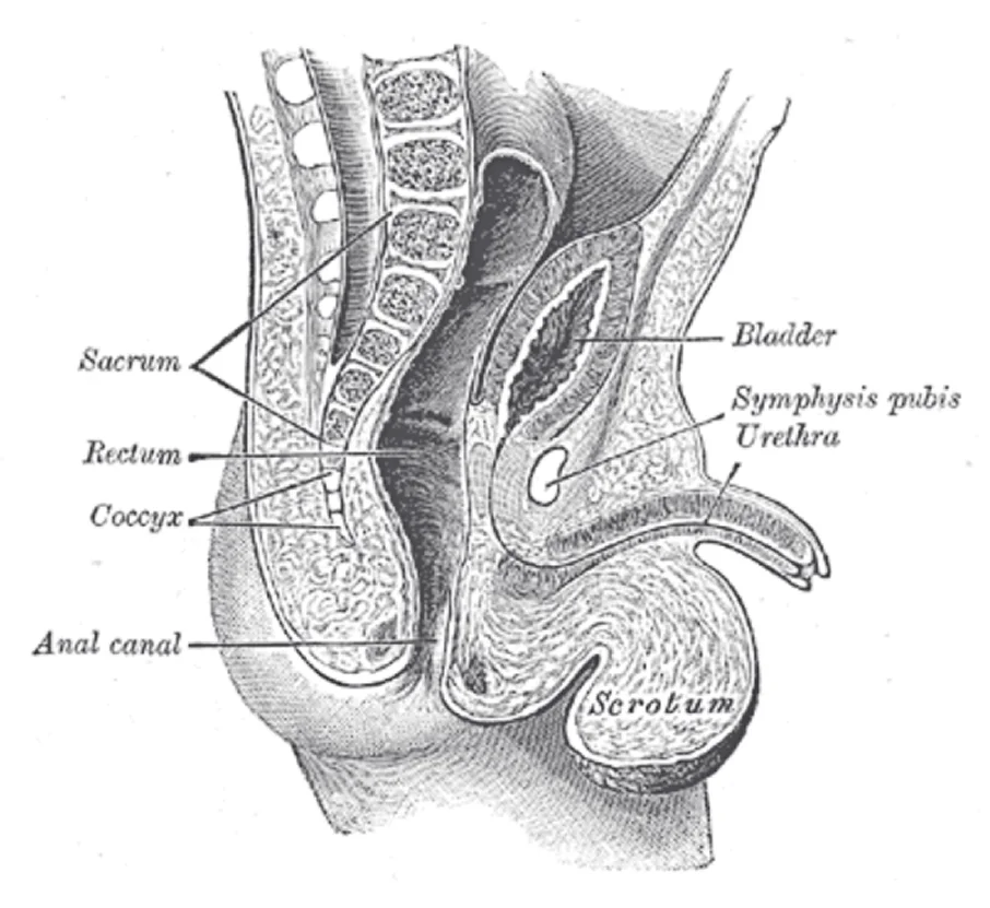
Figure 9Gray1137
Long description
Sagittal section through the pelvis of a newly born male child.

Figure 9: Sagittal section through the pelvis of a newly born male child.
The wall of the bladder is composed of four distinct layers, each with specific histological and functional characteristics. The innermost layer is the mucosa, which is lined by transitional epithelium (urothelium). This specialized epithelium is unique to the urinary tract and allows the bladder to stretch significantly without compromising the barrier function. Beneath the mucosa lies the lamina propria, a layer of loose connective tissue that supports the epithelium and contains blood vessels and nerves. The next layer is the muscularis propria, commonly referred to as the detrusor muscle. This muscle is composed of three layers of smooth muscle fibres: an inner longitudinal layer, a middle circular layer, and an outer longitudinal layer. However, these layers are not sharply defined and blend into one another, allowing for coordinated contraction during voiding. The outermost layer is the adventitia or serosa, depending on the location of the bladder. The superior surface is covered by peritoneum, forming the serosa, while the rest of the bladder is covered by adventitia, which blends with surrounding pelvic structures.
The blood supply to the bladder is derived from the internal iliac artery. The superior vesical arteries, which are branches of the umbilical arteries, supply the upper part of the bladder. The inferior vesical arteries, which arise directly from the internal iliac artery in males or from the vaginal artery in females, supply the lower part of the bladder. Venous drainage follows the arterial supply, with the vesical venous plexus draining into the internal iliac veins. Lymphatic drainage is primarily to the external and internal iliac lymph nodes, with some drainage to the sacral nodes.
Innervation of the bladder is crucial for its function. The parasympathetic nervous system, derived from the S2-S4 spinal segments via the pelvic splanchnic nerves, stimulates contraction of the detrusor muscle and relaxation of the internal urethral sphincter, promoting voiding. The sympathetic nervous system, derived from T11-L2 spinal segments via the hypogastric nerves, inhibits detrusor contraction and stimulates contraction of the internal urethral sphincter, promoting storage. Somatic innervation via the pudendal nerve controls the external urethral sphincter, allowing voluntary control over micturition.
When empty, it lies entirely within the true pelvis below the level of the pelvic brim. As it fills, it expands superiorly into the extraperitoneal space of the anterior abdominal wall, rising out of the true pelvis into the lower part of the abdominal cavity.
The anterior surface relates directly to the retropubic space of Retzius, containing loose fatty tissue separating the bladder wall from the posterior aspect of the pubic bones. In females, the posterior surface relates to the cervix and vagina, separated by loose connective tissue.
DefinitionSpace of Retzius
The retropubic extraperitoneal space situated between the anterior bladder wall and the pubic symphysis, allowing free movement of the bladder during filling and emptying.
| Muscle | Origin | Insertion | Action | Nerve Supply |
|---|---|---|---|---|
| Detrusor muscle | Outer longitudinal, middle circular, and inner longitudinal smooth muscle layers of the bladder wall | Continuous throughout the bladder wall, converging at the internal urethral orifice | Contracts to expel urine from the bladder during micturition | Pelvic splanchnic nerves (parasympathetic S2 to S4) |
| Sphincter urethrae | Ischiopubic rami | Perineal membrane and midline raphe | Provides voluntary and involuntary tone around the urethra to maintain urinary continence | Pudendal nerve (somatic S2 to S4) |
Clinical pearlClinical positioning
The empty bladder lies entirely within the true pelvis, whereas a full bladder expands superiorly into the abdominal cavity, elevating the peritoneal reflection.
Bladder injury typically occurs secondary to blunt pelvic trauma, iatrogenic instrumentation, or penetrating wounds. Lesion sites can be categorised into extraperitoneal and intraperitoneal ruptures.
| Feature | Extraperitoneal rupture | Intraperitoneal rupture |
|---|---|---|
| Anatomical site | Anterolateral wall or base | Dome of the bladder |
| Mechanism | Associated pelvic bone fractures | Blunt trauma to a distended abdomen |
| Extravasation | Confined to perivesical spaces | Free fluid within the peritoneal cavity |
| Management | Usually conservative with catheter drainage | Urgent surgical repair required |
Superiorly, the dome is marked as the primary site for intraperitoneal rupture.
Assessment of Bladder Trauma
- Initial PresentationEvaluate patient with pelvic trauma, gross haematuria, or inability to void.
- Question: Mechanism or signs suggest rupture?
Yes
- Perform retrograde cystography
- Question: Extravasation pattern?
Extraperitoneal
- Catheter drainage alone
Intraperitoneal
- Surgical closure
No
- Monitor output
- The urinary bladder shifts from a flattened tetrahedral shape when empty to a pear-shaped ovoid as it fills and extends superiorly into the retroperitoneal space.
- The superior surface of the bladder is completely covered by peritoneum, whereas the inferior and posterior aspects lack peritoneal coverage and are defined by adventitia.
- The empty bladder fundus in males is separated from the rectum by the rectovesical fascia, seminal vesicles, and terminal ductus deferentes.
- The median umbilical ligament extends from the bladder vertex to the umbilicus as a fibrous remnant of the embryonic urachus.
- The ureters pierce the bladder wall obliquely to form a functional one-way valve, preventing the retrograde flow of urine toward the kidneys during bladder contraction.
- The paravesical fossae are peritoneal depressions situated on either side of the bladder that accommodate loops of the small intestine and sigmoid colon when the bladder is empty.
Exam trapConfusion of storage and excretion
Students frequently assume the urinary bladder only acts as a passive conduit, whereas its primary physiological role is to store variable volumes of fluid safely while protecting delicate surrounding tissues from fluctuations in urinary pH and osmolarity.
A major error in anatomical reasoning is treating the detrusor muscle as a simple smooth muscle lacking complex regulatory inputs. Smooth muscle tone and phasic contractions are governed by distinct pathways, notably phosphatidylinositol-phospholipase C and phosphatidylcholine-phospholipase C signaling cascades.
Another common mistake involves misinterpreting diagnostic marker cut-offs. For instance, the quantitative NMP22BC test relies on a continuous scale converted into a dichotomous variable via a designated threshold estimated at 10 ng/mL. Assuming such biological cut-offs represent absolute clinical boundaries often leads to lower overall diagnostic accuracy. [3]
Students also confuse the performance parameters of screening versus surveillance tests. When evaluating tumour markers, a very high negative predictive value is required if the clinical goal is to avoid an unnecessary cystoscopy when a test result is negative. Conversely, high sensitivity is mandatory during follow-up at 3 months after initial transurethral resection of a bladder tumour or in patients with high-risk disease.
Ignoring the limitations of standard white-light cystoscopy is another frequent pitfall. White-light cystoscopy alone can miss disease, particularly carcinoma in situ, meaning clinicians frequently rely on adjunctive markers or enhanced cystoscopy techniques to identify occult lesions. Finally, assuming that a positive molecular biomarker automatically warrants immediate operative intervention in the same manner as positive urinary cytology is incorrect; positive markers often present complex interpretative challenges regarding anticipatory positives.
At the bedside, anatomical and physiological knowledge of the urinary bladder is essential for interpreting urine markers and surveillance protocols. Clinicians use markers such as the hyaluronic acid and hyaluronidase test, which has demonstrated high performance metrics with a sensitivity of 91 percent, specificity of 70 percent, positive predictive value of 92 percent, and negative predictive value of 67 percent. [3]
In the operating theatre, urological surgeons rely on precise anatomical landmarks during transurethral resection of bladder tumours and surveillance procedures. Decision-making regarding whether to perform an immediate or delayed cystoscopy is guided by risk stratification models and quantitative test outputs. Furthermore, specialized cytokine evaluations, such as the interleukin-6 to interleukin-10 ratio, are utilized to predict bacillus Calmette-Guérin treatment response and recurrence-free survival in patients at intermediate risk, yielding a sensitivity of 83 percent and specificity of 76 percent. [3]
Urinary Bladder Evaluation Pathway
- Initial PresentationAssess patient history and risk factors for bladder pathology.
- Question: Cystoscopy Finding?
Abnormal / Suspicious
- Biopsy & HistologyProceed to transurethral resection and staging.
Normal White-Light
- Biomarker AssessmentEvaluate using quantitative assays or cytology.
On imaging and pathology, understanding the layered muscular architecture and receptor pharmacology aids in interpreting contrast studies and physiological responses. Pharmacological agents target specific muscarinic pathways governing detrusor contraction mechanisms. In examinations, students are frequently tested on their ability to distinguish the diagnostic sensitivities of qualitative assays, such as the NMP22 BladderChek showing low sensitivity rates between 11 and 58 percent, compared to quantitative alternatives. [3]
| Modality | Parameter | Metric / Value |
|---|---|---|
| NMP22 Qualitative Test | Sensitivity range | 11 to 58 percent |
| NMP22BC Quantitative Test | Cut-off threshold | 10 ng/mL |
| HA-HAase Test | Sensitivity | 91 percent |
| HA-HAase Test | Specificity | 70 percent |
- The urinary bladder safely stores variable fluid volumes while protecting tissues from extreme urinary pH and osmolarity.
- Smooth muscle contraction and tone involve complex phospholipase C signaling pathways.
- A very high negative predictive value is required if the clinical goal is to avoid an unnecessary cystoscopy.
- High test sensitivity is essential at 3 months after initial transurethral resection of a bladder tumour.
- White-light cystoscopy alone can miss flat lesions such as carcinoma in situ.
- The quantitative NMP22BC test applies a threshold estimated at 10 ng/mL.
- The hyaluronic acid and hyaluronidase test exhibits a sensitivity of 91 percent and specificity of 70 percent.
- The interleukin-6 to interleukin-10 ratio predicts bacillus Calmette-Guérin response with an 83 percent sensitivity.
Answer each one in your head first, then open the answer.
What is the primary physiological function of the urinary bladder regarding urine?
Show answerHide answer
To store processed liquid until a convenient time for excretion while protecting tissues from variable pH and osmolarity.Which signaling enzymes are implicated in regulating phasic contraction and smooth muscle tone?
Show answerHide answer
Phosphatidylinositol-phospholipase C and phosphatidylcholine-phospholipase C.What diagnostic performance metric is required to safely avoid a cystoscopy when a marker is negative?
Show answerHide answer
A very high negative predictive value.What is the estimated cut-off threshold for the quantitative NMP22BC test?
Show answerHide answer
10 ng/mL.What are the sensitivity and specificity of the interleukin-6 to interleukin-10 ratio in predicting intermediate-risk bacillus Calmette-Guérin response?
Show answerHide answer
83 percent sensitivity and 76 percent specificity.
- Betts JG, Young KA, Wise JA, et al. Gross Anatomy of Urine Transport. OpenStax, Anatomy and Physiology 2e (CC BY-NC-SA 4.0). Accessed .
- Frazier EP, Peters SL, Braverman AS, et al. Signal transduction underlying the control of urinary bladder smooth muscle tone by muscarinic receptors and beta-adrenoceptors. Naunyn-Schmiedeberg's archives of pharmacology 2008;377(4-6):449-462. doi:10.1007/s00210-007-0208-0. Accessed .
- Soria F, Droller MJ, Lotan Y, et al. An up-to-date catalog of available urinary biomarkers for the surveillance of non-muscle invasive bladder cancer. World journal of urology 2018;36(12):1981-1995. doi:10.1007/s00345-018-2380-x. Accessed .
Figure credits
- Figure 1: OpenStax College, via Wikimedia Commons, CC BY 3.0, CC BY 3.0.
- Figure 2: The old versions were from Tsaitgaist. These new images are reworked, changed and improved by Sciencia58 so that there is no problem any more., via Wikimedia Commons, CC BY-SA 4.0, CC BY-SA 4.0.
- Figure 3: Henry Vandyke Carter, via Wikimedia Commons, Public domain, Public domain.
- Figure 4: Cenveo, via Wikimedia Commons, CC BY 4.0, CC BY 4.0.
- Figure 5: Henry Vandyke Carter, via Wikimedia Commons, Public domain, Public domain.
- Figure 6: Henry Vandyke Carter, via Wikimedia Commons, Public domain, Public domain.
- Figure 7: Henry Vandyke Carter, via Wikimedia Commons, Public domain, Public domain.
- Figure 8: Henry Vandyke Carter, via Wikimedia Commons, Public domain, Public domain.
- Figure 9: Henry Vandyke Carter, via Wikimedia Commons, Public domain, Public domain.
Educational content for medical students and doctors. It is not a substitute for clinical judgement or local guidelines.